Movie
Movie Controller

[English] 日本語
 Yorodumi
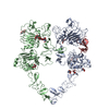
- EMDB-41886: Structure of the HER2/HER4/NRG1b Heterodimer Extracellular Domain -
+ Open data

- Basic information
| Entry |  | ||||||||||||
|---|---|---|---|---|---|---|---|---|---|---|---|---|---|
| Title | Structure of the HER2/HER4/NRG1b Heterodimer Extracellular Domain | ||||||||||||
 Map data Map data | |||||||||||||
 Sample Sample |
| ||||||||||||
 Keywords Keywords | Receptor Tyrosine Kinase / MEMBRANE PROTEIN / TRANSFERASE | ||||||||||||
| Function / homology |  Function and homology information Function and homology informationERBB3 signaling pathway / positive regulation of peptidyl-tyrosine autophosphorylation / establishment of planar polarity involved in nephron morphogenesis / ERBB4 signaling pathway / ERBB4-ERBB4 signaling pathway / olfactory bulb interneuron differentiation / central nervous system morphogenesis / ventricular cardiac muscle cell differentiation / positive regulation of striated muscle cell differentiation / neuregulin receptor activity ...ERBB3 signaling pathway / positive regulation of peptidyl-tyrosine autophosphorylation / establishment of planar polarity involved in nephron morphogenesis / ERBB4 signaling pathway / ERBB4-ERBB4 signaling pathway / olfactory bulb interneuron differentiation / central nervous system morphogenesis / ventricular cardiac muscle cell differentiation / positive regulation of striated muscle cell differentiation / neuregulin receptor activity / cardiac muscle tissue regeneration / negative regulation of secretion / endocardial cell differentiation / negative regulation of neuron migration / negative regulation of immature T cell proliferation in thymus / ERBB3:ERBB2 complex / ERBB2-ERBB4 signaling pathway / immature T cell proliferation in thymus / GRB7 events in ERBB2 signaling / mitochondrial fragmentation involved in apoptotic process / neural crest cell development / ERBB2 signaling pathway / cardiac muscle cell myoblast differentiation / cell communication / RNA polymerase I core binding / semaphorin receptor complex / mammary gland epithelial cell differentiation / transmembrane receptor protein tyrosine kinase activator activity / peripheral nervous system development / Developmental Lineage of Mammary Stem Cells / PI3K events in ERBB4 signaling / embryonic pattern specification / chemorepellent activity / ventricular trabecula myocardium morphogenesis / GABA receptor binding / mammary gland development / ErbB-3 class receptor binding / positive regulation of protein localization to cell surface / cardiac muscle cell differentiation / motor neuron axon guidance / Sema4D induced cell migration and growth-cone collapse / regulation of microtubule-based process / activation of protein kinase B activity / epidermal growth factor receptor activity / positive regulation of tyrosine phosphorylation of STAT protein / ERBB signaling pathway / epidermal growth factor receptor binding / neural crest cell migration / PLCG1 events in ERBB2 signaling / enzyme-linked receptor protein signaling pathway / ERBB2 Activates PTK6 Signaling / regulation of postsynaptic neurotransmitter receptor internalization / ERBB2-EGFR signaling pathway / ERBB2-ERBB3 signaling pathway / Drug-mediated inhibition of ERBB2 signaling / Resistance of ERBB2 KD mutants to trastuzumab / Resistance of ERBB2 KD mutants to sapitinib / Resistance of ERBB2 KD mutants to tesevatinib / Resistance of ERBB2 KD mutants to neratinib / Resistance of ERBB2 KD mutants to osimertinib / Resistance of ERBB2 KD mutants to afatinib / Resistance of ERBB2 KD mutants to AEE788 / Resistance of ERBB2 KD mutants to lapatinib / Drug resistance in ERBB2 TMD/JMD mutants / neurotransmitter receptor localization to postsynaptic specialization membrane / positive regulation of MAP kinase activity / positive regulation of Rho protein signal transduction / neuromuscular junction development / positive regulation of transcription by RNA polymerase I / ERBB2 Regulates Cell Motility / Developmental Lineage of Mammary Gland Myoepithelial Cells / nickel cation binding / oligodendrocyte differentiation / semaphorin-plexin signaling pathway / protein tyrosine kinase activator activity / Signaling by ERBB4 / negative regulation of cardiac muscle cell apoptotic process / mammary gland alveolus development / PI3K events in ERBB2 signaling / Long-term potentiation / regulation of angiogenesis / cell fate commitment / negative regulation of extrinsic apoptotic signaling pathway in absence of ligand / positive regulation of protein targeting to membrane / SHC1 events in ERBB4 signaling / regulation of ERK1 and ERK2 cascade / Nuclear signaling by ERBB4 / Schwann cell development / cell surface receptor signaling pathway via JAK-STAT / positive regulation of cardiac muscle cell proliferation / coreceptor activity / Signaling by ERBB2 / peptidyl-tyrosine phosphorylation / TFAP2 (AP-2) family regulates transcription of growth factors and their receptors / lactation / myelination / transmembrane receptor protein tyrosine kinase activity / synapse assembly / GRB2 events in ERBB2 signaling / positive regulation of cell adhesion Similarity search - Function | ||||||||||||
| Biological species |  Homo sapiens (human) Homo sapiens (human) | ||||||||||||
| Method | single particle reconstruction / cryo EM / Resolution: 3.31 Å | ||||||||||||
 Authors Authors | Trenker R / Diwanji D / Bingham T / Verba KA / Jura N | ||||||||||||
| Funding support |  Germany, Germany,  United States, 3 items United States, 3 items
| ||||||||||||
 Citation Citation | Journal: Elife / Year: 2024 Title: Structural dynamics of the active HER4 and HER2/HER4 complexes is finely tuned by different growth factors and glycosylation. Authors: Raphael Trenker / Devan Diwanji / Tanner Bingham / Kliment A Verba / Natalia Jura / Abstract: Human Epidermal growth factor Receptor 4 (HER4 or ERBB4) carries out essential functions in the development and maintenance of the cardiovascular and nervous systems. HER4 activation is regulated by ...Human Epidermal growth factor Receptor 4 (HER4 or ERBB4) carries out essential functions in the development and maintenance of the cardiovascular and nervous systems. HER4 activation is regulated by a diverse group of extracellular ligands including the neuregulin (NRG) family and betacellulin (BTC), which promote HER4 homodimerization or heterodimerization with other HER receptors. Important cardiovascular functions of HER4 are exerted via heterodimerization with its close homolog and orphan receptor, HER2. To date structural insights into ligand-mediated HER4 activation have been limited to crystallographic studies of HER4 ectodomain homodimers in complex with NRG1β. Here, we report cryo-EM structures of near full-length HER2/HER4 heterodimers and full-length HER4 homodimers bound to NRG1β and BTC. We show that the structures of the heterodimers bound to either ligand are nearly identical and that in both cases the HER2/HER4 heterodimer interface is less dynamic than those observed in structures of HER2/EGFR and HER2/HER3 heterodimers. In contrast, structures of full-length HER4 homodimers bound to NRG1β and BTC display more large-scale dynamics mirroring states previously reported for EGFR homodimers. Our structures also reveal the presence of multiple glycan modifications within HER4 ectodomains, modeled for the first time in HER receptors, that distinctively contribute to the stabilization of HER4 homodimer interfaces over those of HER2/HER4 heterodimers. | ||||||||||||
| History |
|
- Structure visualization
| Supplemental images |
|---|

- Downloads & links
-EMDB archive
| Map data | emd_41886.map.gz | 110.6 MB | EMDB map data format | |
|---|---|---|---|---|
| Header (meta data) | emd-41886-v30.xmlemd-41886.xml | 20 KB 20 KB | Display Display | EMDB header |
| FSC (resolution estimation) | emd_41886_fsc.xml | 13.3 KB | Display | FSC data file |
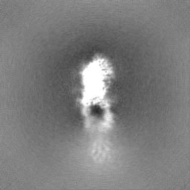
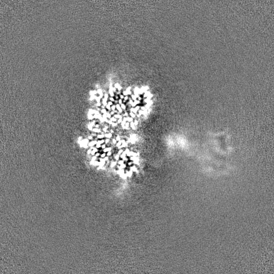
| Images | 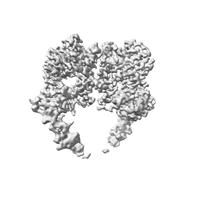 emd_41886.png emd_41886.png | 63.5 KB | ||
| Masks | emd_41886_msk_1.map | 216 MB | Mask map | |
| Filedesc metadata | emd-41886.cif.gz | 7.1 KB | ||
| Others | emd_41886_half_map_1.map.gzemd_41886_half_map_2.map.gz | 200.3 MB 200.3 MB | ||
| Archive directory |  http://ftp.pdbj.org/pub/emdb/structures/EMD-41886ftp://ftp.pdbj.org/pub/emdb/structures/EMD-41886 http://ftp.pdbj.org/pub/emdb/structures/EMD-41886ftp://ftp.pdbj.org/pub/emdb/structures/EMD-41886 | HTTPS FTP |
-Related structure data
| Related structure data | 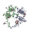 8u4lMC  8u4iC 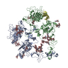 8u4jC  8u4kC C: citing same article ( M: atomic model generated by this map |
|---|---|
| Similar structure data |


-Links
| EMDB pages | EMDB (EBI/PDBe) / EMDataResource |
|---|---|
| Related items in Molecule of the Month |


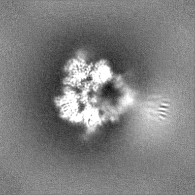
-Map
| File | Download / File: emd_41886.map.gz / Format: CCP4 / Size: 216 MB / Type: IMAGE STORED AS FLOATING POINT NUMBER (4 BYTES) | ||||||||||||||||||||||||||||||||||||
|---|---|---|---|---|---|---|---|---|---|---|---|---|---|---|---|---|---|---|---|---|---|---|---|---|---|---|---|---|---|---|---|---|---|---|---|---|---|
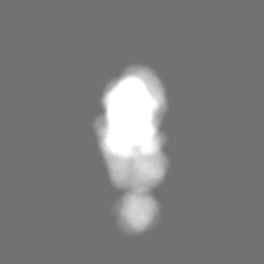
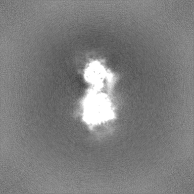
| Projections & slices | Image control
Images are generated by Spider. | ||||||||||||||||||||||||||||||||||||
| Voxel size | X=Y=Z: 0.835 Å | ||||||||||||||||||||||||||||||||||||
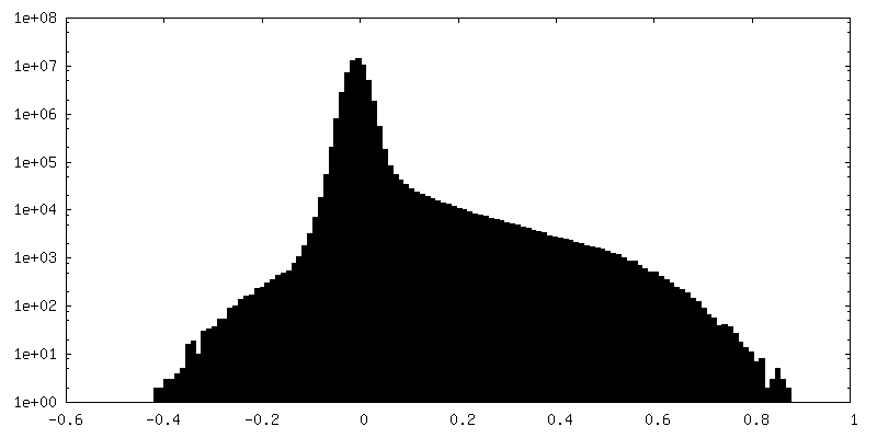
| Density |
| ||||||||||||||||||||||||||||||||||||
| Symmetry | Space group: 1 | ||||||||||||||||||||||||||||||||||||
| Details | EMDB XML:
|
 Z (Sec.)
 Y (Row.)
 X (Col.)


-Supplemental data
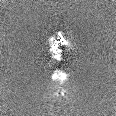
-Mask #1
| File | emd_41886_msk_1.map | ||||||||||||
|---|---|---|---|---|---|---|---|---|---|---|---|---|---|
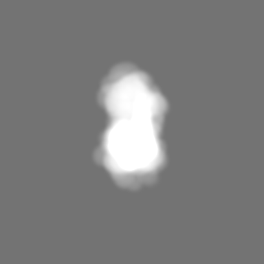
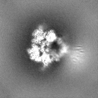
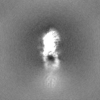
| Projections & Slices |
| ||||||||||||
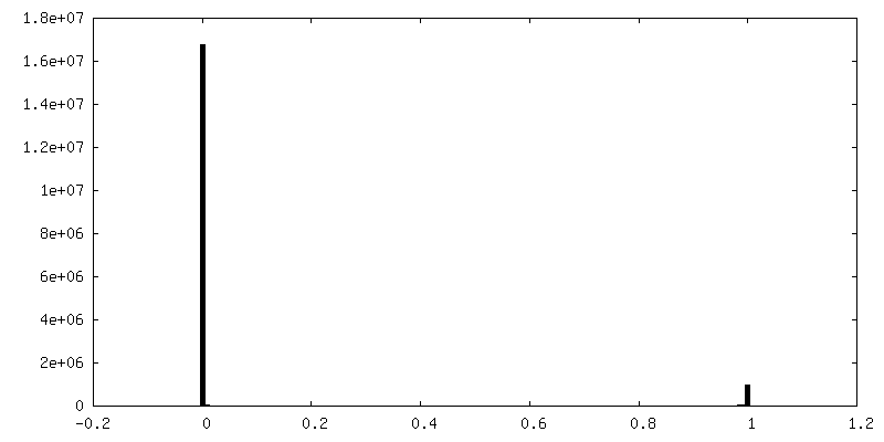
| Density Histograms |


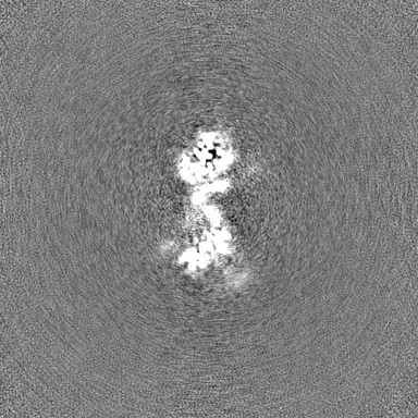
-Half map: #2
| File | emd_41886_half_map_1.map | ||||||||||||
|---|---|---|---|---|---|---|---|---|---|---|---|---|---|
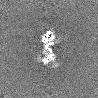
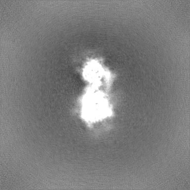
| Projections & Slices |
| ||||||||||||
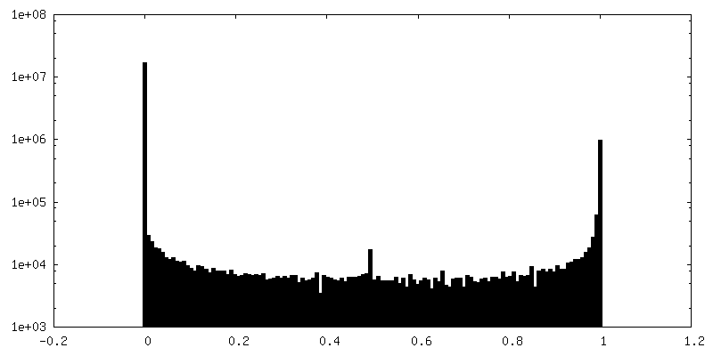
| Density Histograms |


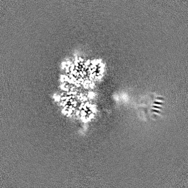
-Half map: #1
| File | emd_41886_half_map_2.map | ||||||||||||
|---|---|---|---|---|---|---|---|---|---|---|---|---|---|
| Projections & Slices |
| ||||||||||||
| Density Histograms |

- Sample components
-Entire : Ternary complex of HER2/HER4/NRG1b
| Entire | Name: Ternary complex of HER2/HER4/NRG1b |
|---|---|
| Components |
|
-Supramolecule #1: Ternary complex of HER2/HER4/NRG1b
| Supramolecule | Name: Ternary complex of HER2/HER4/NRG1b / type: complex / ID: 1 / Parent: 0 / Macromolecule list: #1-#3 |
|---|---|
| Source (natural) | Organism: Homo sapiens (human) |
-Macromolecule #1: Isoform JM-A CYT-1 of Receptor tyrosine-protein kinase erbB-4
| Macromolecule | Name: Isoform JM-A CYT-1 of Receptor tyrosine-protein kinase erbB-4 type: protein_or_peptide / ID: 1 / Number of copies: 1 / Enantiomer: LEVO / EC number: receptor protein-tyrosine kinase |
|---|---|
| Source (natural) | Organism: Homo sapiens (human) |
| Molecular weight | Theoretical: 67.909648 KDa |
| Recombinant expression | Organism: Homo sapiens (human) |
| Sequence | String: QSVCAGTENK LSSLSDLEQQ YRALRKYYEN CEVVMGNLEI TSIEHNRDLS FLRSVREVTG YVLVALNQFR YLPLENLRII RGTKLYEDR YALAIFLNYR KDGNFGLQEL GLKNLTEILN GGVYVDQNKF LCYADTIHWQ DIVRNPWPSN LTLVSTNGSS G CGRCHKSC ...String: QSVCAGTENK LSSLSDLEQQ YRALRKYYEN CEVVMGNLEI TSIEHNRDLS FLRSVREVTG YVLVALNQFR YLPLENLRII RGTKLYEDR YALAIFLNYR KDGNFGLQEL GLKNLTEILN GGVYVDQNKF LCYADTIHWQ DIVRNPWPSN LTLVSTNGSS G CGRCHKSC TGRCWGPTEN HCQTLTRTVC AEQCDGRCYG PYVSDCCHRE CAGGCSGPKD TDCFACMNFN DSGACVTQCP QT FVYNPTT FQLEHNFNAK YTYGAFCVKK CPHNFVVDSS SCVRACPSSK MEVEENGIKM CKPCTDICPK ACDGIGTGSL MSA QTVDSS NIDKFINCTK INGNLIFLVT GIHGDPYNAI EAIDPEKLNV FRTVREITGF LNIQSWPPNM TDFSVFSNLV TIGG RVLYS GLSLLILKQQ GITSLQFQSL KEISAGNIYI TDNSNLCYYH TINWTTLFST INQRIVIRDN RKAENCTAEG MVCNH LCSS DGCWGPGPDQ CLSCRRFSRG RICIESCNLY DGEFREFENG SICVECDPQC EKMEDGLLTC HGPGPDNCTK CSHFKD GPN CVEKCPDGLQ GANSFIFKYA DPDRECHPCH PNCTQGCNGP TSHDCI UniProtKB: Receptor tyrosine-protein kinase erbB-4 |
-Macromolecule #2: Receptor tyrosine-protein kinase erbB-2
| Macromolecule | Name: Receptor tyrosine-protein kinase erbB-2 / type: protein_or_peptide / ID: 2 / Number of copies: 1 / Enantiomer: LEVO / EC number: receptor protein-tyrosine kinase |
|---|---|
| Source (natural) | Organism: Homo sapiens (human) |
| Molecular weight | Theoretical: 66.888008 KDa |
| Recombinant expression | Organism: Homo sapiens (human) |
| Sequence | String: QVCTGTDMKL RLPASPETHL DMLRHLYQGC QVVQGNLELT YLPTNASLSF LQDIQEVQGY VLIAHNQVRQ VPLQRLRIVR GTQLFEDNY ALAVLDNGDP LNNTTPVTGA SPGGLRELQL RSLTEILKGG VLIQRNPQLC YQDTILWKDI FHKNNQLALT L IDTNRSRA ...String: QVCTGTDMKL RLPASPETHL DMLRHLYQGC QVVQGNLELT YLPTNASLSF LQDIQEVQGY VLIAHNQVRQ VPLQRLRIVR GTQLFEDNY ALAVLDNGDP LNNTTPVTGA SPGGLRELQL RSLTEILKGG VLIQRNPQLC YQDTILWKDI FHKNNQLALT L IDTNRSRA CHPCSPMCKG SRCWGESSED CQSLTRTVCA GGCARCKGPL PTDCCHEQCA AGCTGPKHSD CLACLHFNHS GI CELHCPA LVTYNTDTFE SMPNPEGRYT FGASCVTACP YNYLSTDVGS CTLVCPLHNQ EVTAEDGTQR CEKCSKPCAR VCY GLGMEH LREVRAVTSA NIQEFAGCKK IFGSLAFLPE SFDGDPASNT APLQPEQLQV FETLEEITGY LYISAWPDSL PDLS VFQNL QVIRGRILHN GAYSLTLQGL GISWLGLRSL RELGSGLALI HHNTHLCFVH TVPWDQLFRN PHQALLHTAN RPEDE CVGE GLACHQLCAR GHCWGPGPTQ CVNCSQFLRG QECVEECRVL QGLPREYVNA RHCLPCHPEC QPQNGSVTCF GPEADQ CVA CAHYKDPPFC VARCPSGVKP DLSYMPIWKF PDEEGACQPC PIN UniProtKB: Receptor tyrosine-protein kinase erbB-2 |
-Macromolecule #3: Isoform 6 of Pro-neuregulin-1, membrane-bound isoform
| Macromolecule | Name: Isoform 6 of Pro-neuregulin-1, membrane-bound isoform / type: protein_or_peptide / ID: 3 / Number of copies: 1 / Enantiomer: LEVO |
|---|---|
| Source (natural) | Organism: Homo sapiens (human) |
| Molecular weight | Theoretical: 5.890792 KDa |
| Recombinant expression | Organism:  |
| Sequence | String: SHLVKCAEKE KTFCVNGGEC FMVKDLSNPS RYLCKCPNEF TGDRCQNYVM AS UniProtKB: Pro-neuregulin-1, membrane-bound isoform |
-Macromolecule #9: 2-acetamido-2-deoxy-beta-D-glucopyranose
| Macromolecule | Name: 2-acetamido-2-deoxy-beta-D-glucopyranose / type: ligand / ID: 9 / Number of copies: 3 / Formula: NAG |
|---|---|
| Molecular weight | Theoretical: 221.208 Da |
| Chemical component information |  ChemComp-NAG: |
-Experimental details
-Structure determination
| Method | cryo EM |
|---|---|
 Processing Processing | single particle reconstruction |
| Aggregation state | particle |
-Sample preparation
| Buffer | pH: 7.4 Component:
| ||||||||||||
|---|---|---|---|---|---|---|---|---|---|---|---|---|---|
| Grid | Model: Quantifoil R1.2/1.3 / Support film - Material: GRAPHENE OXIDE | ||||||||||||
| Vitrification | Cryogen name: ETHANE / Chamber humidity: 100 % / Chamber temperature: 293 K / Instrument: FEI VITROBOT MARK IV |
- Electron microscopy
| Microscope | TFS KRIOS |
|---|---|
| Image recording | Film or detector model: GATAN K3 (6k x 4k) / Number grids imaged: 1 / Average electron dose: 45.8 e/Å2 |
| Electron beam | Acceleration voltage: 300 kV / Electron source:  FIELD EMISSION GUN FIELD EMISSION GUN |
| Electron optics | Illumination mode: FLOOD BEAM / Imaging mode: BRIGHT FIELD / Nominal defocus max: 2.0 µm / Nominal defocus min: 0.9 µm |
| Experimental equipment |  Model: Titan Krios / Image courtesy: FEI Company |