 Movie
Movie Controller


+ Open data

- Basic information
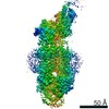
| Entry | Database: PDB / ID: 7tby | ||||||||||||||||||||||||||||||||||||||||||||||||||||||||||||||||||||||||
|---|---|---|---|---|---|---|---|---|---|---|---|---|---|---|---|---|---|---|---|---|---|---|---|---|---|---|---|---|---|---|---|---|---|---|---|---|---|---|---|---|---|---|---|---|---|---|---|---|---|---|---|---|---|---|---|---|---|---|---|---|---|---|---|---|---|---|---|---|---|---|---|---|---|
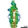
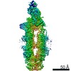
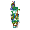
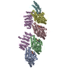
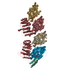
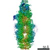
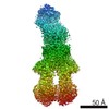
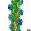
| Title | The structure of human ABCA1 in nanodisc | ||||||||||||||||||||||||||||||||||||||||||||||||||||||||||||||||||||||||
 Components Components | ATP-binding cassette, sub-family A (ABC1), member 1 | ||||||||||||||||||||||||||||||||||||||||||||||||||||||||||||||||||||||||
 Keywords Keywords | TRANSPORT PROTEIN / HDL / cholesterol / Tangier Disease / ABC transporter | ||||||||||||||||||||||||||||||||||||||||||||||||||||||||||||||||||||||||
| Function / homology |  Function and homology information Function and homology informationorganic hydroxy compound transport / : / ATPase-coupled intramembrane lipid carrier activity / P-type phospholipid transporter / phospholipid translocation / ABC-type transporter activity / ATP hydrolysis activity / ATP binding / membrane Similarity search - Function | ||||||||||||||||||||||||||||||||||||||||||||||||||||||||||||||||||||||||
| Biological species |  Homo sapiens (human) Homo sapiens (human) | ||||||||||||||||||||||||||||||||||||||||||||||||||||||||||||||||||||||||
| Method | ELECTRON MICROSCOPY / single particle reconstruction / cryo EM / Resolution: 4 Å | ||||||||||||||||||||||||||||||||||||||||||||||||||||||||||||||||||||||||
 Authors Authors | Sun, Y. / Li, X. | ||||||||||||||||||||||||||||||||||||||||||||||||||||||||||||||||||||||||
| Funding support |  United States, 2items United States, 2items
| ||||||||||||||||||||||||||||||||||||||||||||||||||||||||||||||||||||||||
 Citation Citation | Journal: Nat Cardiovasc Res / Year: 2022 Title: Cholesterol efflux mechanism revealed by structural analysis of human ABCA1 conformational states. Authors: Yingyuan Sun / Xiaochun Li / Abstract: ATP-binding cassette transporter A1 (ABCA1) utilizes energy derived from ATP hydrolysis to export cholesterol and phospholipids from macrophages. ABCA1 plays a central role in the biosynthesis of ...ATP-binding cassette transporter A1 (ABCA1) utilizes energy derived from ATP hydrolysis to export cholesterol and phospholipids from macrophages. ABCA1 plays a central role in the biosynthesis of high-density lipoprotein (HDL), which mediates reverse cholesterol transport and prevents detrimental lipid deposition. Mutations in ABCA1 cause Tangier disease characterized by a remarkable reduction in the amount of HDL in blood. Here we present cryo-electron microscopy structures of human ABCA1 in ATP-bound and nucleotide-free states. Structural comparison reveals that ATP molecules pull the nucleotide-binding domains together, inducing movements of transmembrane helices 1, 2, 7 and 8 through a series of salt-bridge interactions. Subsequently, extracellular domains (ECDs) undergo a rotation and introduce conformational changes in the ECD-transmembrane interface. In addition, while we observe a sterol-like molecule in ECDs, no such density was observed in the structure of an HDL-deficiency mutant ABCA1, demonstrating the physiological importance of ECDs and a putative interaction mode between ABCA1 and its lipid acceptors. Thus, these structures, along with cholesterol efflux assays, advance the understanding ABCA1-mediated reverse cholesterol transport. | ||||||||||||||||||||||||||||||||||||||||||||||||||||||||||||||||||||||||
| History |
|
- Structure visualization
| Movie |
Movie viewer |
|---|---|
| Structure viewer | Molecule: MolmilJmol/JSmol |
- Downloads & links
-Download
| PDBx/mmCIF format | 7tby.cif.gz | 326.9 KB | Display | PDBx/mmCIF format |
|---|---|---|---|---|
| PDB format | pdb7tby.ent.gz | 248.3 KB | Display | PDB format |
| PDBx/mmJSON format | 7tby.json.gz | Tree view | PDBx/mmJSON format | |
| Others |  Other downloads Other downloads |
-Validation report
| Arichive directory | https://data.pdbj.org/pub/pdb/validation_reports/tb/7tbyftp://data.pdbj.org/pub/pdb/validation_reports/tb/7tby | HTTPS FTP |
|---|
-Related structure data
| Related structure data | 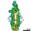 25801MC 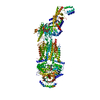 7tbwC  7tbzC 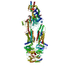 7tc0C M: map data used to model this data C: citing same article ( |
|---|---|
| Similar structure data |

-Links
 PDBj
PDBj


- Assembly
| Deposited unit | 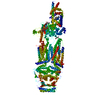
|
|---|---|
| 1 |
|
-Components
| #1: Protein | Mass: 255654.984 Da / Num. of mol.: 1 Source method: isolated from a genetically manipulated source Source: (gene. exp.) Homo sapiens (human) / Gene: ABCA1 / Production host: Homo sapiens (human) / References: UniProt: B2RUU2 | ||||
|---|---|---|---|---|---|
| #2: Polysaccharide | 2-acetamido-2-deoxy-beta-D-glucopyranose-(1-4)-2-acetamido-2-deoxy-beta-D-glucopyranose Source method: isolated from a genetically manipulated source | ||||
| #3: Polysaccharide | beta-D-mannopyranose-(1-3)-[beta-D-mannopyranose-(1-6)]beta-D-mannopyranose-(1-4)-2-acetamido-2- ...beta-D-mannopyranose-(1-3)-[beta-D-mannopyranose-(1-6)]beta-D-mannopyranose-(1-4)-2-acetamido-2-deoxy-beta-D-glucopyranose-(1-4)-2-acetamido-2-deoxy-beta-D-glucopyranose Source method: isolated from a genetically manipulated source | ||||
| #4: Chemical | ChemComp-CLR /   Mass: 386.654 Da / Num. of mol.: 1 / Source method: obtained synthetically / Formula: C27H46O / Feature type: SUBJECT OF INVESTIGATION Mass: 386.654 Da / Num. of mol.: 1 / Source method: obtained synthetically / Formula: C27H46O / Feature type: SUBJECT OF INVESTIGATION | ||||
| #5: Sugar |   Type: D-saccharide, beta linking / Mass: 221.208 Da / Num. of mol.: 2 / Source method: obtained synthetically / Formula: C8H15NO6 Type: D-saccharide, beta linking / Mass: 221.208 Da / Num. of mol.: 2 / Source method: obtained synthetically / Formula: C8H15NO6Has ligand of interest | Y | Has protein modification | Y | |
-Experimental details
-Experiment
| Experiment | Method: ELECTRON MICROSCOPY |
|---|---|
| EM experiment | Aggregation state: PARTICLE / 3D reconstruction method: single particle reconstruction |
- Sample preparation
| Component | Name: human ABCA1 in nanodiscs / Type: COMPLEX / Entity ID: #1 / Source: RECOMBINANT |
|---|---|
| Molecular weight | Value: 0.25 MDa / Experimental value: YES |
| Source (natural) | Organism: Homo sapiens (human) |
| Source (recombinant) | Organism: Homo sapiens (human) |
| Buffer solution | pH: 7.5 |
| Specimen | Embedding applied: NO / Shadowing applied: NO / Staining applied: NO / Vitrification applied: YES |
| Vitrification | Cryogen name: ETHANE / Humidity: 100 % / Chamber temperature: 293 K |
- Electron microscopy imaging
| Experimental equipment |  Model: Titan Krios / Image courtesy: FEI Company |
|---|---|
| Microscopy | Model: FEI TITAN KRIOS |
| Electron gun | Electron source:  FIELD EMISSION GUN / Accelerating voltage: 300 kV / Illumination mode: FLOOD BEAM FIELD EMISSION GUN / Accelerating voltage: 300 kV / Illumination mode: FLOOD BEAM |
| Electron lens | Mode: BRIGHT FIELD / Nominal defocus max: 2000 nm / Nominal defocus min: 1000 nm / Cs: 2.7 mm / C2 aperture diameter: 70 µm |
| Image recording | Electron dose: 60 e/Å2 / Film or detector model: GATAN K3 (6k x 4k) |
- Processing
| Software | Name: PHENIX / Version: 1.17.1_3660: / Classification: refinement | ||||||||||||||||||||||||
|---|---|---|---|---|---|---|---|---|---|---|---|---|---|---|---|---|---|---|---|---|---|---|---|---|---|
| EM software | Name: PHENIX / Category: model refinement | ||||||||||||||||||||||||
| CTF correction | Type: PHASE FLIPPING AND AMPLITUDE CORRECTION | ||||||||||||||||||||||||
| 3D reconstruction | Resolution: 4 Å / Resolution method: FSC 0.143 CUT-OFF / Num. of particles: 82350 / Symmetry type: POINT | ||||||||||||||||||||||||
| Refine LS restraints |
|
