Movie
Movie Controller

[English] 日本語
 Yorodumi
- PDB-7sht: Structure of a partially disrupted IgE high affinity receptor com... -
+ Open data

- Basic information
| Entry | Database: PDB / ID: 7sht | |||||||||
|---|---|---|---|---|---|---|---|---|---|---|
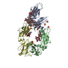
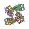
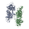
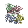
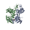
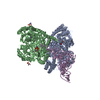
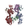
| Title | Structure of a partially disrupted IgE high affinity receptor complex bound to an omalizumab variant | |||||||||
 Components Components |
| |||||||||
 Keywords Keywords | IMMUNE SYSTEM / IgE / allergy / Xolair / Omalizumab | |||||||||
| Function / homology |  Function and homology information Function and homology informationhigh-affinity IgE receptor activity / mononuclear cell differentiation / IgE B cell receptor complex / adaptive immune memory response / primary adaptive immune response / B cell antigen processing and presentation / type I hypersensitivity / Fc receptor-mediated immune complex endocytosis / eosinophil degranulation / IgE immunoglobulin complex ...high-affinity IgE receptor activity / mononuclear cell differentiation / IgE B cell receptor complex / adaptive immune memory response / primary adaptive immune response / B cell antigen processing and presentation / type I hypersensitivity / Fc receptor-mediated immune complex endocytosis / eosinophil degranulation / IgE immunoglobulin complex / macrophage activation / IgE binding / antibody-dependent cellular cytotoxicity / Fc epsilon receptor (FCERI) signaling / type 2 immune response / immunoglobulin complex, circulating / immunoglobulin receptor binding / mast cell degranulation / immunoglobulin mediated immune response / B cell proliferation / macrophage differentiation / B cell activation / Role of LAT2/NTAL/LAB on calcium mobilization / complement activation, classical pathway / antigen binding / FCERI mediated Ca+2 mobilization / B cell receptor signaling pathway / FCERI mediated MAPK activation / FCERI mediated NF-kB activation / antibacterial humoral response / Interleukin-4 and Interleukin-13 signaling / adaptive immune response / cell surface receptor signaling pathway / immune response / inflammatory response / external side of plasma membrane / cell surface / : / extracellular region / plasma membrane Similarity search - Function | |||||||||
| Biological species |  Homo sapiens (human) Homo sapiens (human) | |||||||||
| Method | ELECTRON MICROSCOPY / single particle reconstruction / cryo EM / Resolution: 7.29 Å | |||||||||
 Authors Authors | Pennington, L.F. / Jardetzky, T.S. | |||||||||
| Funding support |  United States, 2items United States, 2items
| |||||||||
 Citation Citation | Journal: Nat Commun / Year: 2021 Title: Directed evolution of and structural insights into antibody-mediated disruption of a stable receptor-ligand complex. Authors: Luke F Pennington / Pascal Gasser / Silke Kleinboelting / Chensong Zhang / Georgios Skiniotis / Alexander Eggel / Theodore S Jardetzky /  Abstract: Antibody drugs exert therapeutic effects via a range of mechanisms, including competitive inhibition, allosteric modulation, and immune effector mechanisms. Facilitated dissociation is an additional ...Antibody drugs exert therapeutic effects via a range of mechanisms, including competitive inhibition, allosteric modulation, and immune effector mechanisms. Facilitated dissociation is an additional mechanism where antibody-mediated "disruption" of stable high-affinity macromolecular complexes can potentially enhance therapeutic efficacy. However, this mechanism is not well understood or utilized therapeutically. Here, we investigate and engineer the weak disruptive activity of an existing therapeutic antibody, omalizumab, which targets IgE antibodies to block the allergic response. We develop a yeast display approach to select for and engineer antibody disruptive efficiency and generate potent omalizumab variants that dissociate receptor-bound IgE. We determine a low resolution cryo-EM structure of a transient disruption intermediate containing the IgE-Fc, its partially dissociated receptor and an antibody inhibitor. Our results provide a conceptual framework for engineering disruptive inhibitors for other targets, insights into the failure in clinical trials of the previous high affinity omalizumab HAE variant and anti-IgE antibodies that safely and rapidly disarm allergic effector cells. | |||||||||
| History |
|
- Structure visualization
| Movie |
Movie viewer |
|---|---|
| Structure viewer | Molecule: MolmilJmol/JSmol |
- Downloads & links
-Download
| PDBx/mmCIF format | 7sht.cif.gz | 269.5 KB | Display | PDBx/mmCIF format |
|---|---|---|---|---|
| PDB format | pdb7sht.ent.gz | 208.9 KB | Display | PDB format |
| PDBx/mmJSON format | 7sht.json.gz | Tree view | PDBx/mmJSON format | |
| Others |  Other downloads Other downloads |
-Validation report
| Arichive directory | https://data.pdbj.org/pub/pdb/validation_reports/sh/7shtftp://data.pdbj.org/pub/pdb/validation_reports/sh/7sht | HTTPS FTP |
|---|
-Related structure data
| Related structure data |  25136MC  7shuC  7shyC  7shzC  7si0C M: map data used to model this data C: citing same article ( |
|---|---|
| Similar structure data |

-Links
 PDBj
PDBj


- Assembly
| Deposited unit | 
|
|---|---|
| 1 |
|
-Components
-Protein , 2 types, 3 molecules ABD
| #1: Protein | Mass: 22541.799 Da / Num. of mol.: 1 / Fragment: extracellular portion / Mutation: W156C Source method: isolated from a genetically manipulated source Source: (gene. exp.) Homo sapiens (human) / Gene: FCER1A, FCE1A / Production host: Homo sapiens (human) / References: UniProt: P12319 |
|---|---|
| #2: Protein | Mass: 35773.160 Da / Num. of mol.: 2 / Mutation: G335C Source method: isolated from a genetically manipulated source Source: (gene. exp.) Homo sapiens (human) / Gene: IGHE / Production host: Homo sapiens (human) / References: UniProt: P01854 |
-Antibody , 2 types, 4 molecules HJKL
| #3: Antibody | Mass: 13451.814 Da / Num. of mol.: 2 Source method: isolated from a genetically manipulated source Source: (gene. exp.) Homo sapiens (human) / Production host: Homo sapiens (human)#4: Antibody | Mass: 14641.863 Da / Num. of mol.: 2 Source method: isolated from a genetically manipulated source Source: (gene. exp.) Homo sapiens (human) / Production host: Homo sapiens (human) |
|---|
-Sugars , 4 types, 7 molecules 
| #5: Polysaccharide | 2-acetamido-2-deoxy-beta-D-glucopyranose-(1-4)-2-acetamido-2-deoxy-beta-D-glucopyranose Source method: isolated from a genetically manipulated source | ||
|---|---|---|---|
| #6: Polysaccharide | beta-D-mannopyranose-(1-4)-2-acetamido-2-deoxy-beta-D-glucopyranose-(1-4)-2-acetamido-2-deoxy-beta- ...beta-D-mannopyranose-(1-4)-2-acetamido-2-deoxy-beta-D-glucopyranose-(1-4)-2-acetamido-2-deoxy-beta-D-glucopyranose Source method: isolated from a genetically manipulated source | ||
| #7: Polysaccharide | Source method: isolated from a genetically manipulated source #8: Sugar |  Type: D-saccharide, beta linking / Mass: 221.208 Da / Num. of mol.: 3 Type: D-saccharide, beta linking / Mass: 221.208 Da / Num. of mol.: 3Source method: isolated from a genetically manipulated source Formula: C8H15NO6 |
-Details
| Has ligand of interest | N |
|---|---|
| Has protein modification | Y |
-Experimental details
-Experiment
| Experiment | Method: ELECTRON MICROSCOPY |
|---|---|
| EM experiment | Aggregation state: PARTICLE / 3D reconstruction method: single particle reconstruction |
- Sample preparation
| Component | Name: Locked complex of IgE-Fc (G335C) and alpha chain of the high affinity IgE receptor (W156C) bound to clone_7 scFV Type: COMPLEX / Entity ID: #1-#4 / Source: RECOMBINANT |
|---|---|
| Molecular weight | Experimental value: NO |
| Source (natural) | Organism: Homo sapiens (human) |
| Source (recombinant) | Organism: Homo sapiens (human) |
| Buffer solution | pH: 7.5 |
| Specimen | Conc.: 4.5 mg/ml / Embedding applied: NO / Shadowing applied: NO / Staining applied: NO / Vitrification applied: YES |
| Specimen support | Grid material: GOLD / Grid mesh size: 200 divisions/in. / Grid type: Quantifoil R1.2/1.3 |
| Vitrification | Cryogen name: ETHANE |
- Electron microscopy imaging
| Experimental equipment |  Model: Titan Krios / Image courtesy: FEI Company |
|---|---|
| Microscopy | Model: TFS KRIOS |
| Electron gun | Electron source:  FIELD EMISSION GUN / Accelerating voltage: 300 kV / Illumination mode: SPOT SCAN FIELD EMISSION GUN / Accelerating voltage: 300 kV / Illumination mode: SPOT SCAN |
| Electron lens | Mode: BRIGHT FIELD |
| Image recording | Average exposure time: 2 sec. / Electron dose: 44 e/Å2 / Film or detector model: GATAN K3 (6k x 4k) / Num. of grids imaged: 1 / Num. of real images: 523 |
- Processing
| Software |
| ||||||||||||||||||||||||
|---|---|---|---|---|---|---|---|---|---|---|---|---|---|---|---|---|---|---|---|---|---|---|---|---|---|
| EM software |
| ||||||||||||||||||||||||
| CTF correction | Type: PHASE FLIPPING AND AMPLITUDE CORRECTION | ||||||||||||||||||||||||
| 3D reconstruction | Resolution: 7.29 Å / Resolution method: FSC 0.143 CUT-OFF / Num. of particles: 24396 / Symmetry type: POINT | ||||||||||||||||||||||||
| Atomic model building | Protocol: FLEXIBLE FIT Details: Initial local fitting done in Chimera, followed by auto dock, and a single round of real space refinement. Carbohydrates were automatically built in phenix when possible, and a handful of ...Details: Initial local fitting done in Chimera, followed by auto dock, and a single round of real space refinement. Carbohydrates were automatically built in phenix when possible, and a handful of flagged outliers in geometry were fixed manually in Phenix. | ||||||||||||||||||||||||
| Atomic model building | 3D fitting-ID: 1 / Source name: PDB / Type: experimental model
| ||||||||||||||||||||||||
| Refinement | Cross valid method: THROUGHOUT | ||||||||||||||||||||||||
| Displacement parameters | Biso max: 756.63 Å2 / Biso mean: 344.0879 Å2 / Biso min: 20 Å2 |