 Movie
Movie Controller


+ Open data

- Basic information
| Entry | Database: PDB / ID: 7shy | ||||||
|---|---|---|---|---|---|---|---|
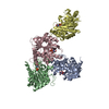
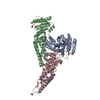
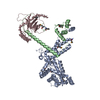
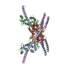
| Title | IgE-Fc in complex with omalizumab scFv | ||||||
 Components Components |
| ||||||
 Keywords Keywords | IMMUNE SYSTEM / IgE / omalizumab / xolair / inhibitor | ||||||
| Function / homology |  Function and homology information Function and homology informationmononuclear cell differentiation / IgE B cell receptor complex / adaptive immune memory response / primary adaptive immune response / B cell antigen processing and presentation / type I hypersensitivity / Fc receptor-mediated immune complex endocytosis / eosinophil degranulation / IgE immunoglobulin complex / macrophage activation ...mononuclear cell differentiation / IgE B cell receptor complex / adaptive immune memory response / primary adaptive immune response / B cell antigen processing and presentation / type I hypersensitivity / Fc receptor-mediated immune complex endocytosis / eosinophil degranulation / IgE immunoglobulin complex / macrophage activation / antibody-dependent cellular cytotoxicity / Fc epsilon receptor (FCERI) signaling / type 2 immune response / immunoglobulin complex, circulating / immunoglobulin receptor binding / mast cell degranulation / B cell proliferation / macrophage differentiation / B cell activation / Role of LAT2/NTAL/LAB on calcium mobilization / complement activation, classical pathway / antigen binding / FCERI mediated Ca+2 mobilization / B cell receptor signaling pathway / FCERI mediated MAPK activation / FCERI mediated NF-kB activation / antibacterial humoral response / Interleukin-4 and Interleukin-13 signaling / adaptive immune response / immune response / inflammatory response / : / extracellular region / plasma membrane Similarity search - Function | ||||||
| Biological species |  Homo sapiens (human) Homo sapiens (human) | ||||||
| Method |  X-RAY DIFFRACTION / SYNCHROTRON / MOLECULAR REPLACEMENT / Resolution: 3 Å X-RAY DIFFRACTION / SYNCHROTRON / MOLECULAR REPLACEMENT / Resolution: 3 Å | ||||||
 Authors Authors | Pennington, L.F. / Jardetzky, T.J. / Kleinboelting, S. | ||||||
| Funding support |  United States, 1items United States, 1items
| ||||||
 Citation Citation | Journal: Nat Commun / Year: 2021 Title: Directed evolution of and structural insights into antibody-mediated disruption of a stable receptor-ligand complex. Authors: Luke F Pennington / Pascal Gasser / Silke Kleinboelting / Chensong Zhang / Georgios Skiniotis / Alexander Eggel / Theodore S Jardetzky /  Abstract: Antibody drugs exert therapeutic effects via a range of mechanisms, including competitive inhibition, allosteric modulation, and immune effector mechanisms. Facilitated dissociation is an additional ...Antibody drugs exert therapeutic effects via a range of mechanisms, including competitive inhibition, allosteric modulation, and immune effector mechanisms. Facilitated dissociation is an additional mechanism where antibody-mediated "disruption" of stable high-affinity macromolecular complexes can potentially enhance therapeutic efficacy. However, this mechanism is not well understood or utilized therapeutically. Here, we investigate and engineer the weak disruptive activity of an existing therapeutic antibody, omalizumab, which targets IgE antibodies to block the allergic response. We develop a yeast display approach to select for and engineer antibody disruptive efficiency and generate potent omalizumab variants that dissociate receptor-bound IgE. We determine a low resolution cryo-EM structure of a transient disruption intermediate containing the IgE-Fc, its partially dissociated receptor and an antibody inhibitor. Our results provide a conceptual framework for engineering disruptive inhibitors for other targets, insights into the failure in clinical trials of the previous high affinity omalizumab HAE variant and anti-IgE antibodies that safely and rapidly disarm allergic effector cells. | ||||||
| History |
|
- Structure visualization
| Structure viewer | Molecule: MolmilJmol/JSmol |
|---|
- Downloads & links
-Download
| PDBx/mmCIF format | 7shy.cif.gz | 724.2 KB | Display | PDBx/mmCIF format |
|---|---|---|---|---|
| PDB format | pdb7shy.ent.gz | 603.2 KB | Display | PDB format |
| PDBx/mmJSON format | 7shy.json.gz | Tree view | PDBx/mmJSON format | |
| Others |  Other downloads Other downloads |
-Validation report
| Arichive directory | https://data.pdbj.org/pub/pdb/validation_reports/sh/7shyftp://data.pdbj.org/pub/pdb/validation_reports/sh/7shy | HTTPS FTP |
|---|
-Related structure data
| Related structure data | 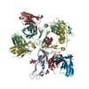 7shtC  7shuC  7shzC  7si0C  5hysS C: citing same article ( S: Starting model for refinement |
|---|---|
| Similar structure data |


-Links
 PDBj
PDBj


- Assembly
| Deposited unit | 
| ||||||||
|---|---|---|---|---|---|---|---|---|---|
| 1 | 
| ||||||||
| 2 | 
| ||||||||
| Unit cell |
|
-Components
-Protein , 1 types, 4 molecules AGHB
| #3: Protein | Mass: 27550.873 Da / Num. of mol.: 4 / Fragment: C3-4 Source method: isolated from a genetically manipulated source Source: (gene. exp.) Homo sapiens (human) / Gene: IGHE / Production host: Homo sapiens (human) / References: UniProt: P01854 |
|---|
-Antibody , 2 types, 8 molecules IKCEJLDF
| #1: Antibody | Mass: 13362.744 Da / Num. of mol.: 4 Source method: isolated from a genetically manipulated source Source: (gene. exp.) Homo sapiens (human) / Production host: Homo sapiens (human)#2: Antibody | Mass: 14665.836 Da / Num. of mol.: 4 Source method: isolated from a genetically manipulated source Source: (gene. exp.) Homo sapiens (human) / Production host: Homo sapiens (human) |
|---|
-Sugars , 6 types, 8 molecules 

| #4: Polysaccharide | alpha-D-mannopyranose-(1-3)-alpha-D-mannopyranose-(2-3)-[alpha-L-fucopyranose-(1-6)]beta-D- ...alpha-D-mannopyranose-(1-3)-alpha-D-mannopyranose-(2-3)-[alpha-L-fucopyranose-(1-6)]beta-D-mannopyranose-(1-4)-2-acetamido-2-deoxy-beta-D-glucopyranose-(1-4)-2-acetamido-2-deoxy-beta-D-glucopyranose Type: oligosaccharide / Mass: 1056.964 Da / Num. of mol.: 1 Source method: isolated from a genetically manipulated source | ||
|---|---|---|---|
| #5: Polysaccharide | beta-D-mannopyranose-(1-4)-alpha-D-mannopyranose-(1-6)-[alpha-D-mannopyranose-(1-3)]beta-D- ...beta-D-mannopyranose-(1-4)-alpha-D-mannopyranose-(1-6)-[alpha-D-mannopyranose-(1-3)]beta-D-mannopyranose-(1-4)-2-acetamido-2-deoxy-beta-D-glucopyranose-(1-4)-2-acetamido-2-deoxy-beta-D-glucopyranose Source method: isolated from a genetically manipulated source | ||
| #6: Polysaccharide | alpha-D-mannopyranose-(1-6)-beta-D-mannopyranose-(1-4)-2-acetamido-2-deoxy-beta-D-glucopyranose-(1- ...alpha-D-mannopyranose-(1-6)-beta-D-mannopyranose-(1-4)-2-acetamido-2-deoxy-beta-D-glucopyranose-(1-4)-2-acetamido-2-deoxy-beta-D-glucopyranose Source method: isolated from a genetically manipulated source | ||
| #7: Polysaccharide | alpha-D-mannopyranose-(1-3)-[alpha-D-mannopyranose-(1-6)]alpha-D-mannopyranose-(1-6)-[alpha-D- ...alpha-D-mannopyranose-(1-3)-[alpha-D-mannopyranose-(1-6)]alpha-D-mannopyranose-(1-6)-[alpha-D-mannopyranose-(1-3)]beta-D-mannopyranose-(1-4)-2-acetamido-2-deoxy-beta-D-glucopyranose-(1-4)-2-acetamido-2-deoxy-beta-D-glucopyranose Source method: isolated from a genetically manipulated source | ||
| #9: Sugar |  Type: D-saccharide, beta linking / Mass: 221.208 Da / Num. of mol.: 3 Type: D-saccharide, beta linking / Mass: 221.208 Da / Num. of mol.: 3Source method: isolated from a genetically manipulated source Formula: C8H15NO6 #10: Sugar | ChemComp-MAN / |  Type: D-saccharide, alpha linking / Mass: 180.156 Da / Num. of mol.: 1 Type: D-saccharide, alpha linking / Mass: 180.156 Da / Num. of mol.: 1Source method: isolated from a genetically manipulated source Formula: C6H12O6 |
-Non-polymers , 2 types, 7 molecules 

| #8: Chemical | ChemComp-GOL /  Mass: 92.094 Da / Num. of mol.: 1 / Source method: obtained synthetically / Formula: C3H8O3 Mass: 92.094 Da / Num. of mol.: 1 / Source method: obtained synthetically / Formula: C3H8O3 |
|---|---|
| #11: Water | ChemComp-HOH / Mass: 18.015 Da / Num. of mol.: 6 / Source method: isolated from a natural source / Formula: H2O |
-Details
| Has ligand of interest | N |
|---|---|
| Has protein modification | Y |
-Experimental details
-Experiment
| Experiment | Method: X-RAY DIFFRACTION / Number of used crystals: 1 |
|---|
- Sample preparation
| Crystal | Density Matthews: 2.7 Å3/Da / Density % sol: 54.41 % |
|---|---|
| Crystal grow | Temperature: 293 K / Method: vapor diffusion, hanging drop Details: 0.02 M Magnesium chloride, 0.1M HEPES pH 7.5, 22% (w/v) Polyacrylic acid 5100 sodium salt |
-Data collection
| Diffraction | Mean temperature: 100 K / Serial crystal experiment: N |
|---|---|
| Diffraction source | Source: SYNCHROTRON / Site: SSRL / Beamline: BL9-2 / Wavelength: 0.97946 Å |
| Detector | Type: DECTRIS PILATUS 6M / Detector: PIXEL / Date: Jan 30, 2019 |
| Radiation | Protocol: SINGLE WAVELENGTH / Monochromatic (M) / Laue (L): M / Scattering type: x-ray |
| Radiation wavelength | Wavelength: 0.97946 Å / Relative weight: 1 |
| Reflection | Resolution: 3→39.93 Å / Num. obs: 160286 / % possible obs: 98 % / Redundancy: 3.5 % / CC1/2: 0.998 / Net I/σ(I): 11.78 |
| Reflection shell | Resolution: 3→3.107 Å / Mean I/σ(I) obs: 1.29 / Num. unique obs: 16540 / CC1/2: 0.635 |
- Processing
| Software |
| ||||||||||||||||||
|---|---|---|---|---|---|---|---|---|---|---|---|---|---|---|---|---|---|---|---|
| Refinement | Method to determine structure: MOLECULAR REPLACEMENT Starting model: 5hys Resolution: 3→39.93 Å / Cross valid method: THROUGHOUT
| ||||||||||||||||||
| Displacement parameters | Biso max: 266.83 Å2 / Biso mean: 99.1952 Å2 / Biso min: 39 Å2 | ||||||||||||||||||
| Refinement step | Cycle: LAST / Resolution: 3→39.93 Å
|
