 Movie
Movie Controller

[English] 日本語
 Yorodumi
- PDB-3dmd: Structures and Conformations in Solution of the Signal Recognitio... -
+ Open data

- Basic information
| Entry | Database: PDB / ID: 3dmd | ||||||
|---|---|---|---|---|---|---|---|
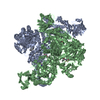
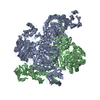
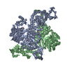
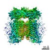
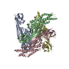
| Title | Structures and Conformations in Solution of the Signal Recognition Particle Receptor from the archaeon Pyrococcus furiosus | ||||||
 Components Components | Signal recognition particle receptor | ||||||
 Keywords Keywords | TRANSPORT PROTEIN / SIGNAL RECOGNITION PARTICLE RECEPTOR / FTSY / SRP-GTPase / PROTEIN-TARGETING | ||||||
| Function / homology |  Function and homology information Function and homology informationsignal recognition particle binding / signal-recognition-particle GTPase / SRP-dependent cotranslational protein targeting to membrane / GTPase activity / GTP binding / metal ion binding / plasma membrane / cytoplasm Similarity search - Function | ||||||
| Biological species |   Pyrococcus furiosus (archaea) Pyrococcus furiosus (archaea) | ||||||
| Method |  X-RAY DIFFRACTION / SYNCHROTRON / MOLECULAR REPLACEMENT / Resolution: 2.21 Å X-RAY DIFFRACTION / SYNCHROTRON / MOLECULAR REPLACEMENT / Resolution: 2.21 Å | ||||||
 Authors Authors | Egea, P.F. / Tsuruta, H. / Napetschnig, J. / Walter, P. / Stroud, R.M. | ||||||
 Citation Citation | Journal: Plos One / Year: 2008 Title: Structures of the Signal Recognition Particle Receptor from the Archaeon Pyrococcus furiosus: Implications for the Targeting Step at the Membrane. Authors: Egea, P.F. / Tsuruta, H. / de Leon, G.P. / Napetschnig, J. / Walter, P. / Stroud, R.M. | ||||||
| History |
|
- Structure visualization
| Structure viewer | Molecule: MolmilJmol/JSmol |
|---|
- Downloads & links
-Download
| PDBx/mmCIF format | 3dmd.cif.gz | 206.9 KB | Display | PDBx/mmCIF format |
|---|---|---|---|---|
| PDB format | pdb3dmd.ent.gz | 165.4 KB | Display | PDB format |
| PDBx/mmJSON format | 3dmd.json.gz | Tree view | PDBx/mmJSON format | |
| Others |  Other downloads Other downloads |
-Validation report
| Arichive directory | https://data.pdbj.org/pub/pdb/validation_reports/dm/3dmdftp://data.pdbj.org/pub/pdb/validation_reports/dm/3dmd | HTTPS FTP |
|---|
-Related structure data
| Related structure data |  3dm9SC  3e70C S: Starting model for refinement C: citing same article ( |
|---|---|
| Similar structure data |

-Links
 PDBj
PDBj- Assembly
| Deposited unit | 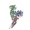
| ||||||||
|---|---|---|---|---|---|---|---|---|---|
| 1 | 
| ||||||||
| 2 | 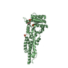
| ||||||||
| 3 | 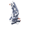
| ||||||||
| 4 | 
| ||||||||
| Unit cell |
|
-Components
| #1: Protein | Mass: 36439.906 Da / Num. of mol.: 3 Source method: isolated from a genetically manipulated source Source: (gene. exp.) Pyrococcus furiosus (archaea) / Strain: DSM3638 / Gene: PF1766 / Plasmid: pET28b / Production host:  #2: Chemical | ChemComp-SO4 /   Mass: 96.063 Da / Num. of mol.: 10 / Source method: obtained synthetically / Formula: SO4 Mass: 96.063 Da / Num. of mol.: 10 / Source method: obtained synthetically / Formula: SO4#3: Chemical | ChemComp-GOL /   Mass: 92.094 Da / Num. of mol.: 6 / Source method: obtained synthetically / Formula: C3H8O3 Mass: 92.094 Da / Num. of mol.: 6 / Source method: obtained synthetically / Formula: C3H8O3#4: Water | ChemComp-HOH / |  Mass: 18.015 Da / Num. of mol.: 469 / Source method: isolated from a natural source / Formula: H2O Mass: 18.015 Da / Num. of mol.: 469 / Source method: isolated from a natural source / Formula: H2O |
|---|
-Experimental details
-Experiment
| Experiment | Method: X-RAY DIFFRACTION / Number of used crystals: 1 |
|---|
- Sample preparation
| Crystal | Density Matthews: 3.1 Å3/Da / Density % sol: 60.32 % |
|---|---|
| Crystal grow | Temperature: 293 K / Method: vapor diffusion, hanging drop / pH: 5 Details: 0.9-1.2M Lithium Sulfate, 0.4-0.6M Ammonium Sulfate, 100 mM Na Citrate, pH 5.0, VAPOR DIFFUSION, HANGING DROP, temperature 293K |
-Data collection
| Diffraction | Mean temperature: 80 K |
|---|---|
| Diffraction source | Source: SYNCHROTRON / Site: ALS  / Beamline: 8.3.1 / Beamline: 8.3.1 |
| Detector | Type: ADSC QUANTUM 210 / Detector: CCD / Date: Aug 31, 2006 |
| Radiation | Protocol: SINGLE WAVELENGTH / Monochromatic (M) / Laue (L): M / Scattering type: x-ray |
| Radiation wavelength | Relative weight: 1 |
| Reflection | Resolution: 2.21→88.4 Å / Num. all: 65992 / Num. obs: 65992 / % possible obs: 98.8 % / Observed criterion σ(F): 1 / Redundancy: 3.5 % / Biso Wilson estimate: 43 Å2 / Rmerge(I) obs: 0.074 / Rsym value: 0.074 / Net I/σ(I): 8.9 |
| Reflection shell | Resolution: 2.21→2.33 Å / Redundancy: 3.1 % / Rmerge(I) obs: 0.766 / Mean I/σ(I) obs: 1.4 / Num. unique all: 9460 / Rsym value: 0.766 / % possible all: 97.1 |
- Processing
| Software |
| |||||||||||||||||||||||||||||||||||||||||||||||||
|---|---|---|---|---|---|---|---|---|---|---|---|---|---|---|---|---|---|---|---|---|---|---|---|---|---|---|---|---|---|---|---|---|---|---|---|---|---|---|---|---|---|---|---|---|---|---|---|---|---|---|
| Refinement | Method to determine structure: MOLECULAR REPLACEMENT Starting model: PDB ENTRY 3DM9 Resolution: 2.21→65.06 Å / SU ML: 0.35 / Cross valid method: THROUGHOUT / σ(F): 1.34 / Phase error: 26.17 / Stereochemistry target values: ML
| |||||||||||||||||||||||||||||||||||||||||||||||||
| Solvent computation | Shrinkage radii: 0.9 Å / VDW probe radii: 1.11 Å / Solvent model: FLAT BULK SOLVENT MODEL / Bsol: 51.919 Å2 / ksol: 0.36 e/Å3 | |||||||||||||||||||||||||||||||||||||||||||||||||
| Displacement parameters | Biso mean: 42 Å2
| |||||||||||||||||||||||||||||||||||||||||||||||||
| Refinement step | Cycle: LAST / Resolution: 2.21→65.06 Å
| |||||||||||||||||||||||||||||||||||||||||||||||||
| Refine LS restraints |
| |||||||||||||||||||||||||||||||||||||||||||||||||
| LS refinement shell | Refine-ID: X-RAY DIFFRACTION / Total num. of bins used: 14
|
