 Movie
Movie Controller

[English] 日本語
 Yorodumi
- PDB-3khs: Crystal structure of grouper iridovirus purine nucleoside phospho... -
+ Open data

- Basic information
| Entry | Database: PDB / ID: 3khs | ||||||
|---|---|---|---|---|---|---|---|
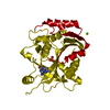
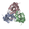
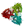
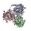
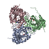
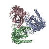
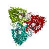
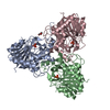
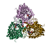
| Title | Crystal structure of grouper iridovirus purine nucleoside phosphorylase | ||||||
 Components Components | Purine nucleoside phosphorylase | ||||||
 Keywords Keywords | HYDROLASE / alpha-beta structure / mixed beta-barrel | ||||||
| Function / homology |  Function and homology information Function and homology informationnucleoside metabolic process / purine-nucleoside phosphorylase / purine-nucleoside phosphorylase activity Similarity search - Function | ||||||
| Biological species |  Grouper iridovirus Grouper iridovirus | ||||||
| Method |  X-RAY DIFFRACTION / MOLECULAR REPLACEMENT / Resolution: 2.38 Å X-RAY DIFFRACTION / MOLECULAR REPLACEMENT / Resolution: 2.38 Å | ||||||
 Authors Authors | Kang, Y.N. / Zhang, Y. / Allan, P.W. / Parker, W.B. / Ting, J.W. / Chang, C.Y. / Ealick, S.E. | ||||||
 Citation Citation | Journal: Acta Crystallogr.,Sect.D / Year: 2010 Title: Structure of grouper iridovirus purine nucleoside phosphorylase Authors: Kang, Y.N. / Zhang, Y. / Allan, P.W. / Parker, W.B. / Ting, J.W. / Chang, C.Y. / Ealick, S.E. | ||||||
| History |
|
- Structure visualization
| Structure viewer | Molecule: MolmilJmol/JSmol |
|---|
- Downloads & links
-Download
| PDBx/mmCIF format | 3khs.cif.gz | 209.4 KB | Display | PDBx/mmCIF format |
|---|---|---|---|---|
| PDB format | pdb3khs.ent.gz | 169.1 KB | Display | PDB format |
| PDBx/mmJSON format | 3khs.json.gz | Tree view | PDBx/mmJSON format | |
| Others |  Other downloads Other downloads |
-Validation report
| Arichive directory | https://data.pdbj.org/pub/pdb/validation_reports/kh/3khsftp://data.pdbj.org/pub/pdb/validation_reports/kh/3khs | HTTPS FTP |
|---|
-Related structure data
| Related structure data | 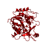 1ulaS S: Starting model for refinement |
|---|---|
| Similar structure data |

-Links
 PDBj
PDBj- Assembly
| Deposited unit | 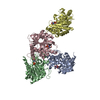
| ||||||||
|---|---|---|---|---|---|---|---|---|---|
| 1 | 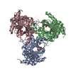
| ||||||||
| 2 | 
| ||||||||
| Unit cell |
|
-Components
| #1: Protein | Mass: 30366.969 Da / Num. of mol.: 4 Source method: isolated from a genetically manipulated source Source: (gene. exp.) Grouper iridovirus / Gene: GIV43 / Plasmid: pET-20b(+) / Production host:  References: UniProt: Q5YBA4, purine-nucleoside phosphorylase #2: Chemical | ChemComp-PO4 /   Mass: 94.971 Da / Num. of mol.: 4 / Source method: obtained synthetically / Formula: PO4 Mass: 94.971 Da / Num. of mol.: 4 / Source method: obtained synthetically / Formula: PO4#3: Chemical | ChemComp-TRS /   Mass: 122.143 Da / Num. of mol.: 4 / Source method: obtained synthetically / Formula: C4H12NO3 / Comment: pH buffer*YM Mass: 122.143 Da / Num. of mol.: 4 / Source method: obtained synthetically / Formula: C4H12NO3 / Comment: pH buffer*YM#4: Water | ChemComp-HOH / |  Mass: 18.015 Da / Num. of mol.: 194 / Source method: isolated from a natural source / Formula: H2O Mass: 18.015 Da / Num. of mol.: 194 / Source method: isolated from a natural source / Formula: H2OHas protein modification | Y | |
|---|
-Experimental details
-Experiment
| Experiment | Method: X-RAY DIFFRACTION / Number of used crystals: 1 |
|---|
- Sample preparation
| Crystal | Density Matthews: 3.12 Å3/Da / Density % sol: 60.56 % |
|---|---|
| Crystal grow | Temperature: 275 K / Method: vapor diffusion, hanging drop / pH: 8 Details: 15-17% PEG 1000, 0.1 M Tris-HCl, pH 8.0, VAPOR DIFFUSION, HANGING DROP, temperature 275K |
-Data collection
| Diffraction | Mean temperature: 100 K |
|---|---|
| Diffraction source | Source: ROTATING ANODE / Type: RIGAKU / Wavelength: 1.5418 Å |
| Detector | Type: RIGAKU RAXIS IV++ / Detector: IMAGE PLATE / Date: Jan 21, 2005 |
| Radiation | Protocol: SINGLE WAVELENGTH / Monochromatic (M) / Laue (L): M / Scattering type: x-ray |
| Radiation wavelength | Wavelength: 1.5418 Å / Relative weight: 1 |
| Reflection | Resolution: 2.38→50 Å / Num. all: 58889 / Num. obs: 56658 / % possible obs: 96.2 % / Observed criterion σ(F): 0 / Observed criterion σ(I): 0 / Redundancy: 3.2 % / Biso Wilson estimate: 37.7 Å2 / Rmerge(I) obs: 0.052 / Rsym value: 0.052 / Net I/σ(I): 24.8 |
| Reflection shell | Resolution: 2.38→2.47 Å / Redundancy: 3.1 % / Rmerge(I) obs: 0.404 / Mean I/σ(I) obs: 3.1 / Num. unique all: 5946 / Rsym value: 0.404 / % possible all: 99.9 |
- Processing
| Software |
| ||||||||||||||||||||||||||||||||||||||||||||||||||||||||||||||||||||||||||||||||
|---|---|---|---|---|---|---|---|---|---|---|---|---|---|---|---|---|---|---|---|---|---|---|---|---|---|---|---|---|---|---|---|---|---|---|---|---|---|---|---|---|---|---|---|---|---|---|---|---|---|---|---|---|---|---|---|---|---|---|---|---|---|---|---|---|---|---|---|---|---|---|---|---|---|---|---|---|---|---|---|---|---|
| Refinement | Method to determine structure: MOLECULAR REPLACEMENT Starting model: PDB 1ULA Resolution: 2.38→44.65 Å / Rfactor Rfree error: 0.003 / Data cutoff high absF: 340179.91 / Data cutoff low absF: 0 / Isotropic thermal model: RESTRAINED / Cross valid method: THROUGHOUT / σ(F): 0 / σ(I): 0 / Stereochemistry target values: Engh & Huber
| ||||||||||||||||||||||||||||||||||||||||||||||||||||||||||||||||||||||||||||||||
| Solvent computation | Solvent model: FLAT MODEL / Bsol: 40.8847 Å2 / ksol: 0.336308 e/Å3 | ||||||||||||||||||||||||||||||||||||||||||||||||||||||||||||||||||||||||||||||||
| Displacement parameters | Biso mean: 47.7 Å2
| ||||||||||||||||||||||||||||||||||||||||||||||||||||||||||||||||||||||||||||||||
| Refine analyze |
| ||||||||||||||||||||||||||||||||||||||||||||||||||||||||||||||||||||||||||||||||
| Refinement step | Cycle: LAST / Resolution: 2.38→44.65 Å
| ||||||||||||||||||||||||||||||||||||||||||||||||||||||||||||||||||||||||||||||||
| Refine LS restraints |
| ||||||||||||||||||||||||||||||||||||||||||||||||||||||||||||||||||||||||||||||||
| Refine LS restraints NCS | NCS model details: NONE | ||||||||||||||||||||||||||||||||||||||||||||||||||||||||||||||||||||||||||||||||
| LS refinement shell | Resolution: 2.38→2.53 Å / Rfactor Rfree error: 0.011 / Total num. of bins used: 6
| ||||||||||||||||||||||||||||||||||||||||||||||||||||||||||||||||||||||||||||||||
| Xplor file |
|
