 Movie
Movie Controller


+ Open data

- Basic information
| Entry | Database: PDB / ID: 7p3r | |||||||||||||||
|---|---|---|---|---|---|---|---|---|---|---|---|---|---|---|---|---|
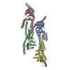
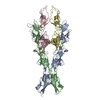
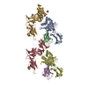
| Title | Helical structure of the toxin MakA from Vibrio cholera | |||||||||||||||
 Components Components | MakA tetramer | |||||||||||||||
 Keywords Keywords | TOXIN / Pore-forming toxin / Vibrio cholerae | |||||||||||||||
| Function / homology | Hemolysin BL-binding component / Bacillus haemolytic enterotoxin (HBL) / : / Hemolysin E; Chain: A; / Hemolysin E; Chain: A; - #10 / Up-down Bundle / Mainly Alpha / membrane / Non-hemolytic enterotoxin lytic component L1 Function and homology information Function and homology information | |||||||||||||||
| Biological species |  Vibrio cholerae O1 biovar El Tor str. N16961 (bacteria) Vibrio cholerae O1 biovar El Tor str. N16961 (bacteria) | |||||||||||||||
| Method | ELECTRON MICROSCOPY / helical reconstruction / cryo EM / Resolution: 3.65 Å | |||||||||||||||
 Authors Authors | Berg, A. / Nadeem, A. / Uhlin, B.E. / Wai, S.N. / Barandun, J. | |||||||||||||||
| Funding support |  Sweden, European Union, 4items Sweden, European Union, 4items
| |||||||||||||||
 Citation Citation | Journal: Elife / Year: 2022 Title: Protein-lipid interaction at low pH induces oligomerization of the MakA cytotoxin from . Authors: Aftab Nadeem / Alexandra Berg / Hudson Pace / Athar Alam / Eric Toh / Jörgen Ådén / Nikola Zlatkov / Si Lhyam Myint / Karina Persson / Gerhard Gröbner / Anders Sjöstedt / Marta Bally / ...Authors: Aftab Nadeem / Alexandra Berg / Hudson Pace / Athar Alam / Eric Toh / Jörgen Ådén / Nikola Zlatkov / Si Lhyam Myint / Karina Persson / Gerhard Gröbner / Anders Sjöstedt / Marta Bally / Jonas Barandun / Bernt Eric Uhlin / Sun Nyunt Wai / Abstract: The α-pore-forming toxins (α-PFTs) from pathogenic bacteria damage host cell membranes by pore formation. We demonstrate a remarkable, hitherto unknown mechanism by an α-PFT protein from . As part ...The α-pore-forming toxins (α-PFTs) from pathogenic bacteria damage host cell membranes by pore formation. We demonstrate a remarkable, hitherto unknown mechanism by an α-PFT protein from . As part of the MakA/B/E tripartite toxin, MakA is involved in membrane pore formation similar to other α-PFTs. In contrast, MakA in isolation induces tube-like structures in acidic endosomal compartments of epithelial cells in vitro. The present study unravels the dynamics of tubular growth, which occurs in a pH-, lipid-, and concentration-dependent manner. Within acidified organelle lumens or when incubated with cells in acidic media, MakA forms oligomers and remodels membranes into high-curvature tubes leading to loss of membrane integrity. A 3.7 Å cryo-electron microscopy structure of MakA filaments reveals a unique protein-lipid superstructure. MakA forms a pinecone-like spiral with a central cavity and a thin annular lipid bilayer embedded between the MakA transmembrane helices in its active α-PFT conformation. Our study provides insights into a novel tubulation mechanism of an α-PFT protein and a new mode of action by a secreted bacterial toxin. | |||||||||||||||
| History |
|
- Structure visualization
| Movie |
Movie viewer |
|---|---|
| Structure viewer | Molecule: MolmilJmol/JSmol |
- Downloads & links
-Download
| PDBx/mmCIF format | 7p3r.cif.gz | 195.9 KB | Display | PDBx/mmCIF format |
|---|---|---|---|---|
| PDB format | pdb7p3r.ent.gz | 148.4 KB | Display | PDB format |
| PDBx/mmJSON format | 7p3r.json.gz | Tree view | PDBx/mmJSON format | |
| Others |  Other downloads Other downloads |
-Validation report
| Arichive directory | https://data.pdbj.org/pub/pdb/validation_reports/p3/7p3rftp://data.pdbj.org/pub/pdb/validation_reports/p3/7p3r | HTTPS FTP |
|---|
-Related structure data
| Related structure data |  13185MC M: map data used to model this data C: citing same article ( |
|---|---|
| Similar structure data | |
| EM raw data | EMPIAR-10869 (Title: Helical structure of the toxin MakA from Vibrio cholera Data size: 195.8 Data #1: Unaligned multi-frame micrographs of the oligomerized Vibrio cholerae toxin MakA [micrographs - multiframe]) |


-Links
 PDBj
PDBj- Assembly
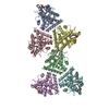
| Deposited unit | 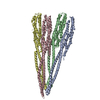
|
|---|---|
| 1 |
|
-Components
| #1: Protein | Mass: 39028.582 Da / Num. of mol.: 4 Source method: isolated from a genetically manipulated source Source: (gene. exp.) Vibrio cholerae O1 biovar El Tor str. N16961 Gene: VC_A0883 / Production host: |
|---|
-Experimental details
-Experiment
| Experiment | Method: ELECTRON MICROSCOPY |
|---|---|
| EM experiment | Aggregation state: HELICAL ARRAY / 3D reconstruction method: helical reconstruction |
- Sample preparation
| Component | Name: Vibrio cholera MakA filament / Type: COMPLEX / Entity ID: all / Source: RECOMBINANT |
|---|---|
| Molecular weight | Experimental value: NO |
| Source (natural) | Organism: Vibrio cholerae O1 biovar El Tor str. N16961 (bacteria) |
| Source (recombinant) | Organism: |
| Buffer solution | pH: 6.5 |
| Buffer component | Conc.: 120 mM / Name: Sodium Citrate |
| Specimen | Embedding applied: NO / Shadowing applied: NO / Staining applied: NO / Vitrification applied: YES |
| Specimen support | Grid material: COPPER / Grid mesh size: 200 divisions/in. / Grid type: Quantifoil R2/1 |
| Vitrification | Instrument: FEI VITROBOT MARK I / Cryogen name: ETHANE |
- Electron microscopy imaging
| Experimental equipment |  Model: Titan Krios / Image courtesy: FEI Company |
|---|---|
| Microscopy | Model: FEI TITAN KRIOS |
| Electron gun | Electron source:  FIELD EMISSION GUN / Accelerating voltage: 300 kV / Illumination mode: FLOOD BEAM FIELD EMISSION GUN / Accelerating voltage: 300 kV / Illumination mode: FLOOD BEAM |
| Electron lens | Mode: BRIGHT FIELD / Nominal defocus max: 2500 nm / Nominal defocus min: 750 nm / Cs: 2.7 mm |
| Specimen holder | Cryogen: NITROGEN / Specimen holder model: FEI TITAN KRIOS AUTOGRID HOLDER |
| Image recording | Electron dose: 43 e/Å2 / Detector mode: COUNTING / Film or detector model: GATAN K2 QUANTUM (4k x 4k) / Num. of grids imaged: 1 / Num. of real images: 2476 |
- Processing
| EM software |
| ||||||||||||||||||||||||||||||||||||
|---|---|---|---|---|---|---|---|---|---|---|---|---|---|---|---|---|---|---|---|---|---|---|---|---|---|---|---|---|---|---|---|---|---|---|---|---|---|
| CTF correction | Type: NONE | ||||||||||||||||||||||||||||||||||||
| Helical symmerty | Angular rotation/subunit: 48.5901 ° / Axial rise/subunit: 5.84149 Å / Axial symmetry: C1 | ||||||||||||||||||||||||||||||||||||
| Particle selection | Num. of particles selected: 95603 | ||||||||||||||||||||||||||||||||||||
| 3D reconstruction | Resolution: 3.65 Å / Resolution method: FSC 0.143 CUT-OFF / Num. of particles: 65485 / Symmetry type: HELICAL | ||||||||||||||||||||||||||||||||||||
| Atomic model building | Protocol: OTHER / Space: REAL | ||||||||||||||||||||||||||||||||||||
| Atomic model building | PDB-ID: 6EZV Accession code: 6EZV / Source name: PDB / Type: experimental model |

