 Movie
Movie Controller


+ Open data

- Basic information
| Entry | Database: PDB / ID: 1hg4 | ||||||
|---|---|---|---|---|---|---|---|
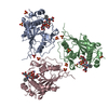
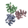
| Title | Ultraspiracle ligand binding domain from Drosophila melanogaster | ||||||
 Components Components | ULTRASPIRACLE | ||||||
 Keywords Keywords | NUCLEAR HORMONE RECEPTOR / TRANSCRIPTION FACTOR / LIGAND BINDING | ||||||
| Function / homology |  Function and homology information Function and homology informationecdysone biosynthetic process / cellular response to ecdysone / Transcriptional regulation of white adipocyte differentiation / Regulation of lipid metabolism by PPARalpha / Signaling by Retinoic Acid / Transcriptional regulation of granulopoiesis / Cytoprotection by HMOX1 / ecdysone receptor holocomplex / activator ecdysone receptor complex / Recycling of bile acids and salts ...ecdysone biosynthetic process / cellular response to ecdysone / Transcriptional regulation of white adipocyte differentiation / Regulation of lipid metabolism by PPARalpha / Signaling by Retinoic Acid / Transcriptional regulation of granulopoiesis / Cytoprotection by HMOX1 / ecdysone receptor holocomplex / activator ecdysone receptor complex / Recycling of bile acids and salts / Carnitine shuttle / NR1H3 & NR1H2 regulate gene expression linked to cholesterol transport and efflux / NR1H2 & NR1H3 regulate gene expression to control bile acid homeostasis / Nuclear Receptor transcription pathway / response to ecdysone / ecdysone receptor signaling pathway / pupariation / regulation of development, heterochronic / mushroom body development / metamorphosis / border follicle cell migration / MLL4 and MLL3 complexes regulate expression of PPARG target genes in adipogenesis and hepatic steatosis / polytene chromosome / regulation of organ growth / dendrite morphogenesis / response to starvation / neuron remodeling / nuclear steroid receptor activity / germ cell development / hormone binding / negative regulation of cell differentiation / core promoter sequence-specific DNA binding / retinoic acid receptor signaling pathway / nuclear receptor activity / nervous system development / cell differentiation / transcription cis-regulatory region binding / RNA polymerase II cis-regulatory region sequence-specific DNA binding / protein heterodimerization activity / signaling receptor binding / negative regulation of DNA-templated transcription / lipid binding / dendrite / positive regulation of DNA-templated transcription / positive regulation of transcription by RNA polymerase II / DNA binding / zinc ion binding / nucleus Similarity search - Function | ||||||
| Biological species |  | ||||||
| Method |  X-RAY DIFFRACTION / SYNCHROTRON / MOLECULAR REPLACEMENT / Resolution: 2.4 Å X-RAY DIFFRACTION / SYNCHROTRON / MOLECULAR REPLACEMENT / Resolution: 2.4 Å | ||||||
 Authors Authors | Schwabe, J.W.R. / Clayton, G.M. | ||||||
 Citation Citation | Journal: Proc.Natl.Acad.Sci.USA / Year: 2001 Title: The Structure of the Ultraspiracle Ligand-Binding Domain Reveals a Nuclear Receptor Locked in an Inactive Conformation Authors: Clayton, G.M. / Peak-Chew, S.Y. / Evans, R.M. / Schwabe, J.W.R. | ||||||
| History |
|
- Structure visualization
| Structure viewer | Molecule: MolmilJmol/JSmol |
|---|
- Downloads & links
-Download
| PDBx/mmCIF format | 1hg4.cif.gz | 297.4 KB | Display | PDBx/mmCIF format |
|---|---|---|---|---|
| PDB format | pdb1hg4.ent.gz | 241.7 KB | Display | PDB format |
| PDBx/mmJSON format | 1hg4.json.gz | Tree view | PDBx/mmJSON format | |
| Others |  Other downloads Other downloads |
-Validation report
| Arichive directory | https://data.pdbj.org/pub/pdb/validation_reports/hg/1hg4ftp://data.pdbj.org/pub/pdb/validation_reports/hg/1hg4 | HTTPS FTP |
|---|
-Related structure data
| Related structure data | 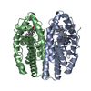 1ereS S: Starting model for refinement |
|---|---|
| Similar structure data |


-Links
 PDBj
PDBj
- Assembly
| Deposited unit | 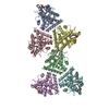
| ||||||||
|---|---|---|---|---|---|---|---|---|---|
| 1 | 
| ||||||||
| 2 | 
| ||||||||
| Unit cell |
| ||||||||
| Details | BUT THE BIOLOGICALLY ACTIVE MOLECULE IS HETERODIMER |
-Components
| #1: Protein | Mass: 31355.182 Da / Num. of mol.: 6 / Fragment: LIGAND-BINDING DOMAIN RESIDUES 230-508 Source method: isolated from a genetically manipulated source Details: PHOSPHATIDIC ACID MODELLED IN LIGAND BINDING POCKET OF 6 PROTEIN MONOMERS Source: (gene. exp.)  #2: Chemical | ChemComp-LPP /   Mass: 648.891 Da / Num. of mol.: 6 / Source method: obtained synthetically / Formula: C35H69O8P / Comment: phospholipid*YM Mass: 648.891 Da / Num. of mol.: 6 / Source method: obtained synthetically / Formula: C35H69O8P / Comment: phospholipid*YM#3: Water | ChemComp-HOH / |  Mass: 18.015 Da / Num. of mol.: 188 / Source method: isolated from a natural source / Formula: H2O Mass: 18.015 Da / Num. of mol.: 188 / Source method: isolated from a natural source / Formula: H2OCompound details | THE C-TERMINAL STEROID-BINDING DOMAIN OF RECEPTOR FOR ECDYSONE. MAY PLAY AN IMPORTANT ROLE AS A ...THE C-TERMINAL STEROID-BINDING DOMAIN OF RECEPTOR FOR ECDYSONE. MAY PLAY AN IMPORTANT ROLE AS A MODULATOR IN INSECT METAMORPHO | |
|---|
-Experimental details
-Experiment
| Experiment | Method: X-RAY DIFFRACTION / Number of used crystals: 2 |
|---|
- Sample preparation
| Crystal | Density Matthews: 2.35 Å3/Da / Density % sol: 47.77 % | |||||||||||||||||||||||||
|---|---|---|---|---|---|---|---|---|---|---|---|---|---|---|---|---|---|---|---|---|---|---|---|---|---|---|
| Crystal grow | pH: 7 / Details: pH 7.00 | |||||||||||||||||||||||||
| Crystal grow | *PLUS Temperature: 18 ℃ / pH: 4.6 / Method: vapor diffusion / Details: used streak-seeding | |||||||||||||||||||||||||
| Components of the solutions | *PLUS
|
-Data collection
| Diffraction | Mean temperature: 100 K |
|---|---|
| Diffraction source | Source: SYNCHROTRON / Site: SRS  / Beamline: PX9.6 / Wavelength: 0.87 / Beamline: PX9.6 / Wavelength: 0.87 |
| Detector | Type: ADSC CCD / Detector: CCD |
| Radiation | Protocol: SINGLE WAVELENGTH / Monochromatic (M) / Laue (L): M / Scattering type: x-ray |
| Radiation wavelength | Wavelength: 0.87 Å / Relative weight: 1 |
| Reflection | Resolution: 2.2→34 Å / Num. obs: 60384 / % possible obs: 90 % / Observed criterion σ(I): 2 / Redundancy: 2.9 % / Rmerge(I) obs: 0.126 / Net I/σ(I): 4.4 |
| Reflection | *PLUS Lowest resolution: 34 Å / % possible obs: 90 % / Num. measured all: 175324 |
| Reflection shell | *PLUS Highest resolution: 2.4 Å / Lowest resolution: 2.6 Å / Rmerge(I) obs: 0.305 / Mean I/σ(I) obs: 2.1 |
- Processing
| Software |
| ||||||||||||||||||||||||||||||||||||||||||||||||||||||||||||
|---|---|---|---|---|---|---|---|---|---|---|---|---|---|---|---|---|---|---|---|---|---|---|---|---|---|---|---|---|---|---|---|---|---|---|---|---|---|---|---|---|---|---|---|---|---|---|---|---|---|---|---|---|---|---|---|---|---|---|---|---|---|
| Refinement | Method to determine structure: MOLECULAR REPLACEMENT Starting model: 1ERE Resolution: 2.4→34 Å / Cross valid method: THROUGHOUT / σ(F): 2
| ||||||||||||||||||||||||||||||||||||||||||||||||||||||||||||
| Refinement step | Cycle: LAST / Resolution: 2.4→34 Å
| ||||||||||||||||||||||||||||||||||||||||||||||||||||||||||||
| Refine LS restraints |
| ||||||||||||||||||||||||||||||||||||||||||||||||||||||||||||
| Refine LS restraints NCS | NCS model details: RESTRAINTS |
