 Movie
Movie Controller


+ Open data

- Basic information
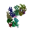
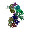
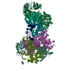
| Entry | Database: PDB / ID: 7npf | ||||||||||||||||||||||||
|---|---|---|---|---|---|---|---|---|---|---|---|---|---|---|---|---|---|---|---|---|---|---|---|---|---|
| Title | Vibrio cholerae ParA2-ATPyS-DNA filament | ||||||||||||||||||||||||
 Components Components |
| ||||||||||||||||||||||||
 Keywords Keywords | DNA BINDING PROTEIN / ATPase / Chromosome segregation / Bacterial cell division / filament | ||||||||||||||||||||||||
| Function / homology |  Function and homology information Function and homology informationParA helix turn helix domain / ParA helix turn helix domain / : / AAA domain / AAA domain / P-loop containing nucleoside triphosphate hydrolase Similarity search - Domain/homology | ||||||||||||||||||||||||
| Biological species |   Vibrio cholerae (bacteria) Vibrio cholerae (bacteria) Neoarius leptaspis (salmon catfish) Neoarius leptaspis (salmon catfish) | ||||||||||||||||||||||||
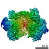
| Method | ELECTRON MICROSCOPY / helical reconstruction / cryo EM / Resolution: 4.5 Å | ||||||||||||||||||||||||
 Authors Authors | Parker, A.V. / Bergeron, J.R.C. | ||||||||||||||||||||||||
| Funding support |  United Kingdom, 1items United Kingdom, 1items
| ||||||||||||||||||||||||
 Citation Citation | Journal: Nat Commun / Year: 2021 Title: The structure of the bacterial DNA segregation ATPase filament reveals the conformational plasticity of ParA upon DNA binding. Authors: Alexandra V Parker / Daniel Mann / Svetomir B Tzokov / Ling C Hwang / Julien R C Bergeron /  Abstract: The efficient segregation of replicated genetic material is an essential step for cell division. Bacterial cells use several evolutionarily-distinct genome segregation systems, the most common of ...The efficient segregation of replicated genetic material is an essential step for cell division. Bacterial cells use several evolutionarily-distinct genome segregation systems, the most common of which is the type I Par system. It consists of an adapter protein, ParB, that binds to the DNA cargo via interaction with the parS DNA sequence; and an ATPase, ParA, that binds nonspecific DNA and mediates cargo transport. However, the molecular details of how this system functions are not well understood. Here, we report the cryo-EM structure of the Vibrio cholerae ParA2 filament bound to DNA, as well as the crystal structures of this protein in various nucleotide states. These structures show that ParA forms a left-handed filament on DNA, stabilized by nucleotide binding, and that ParA undergoes profound structural rearrangements upon DNA binding and filament assembly. Collectively, our data suggest the structural basis for ParA's cooperative binding to DNA and the formation of high ParA density regions on the nucleoid. | ||||||||||||||||||||||||
| History |
|
- Structure visualization
| Movie |
Movie viewer |
|---|---|
| Structure viewer | Molecule: MolmilJmol/JSmol |
- Downloads & links
-Download
| PDBx/mmCIF format | 7npf.cif.gz | 590.3 KB | Display | PDBx/mmCIF format |
|---|---|---|---|---|
| PDB format | pdb7npf.ent.gz | 483.1 KB | Display | PDB format |
| PDBx/mmJSON format | 7npf.json.gz | Tree view | PDBx/mmJSON format | |
| Others |  Other downloads Other downloads |
-Validation report
| Arichive directory | https://data.pdbj.org/pub/pdb/validation_reports/np/7npfftp://data.pdbj.org/pub/pdb/validation_reports/np/7npf | HTTPS FTP |
|---|
-Related structure data
| Related structure data |  12515MC  7npdC M: map data used to model this data C: citing same article ( |
|---|---|
| Similar structure data |


-Links
 PDBj
PDBj


- Assembly
| Deposited unit | 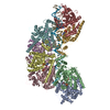
|
|---|---|
| 1 |
|
-Components
| #1: Protein | Mass: 46440.969 Da / Num. of mol.: 8 Source method: isolated from a genetically manipulated source Source: (gene. exp.) Vibrio cholerae (bacteria)Gene: parA, BC353_10845, C9J66_03480, D6U24_01990, ERS013138_01197, ERS013166_00021, ERS013186_00500, ERS013193_00027, ERS013198_00323, ERS013199_01186, ERS013200_00295, ERS013202_00851, ERS013206_ ...Gene: parA, BC353_10845, C9J66_03480, D6U24_01990, ERS013138_01197, ERS013166_00021, ERS013186_00500, ERS013193_00027, ERS013198_00323, ERS013199_01186, ERS013200_00295, ERS013202_00851, ERS013206_01885, EYB64_07940, F0315_18570, FLM02_03395, FXF03_20770, HPY05_13545 Production host: #2: DNA chain | | Mass: 15302.170 Da / Num. of mol.: 1 / Source method: isolated from a natural source / Source: (natural) Neoarius leptaspis (salmon catfish)#3: DNA chain | | Mass: 14860.490 Da / Num. of mol.: 1 / Source method: isolated from a natural source / Source: (natural) Neoarius leptaspis (salmon catfish)#4: Chemical | ChemComp-MG /   Mass: 24.305 Da / Num. of mol.: 8 / Source method: obtained synthetically / Formula: Mg / Feature type: SUBJECT OF INVESTIGATION Mass: 24.305 Da / Num. of mol.: 8 / Source method: obtained synthetically / Formula: Mg / Feature type: SUBJECT OF INVESTIGATION#5: Chemical | ChemComp-AGS /   Mass: 523.247 Da / Num. of mol.: 8 / Source method: obtained synthetically / Formula: C10H16N5O12P3S / Feature type: SUBJECT OF INVESTIGATION / Comment: ATP-gamma-S, energy-carrying molecule analogue*YM Mass: 523.247 Da / Num. of mol.: 8 / Source method: obtained synthetically / Formula: C10H16N5O12P3S / Feature type: SUBJECT OF INVESTIGATION / Comment: ATP-gamma-S, energy-carrying molecule analogue*YMHas ligand of interest | Y | Has protein modification | N | |
|---|
-Experimental details
-Experiment
| Experiment | Method: ELECTRON MICROSCOPY |
|---|---|
| EM experiment | Aggregation state: FILAMENT / 3D reconstruction method: helical reconstruction |
- Sample preparation
| Component | Name: ParA2-ATPgS-DNA / Type: COMPLEX / Entity ID: #1-#3 / Source: MULTIPLE SOURCES |
|---|---|
| Molecular weight | Experimental value: NO |
| Buffer solution | pH: 7 |
| Specimen | Embedding applied: NO / Shadowing applied: NO / Staining applied: NO / Vitrification applied: YES |
| Vitrification | Cryogen name: ETHANE |
- Electron microscopy imaging
| Experimental equipment |  Model: Titan Krios / Image courtesy: FEI Company |
|---|---|
| Microscopy | Model: FEI TITAN KRIOS |
| Electron gun | Electron source:  FIELD EMISSION GUN / Accelerating voltage: 300 kV / Illumination mode: FLOOD BEAM FIELD EMISSION GUN / Accelerating voltage: 300 kV / Illumination mode: FLOOD BEAM |
| Electron lens | Mode: BRIGHT FIELD |
| Image recording | Electron dose: 52.02 e/Å2 / Film or detector model: GATAN K2 SUMMIT (4k x 4k) |
- Processing
| CTF correction | Type: PHASE FLIPPING AND AMPLITUDE CORRECTION |
|---|---|
| Helical symmerty | Angular rotation/subunit: -80.57 ° / Axial rise/subunit: 28.68 Å / Axial symmetry: C1 |
| 3D reconstruction | Resolution: 4.5 Å / Resolution method: FSC 0.143 CUT-OFF / Num. of particles: 182997 / Symmetry type: HELICAL |
| Atomic model building | Protocol: OTHER |
