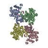
Movie
Movie Controller

[English] 日本語
 Yorodumi
- PDB-7nj0: CryoEM structure of the human Separase-Cdk1-cyclin B1-Cks1 complex -
+ Open data

- Basic information
| Entry | Database: PDB / ID: 7nj0 | |||||||||
|---|---|---|---|---|---|---|---|---|---|---|
| Title | CryoEM structure of the human Separase-Cdk1-cyclin B1-Cks1 complex | |||||||||
 Components Components |
| |||||||||
 Keywords Keywords | HYDROLASE / autoinhibition phosphate-binding pocket pseudosubstrate Scc1 | |||||||||
| Function / homology |  Function and homology information Function and homology informationregulation of Schwann cell differentiation / regulation of attachment of mitotic spindle microtubules to kinetochore / pronuclear fusion / response to DDT / regulation of chromosome condensation / negative regulation of sister chromatid cohesion / cyclin B1-CDK1 complex / separase / positive regulation of mitochondrial ATP synthesis coupled electron transport / Mitotic Prophase ...regulation of Schwann cell differentiation / regulation of attachment of mitotic spindle microtubules to kinetochore / pronuclear fusion / response to DDT / regulation of chromosome condensation / negative regulation of sister chromatid cohesion / cyclin B1-CDK1 complex / separase / positive regulation of mitochondrial ATP synthesis coupled electron transport / Mitotic Prophase / positive regulation of mitotic sister chromatid segregation / ventricular cardiac muscle cell development / histone kinase activity / meiotic chromosome separation / Golgi disassembly / homologous chromosome segregation / E2F-enabled inhibition of pre-replication complex formation / microtubule cytoskeleton organization involved in mitosis / Depolymerization of the Nuclear Lamina / positive regulation of mRNA 3'-end processing / regulation of mitotic cell cycle spindle assembly checkpoint / mitotic sister chromatid separation / positive regulation of attachment of spindle microtubules to kinetochore / MASTL Facilitates Mitotic Progression / Activation of NIMA Kinases NEK9, NEK6, NEK7 / tissue regeneration / patched binding / Phosphorylation of Emi1 / oocyte maturation / establishment of mitotic spindle localization / Transcriptional regulation by RUNX2 / mitotic nuclear membrane disassembly / Nuclear Pore Complex (NPC) Disassembly / Phosphorylation of the APC/C / G2/M DNA replication checkpoint / outer kinetochore / protein localization to kinetochore / Transcription of E2F targets under negative control by p107 (RBL1) and p130 (RBL2) in complex with HDAC1 / digestive tract development / Initiation of Nuclear Envelope (NE) Reformation / Polo-like kinase mediated events / positive regulation of mitotic metaphase/anaphase transition / Golgi Cisternae Pericentriolar Stack Reorganization / cellular response to fatty acid / cyclin-dependent protein serine/threonine kinase activator activity / [RNA-polymerase]-subunit kinase / cellular response to iron(III) ion / chromosome condensation / Condensation of Prometaphase Chromosomes / response to amine / centrosome cycle / positive regulation of ubiquitin-dependent protein catabolic process / cyclin-dependent protein serine/threonine kinase regulator activity / response to copper ion / SCF ubiquitin ligase complex / cyclin-dependent protein kinase activity / regulation of heterochromatin organization / peptidyl-threonine phosphorylation / MAPK3 (ERK1) activation / G1/S-Specific Transcription / mitotic G2 DNA damage checkpoint signaling / Regulation of APC/C activators between G1/S and early anaphase / mitotic sister chromatid segregation / microtubule organizing center / mitotic cytokinesis / catalytic activity / response to axon injury / ubiquitin-like protein ligase binding / chromosome organization / regulation of embryonic development / response to cadmium ion / positive regulation of G2/M transition of mitotic cell cycle / ERK1 and ERK2 cascade / response to mechanical stimulus / cyclin-dependent kinase / mitotic metaphase chromosome alignment / cyclin-dependent protein serine/threonine kinase activity / positive regulation of cardiac muscle cell proliferation / protein deubiquitination / cysteine-type endopeptidase inhibitor activity / Regulation of MITF-M-dependent genes involved in cell cycle and proliferation / cyclin-dependent protein kinase holoenzyme complex / Chk1/Chk2(Cds1) mediated inactivation of Cyclin B:Cdk1 complex / Cyclin A/B1/B2 associated events during G2/M transition / positive regulation of mitotic cell cycle / Nuclear events stimulated by ALK signaling in cancer / epithelial cell differentiation / Loss of Nlp from mitotic centrosomes / Loss of proteins required for interphase microtubule organization from the centrosome / RNA polymerase II CTD heptapeptide repeat kinase activity / cysteine-type peptidase activity / negative regulation of protein ubiquitination / Recruitment of mitotic centrosome proteins and complexes / Recruitment of NuMA to mitotic centrosomes / Anchoring of the basal body to the plasma membrane / TP53 Regulates Transcription of Genes Involved in G2 Cell Cycle Arrest / regulation of mitotic cell cycle / Hsp70 protein binding / APC/C:Cdc20 mediated degradation of Cyclin B / AURKA Activation by TPX2 Similarity search - Function | |||||||||
| Biological species |  Homo sapiens (human) Homo sapiens (human) | |||||||||
| Method | ELECTRON MICROSCOPY / single particle reconstruction / cryo EM / Resolution: 3.6 Å | |||||||||
 Authors Authors | Yu, J. / Raia, P. / Ghent, C.M. / Raisch, T. / Sadian, Y. / Barford, D. / Raunser, S. / Morgan, D.O. / Boland, A. | |||||||||
| Funding support |  Switzerland, Switzerland,  United States, 2items United States, 2items
| |||||||||
 Citation Citation | Journal: Nature / Year: 2021 Title: Structural basis of human separase regulation by securin and CDK1-cyclin B1. Authors: Jun Yu / Pierre Raia / Chloe M Ghent / Tobias Raisch / Yashar Sadian / Simone Cavadini / Pramod M Sabale / David Barford / Stefan Raunser / David O Morgan / Andreas Boland /   Abstract: In early mitosis, the duplicated chromosomes are held together by the ring-shaped cohesin complex. Separation of chromosomes during anaphase is triggered by separase-a large cysteine endopeptidase ...In early mitosis, the duplicated chromosomes are held together by the ring-shaped cohesin complex. Separation of chromosomes during anaphase is triggered by separase-a large cysteine endopeptidase that cleaves the cohesin subunit SCC1 (also known as RAD21). Separase is activated by degradation of its inhibitors, securin and cyclin B, but the molecular mechanisms of separase regulation are not clear. Here we used cryogenic electron microscopy to determine the structures of human separase in complex with either securin or CDK1-cyclin B1-CKS1. In both complexes, separase is inhibited by pseudosubstrate motifs that block substrate binding at the catalytic site and at nearby docking sites. As in Caenorhabditis elegans and yeast, human securin contains its own pseudosubstrate motifs. By contrast, CDK1-cyclin B1 inhibits separase by deploying pseudosubstrate motifs from intrinsically disordered loops in separase itself. One autoinhibitory loop is oriented by CDK1-cyclin B1 to block the catalytic sites of both separase and CDK1. Another autoinhibitory loop blocks substrate docking in a cleft adjacent to the separase catalytic site. A third separase loop contains a phosphoserine that promotes complex assembly by binding to a conserved phosphate-binding pocket in cyclin B1. Our study reveals the diverse array of mechanisms by which securin and CDK1-cyclin B1 bind and inhibit separase, providing the molecular basis for the robust control of chromosome segregation. | |||||||||
| History |
|
- Structure visualization
| Movie |
Movie viewer |
|---|---|
| Structure viewer | Molecule: MolmilJmol/JSmol |
- Downloads & links
-Download
| PDBx/mmCIF format | 7nj0.cif.gz | 358.5 KB | Display | PDBx/mmCIF format |
|---|---|---|---|---|
| PDB format | pdb7nj0.ent.gz | 267.2 KB | Display | PDB format |
| PDBx/mmJSON format | 7nj0.json.gz | Tree view | PDBx/mmJSON format | |
| Others |  Other downloads Other downloads |
-Validation report
| Arichive directory | https://data.pdbj.org/pub/pdb/validation_reports/nj/7nj0ftp://data.pdbj.org/pub/pdb/validation_reports/nj/7nj0 | HTTPS FTP |
|---|
-Related structure data
| Related structure data | 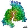 12368MC  7nj1C M: map data used to model this data C: citing same article ( |
|---|---|
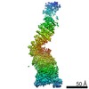
| Similar structure data |
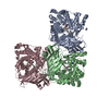
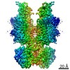

-Links
 PDBj
PDBj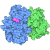













- Assembly
| Deposited unit | 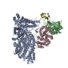
|
|---|---|
| 1 |
|
-Components
| #1: Protein | Mass: 244677.328 Da / Num. of mol.: 1 Source method: isolated from a genetically manipulated source Source: (gene. exp.) Homo sapiens (human) / Gene: PTTG1, EAP1, PTTG, TUTR1, ESPL1, ESP1, KIAA0165 / Production host:   Spodoptera frugiperda (fall armyworm) / References: UniProt: O95997, UniProt: Q14674, separase Spodoptera frugiperda (fall armyworm) / References: UniProt: O95997, UniProt: Q14674, separase |
|---|---|
| #2: Protein | Mass: 36667.098 Da / Num. of mol.: 1 Source method: isolated from a genetically manipulated source Source: (gene. exp.) Homo sapiens (human) / Gene: CDK1, CDC2, CDC28A, CDKN1, P34CDC2 / Production host: Spodoptera frugiperda (fall armyworm)References: UniProt: P06493, cyclin-dependent kinase, [RNA-polymerase]-subunit kinase |
| #3: Protein | Mass: 52625.723 Da / Num. of mol.: 1 Source method: isolated from a genetically manipulated source Source: (gene. exp.) Homo sapiens (human) / Gene: CCNB1, CCNB / Production host: Spodoptera frugiperda (fall armyworm) / References: UniProt: P14635 |
| #4: Protein | Mass: 9679.211 Da / Num. of mol.: 1 Source method: isolated from a genetically manipulated source Source: (gene. exp.) Homo sapiens (human) / Gene: CKS1B, CKS1, PNAS-143, PNAS-16 / Production host: Spodoptera frugiperda (fall armyworm) / References: UniProt: P61024 |
| #5: Chemical | ChemComp-PO4 /  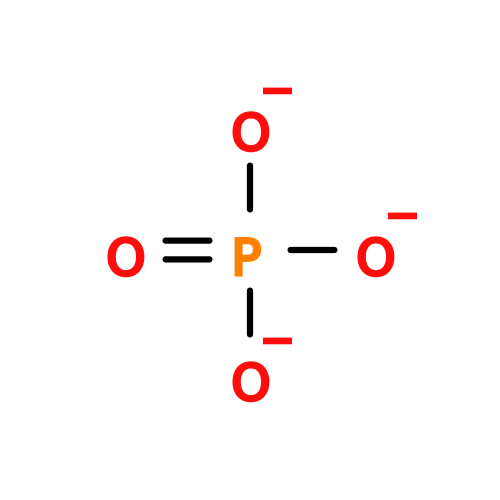 Mass: 94.971 Da / Num. of mol.: 1 / Source method: obtained synthetically / Formula: PO4 Mass: 94.971 Da / Num. of mol.: 1 / Source method: obtained synthetically / Formula: PO4 |
| Has ligand of interest | Y |
| Has protein modification | Y |
-Experimental details
-Experiment
| Experiment | Method: ELECTRON MICROSCOPY |
|---|---|
| EM experiment | Aggregation state: PARTICLE / 3D reconstruction method: single particle reconstruction |
- Sample preparation
| Component | Name: Mutual inhibitory complex of human separase-Cdk1-cyclin B1-Cks1 (CCC) complex. Type: COMPLEX / Entity ID: #1-#4 / Source: RECOMBINANT |
|---|---|
| Molecular weight | Experimental value: NO |
| Source (natural) | Organism: Homo sapiens (human) |
| Source (recombinant) | Organism: Spodoptera frugiperda (fall armyworm) |
| Buffer solution | pH: 7.8 |
| Specimen | Conc.: 0.05 mg/ml / Embedding applied: NO / Shadowing applied: NO / Staining applied: NO / Vitrification applied: YES Details: The sample was monodisperse. We use graphene oxide-coated EM grids. |
| Specimen support | Grid material: GOLD / Grid type: Quantifoil R1.2/1.3 |
| Vitrification | Instrument: LEICA EM GP / Cryogen name: ETHANE / Humidity: 90 % / Chamber temperature: 293 K |
- Electron microscopy imaging
| Experimental equipment |  Model: Titan Krios / Image courtesy: FEI Company |
|---|---|
| Microscopy | Model: FEI TITAN KRIOS |
| Electron gun | Electron source:  FIELD EMISSION GUN / Accelerating voltage: 300 kV / Illumination mode: FLOOD BEAM FIELD EMISSION GUN / Accelerating voltage: 300 kV / Illumination mode: FLOOD BEAM |
| Electron lens | Mode: BRIGHT FIELD / Nominal magnification: 105000 X / Nominal defocus max: 2500 nm / Nominal defocus min: 1300 nm / Calibrated defocus min: 1300 nm / Calibrated defocus max: 2500 nm / C2 aperture diameter: 50 µm / Alignment procedure: COMA FREE |
| Specimen holder | Cryogen: NITROGEN / Specimen holder model: FEI TITAN KRIOS AUTOGRID HOLDER |
| Image recording | Average exposure time: 3 sec. / Electron dose: 78 e/Å2 / Film or detector model: GATAN K3 BIOQUANTUM (6k x 4k) / Num. of grids imaged: 5 / Num. of real images: 13640 |
- Processing
| Software | Name: PHENIX / Version: 1.18rc5_3822: / Classification: refinement | |||||||||||||||||||||||||||
|---|---|---|---|---|---|---|---|---|---|---|---|---|---|---|---|---|---|---|---|---|---|---|---|---|---|---|---|---|
| EM software |
| |||||||||||||||||||||||||||
| CTF correction | Type: PHASE FLIPPING AND AMPLITUDE CORRECTION | |||||||||||||||||||||||||||
| Symmetry | Point symmetry: C1 (asymmetric) | |||||||||||||||||||||||||||
| 3D reconstruction | Resolution: 3.6 Å / Resolution method: FSC 0.143 CUT-OFF / Num. of particles: 312836 / Symmetry type: POINT | |||||||||||||||||||||||||||
| Atomic model building | Protocol: AB INITIO MODEL | |||||||||||||||||||||||||||
| Atomic model building |
| |||||||||||||||||||||||||||
| Refine LS restraints |
|