 Movie
Movie Controller


+ Open data

- Basic information
| Entry | Database: PDB / ID: 7mtd | ||||||
|---|---|---|---|---|---|---|---|
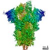
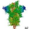
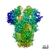
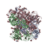
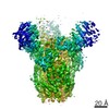
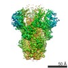
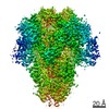
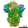
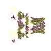
| Title | Structure of aged SARS-CoV-2 S2P spike at pH 7.4 | ||||||
 Components Components | Spike glycoprotein | ||||||
 Keywords Keywords | VIRAL PROTEIN / COVID-19 / SARS-CoV-2 spike / S2P | ||||||
| Function / homology |  Function and homology information Function and homology informationMaturation of spike protein / viral translation / Translation of Structural Proteins / Virion Assembly and Release / host cell surface / host extracellular space / suppression by virus of host tetherin activity / Induction of Cell-Cell Fusion / structural constituent of virion / entry receptor-mediated virion attachment to host cell ...Maturation of spike protein / viral translation / Translation of Structural Proteins / Virion Assembly and Release / host cell surface / host extracellular space / suppression by virus of host tetherin activity / Induction of Cell-Cell Fusion / structural constituent of virion / entry receptor-mediated virion attachment to host cell / host cell endoplasmic reticulum-Golgi intermediate compartment membrane / membrane fusion / receptor-mediated endocytosis of virus by host cell / Attachment and Entry / positive regulation of viral entry into host cell / receptor-mediated virion attachment to host cell / receptor ligand activity / symbiont-mediated suppression of host innate immune response / host cell surface receptor binding / fusion of virus membrane with host plasma membrane / fusion of virus membrane with host endosome membrane / viral envelope / virion attachment to host cell / SARS-CoV-2 activates/modulates innate and adaptive immune responses / host cell plasma membrane / virion membrane / identical protein binding / membrane / plasma membrane Similarity search - Function | ||||||
| Biological species |   Severe acute respiratory syndrome coronavirus 2 Severe acute respiratory syndrome coronavirus 2 | ||||||
| Method | ELECTRON MICROSCOPY / single particle reconstruction / cryo EM / Resolution: 3.5 Å | ||||||
 Authors Authors | Tsybovsky, Y. / Olia, A.S. / Kwong, P.D. | ||||||
 Citation Citation | Journal: J Biol Chem / Year: 2021 Title: SARS-CoV-2 S2P spike ages through distinct states with altered immunogenicity. Authors: Adam S Olia / Yaroslav Tsybovsky / Steven J Chen / Cuiping Liu / Alexandra F Nazzari / Li Ou / Lingshu Wang / Wing-Pui Kong / Kwan Leung / Tracy Liu / Tyler Stephens / I-Ting Teng / Shuishu ...Authors: Adam S Olia / Yaroslav Tsybovsky / Steven J Chen / Cuiping Liu / Alexandra F Nazzari / Li Ou / Lingshu Wang / Wing-Pui Kong / Kwan Leung / Tracy Liu / Tyler Stephens / I-Ting Teng / Shuishu Wang / Eun Sung Yang / Baoshan Zhang / Yi Zhang / Tongqing Zhou / John R Mascola / Peter D Kwong /  Abstract: The SARS-CoV-2 spike is the primary target of virus-neutralizing antibodies and critical to the development of effective vaccines against COVID-19. Here, we demonstrate that the prefusion-stabilized ...The SARS-CoV-2 spike is the primary target of virus-neutralizing antibodies and critical to the development of effective vaccines against COVID-19. Here, we demonstrate that the prefusion-stabilized two-proline "S2P" spike-widely employed for laboratory work and clinical studies-unfolds when stored at 4 °C, physiological pH, as observed by electron microscopy (EM) and differential scanning calorimetry, but that its trimeric, native-like conformation can be reacquired by low pH treatment. When stored for approximately 1 week, this unfolding does not significantly alter antigenic characteristics; however, longer storage diminishes antibody binding, and month-old spike elicits virtually no neutralization in mice despite inducing high ELISA-binding titers. Cryo-EM structures reveal the folded fraction of spike to decrease with aging; however, its structure remains largely similar, although with varying mobility of the receptor-binding domain. Thus, the SARS-CoV-2 spike is susceptible to unfolding, which affects immunogenicity, highlighting the need to monitor its integrity. | ||||||
| History |
|
- Structure visualization
| Movie |
Movie viewer |
|---|---|
| Structure viewer | Molecule: MolmilJmol/JSmol |
- Downloads & links
-Download
| PDBx/mmCIF format | 7mtd.cif.gz | 448 KB | Display | PDBx/mmCIF format |
|---|---|---|---|---|
| PDB format | pdb7mtd.ent.gz | 359.7 KB | Display | PDB format |
| PDBx/mmJSON format | 7mtd.json.gz | Tree view | PDBx/mmJSON format | |
| Others |  Other downloads Other downloads |
-Validation report
| Summary document | 7mtd_validation.pdf.gz | 1.8 MB | Display | wwPDB validaton report |
|---|---|---|---|---|
| Full document | 7mtd_full_validation.pdf.gz | 1.8 MB | Display | |
| Data in XML | 7mtd_validation.xml.gz | 80.1 KB | Display | |
| Data in CIF | 7mtd_validation.cif.gz | 120.2 KB | Display | |
| Arichive directory | https://data.pdbj.org/pub/pdb/validation_reports/mt/7mtdftp://data.pdbj.org/pub/pdb/validation_reports/mt/7mtd | HTTPS FTP |
-Related structure data
| Related structure data | 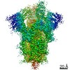 23983MC 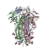 7mtcC 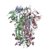 7mteC M: map data used to model this data C: citing same article ( |
|---|---|
| Similar structure data |

-Links
 PDBj
PDBj


- Assembly
| Deposited unit | 
|
|---|---|
| 1 |
|
-Components
| #1: Protein | Mass: 141532.797 Da / Num. of mol.: 3 Source method: isolated from a genetically manipulated source Source: (gene. exp.) Severe acute respiratory syndrome coronavirus 2Gene: S, 2 / Production host:  Homo sapiens (human) / References: UniProt: P0DTC2 Homo sapiens (human) / References: UniProt: P0DTC2#2: Polysaccharide | 2-acetamido-2-deoxy-beta-D-glucopyranose-(1-4)-2-acetamido-2-deoxy-beta-D-glucopyranose Source method: isolated from a genetically manipulated source #3: Sugar | ChemComp-NAG /   Type: D-saccharide, beta linking / Mass: 221.208 Da / Num. of mol.: 13 / Source method: obtained synthetically / Formula: C8H15NO6 Type: D-saccharide, beta linking / Mass: 221.208 Da / Num. of mol.: 13 / Source method: obtained synthetically / Formula: C8H15NO6Has ligand of interest | N | |
|---|
-Experimental details
-Experiment
| Experiment | Method: ELECTRON MICROSCOPY |
|---|---|
| EM experiment | Aggregation state: PARTICLE / 3D reconstruction method: single particle reconstruction |
- Sample preparation
| Component | Name: SARS-CoV-2 S2P spike trimer / Type: COMPLEX / Entity ID: #1 / Source: RECOMBINANT |
|---|---|
| Molecular weight | Value: 0.42 MDa / Experimental value: NO |
| Source (natural) | Organism: Severe acute respiratory syndrome coronavirus 2 |
| Source (recombinant) | Organism: Homo sapiens (human) / Strain: 293-F |
| Buffer solution | pH: 7.4 |
| Buffer component | Formula: PBS |
| Specimen | Conc.: 0.5 mg/ml / Embedding applied: NO / Shadowing applied: NO / Staining applied: NO / Vitrification applied: YES |
| Vitrification | Instrument: FEI VITROBOT MARK IV / Cryogen name: ETHANE / Humidity: 90 % / Chamber temperature: 277 K |
- Electron microscopy imaging
| Experimental equipment |  Model: Titan Krios / Image courtesy: FEI Company |
|---|---|
| Microscopy | Model: FEI TITAN KRIOS |
| Electron gun | Electron source:  FIELD EMISSION GUN / Accelerating voltage: 300 kV / Illumination mode: FLOOD BEAM FIELD EMISSION GUN / Accelerating voltage: 300 kV / Illumination mode: FLOOD BEAM |
| Electron lens | Mode: BRIGHT FIELD |
| Image recording | Electron dose: 40 e/Å2 / Film or detector model: GATAN K3 BIOQUANTUM (6k x 4k) |
- Processing
| Software | Name: PHENIX / Version: 1.12_2829: / Classification: refinement | ||||||||||||||||||||||||
|---|---|---|---|---|---|---|---|---|---|---|---|---|---|---|---|---|---|---|---|---|---|---|---|---|---|
| EM software | Name: RELION / Version: 3.1 / Category: 3D reconstruction | ||||||||||||||||||||||||
| CTF correction | Type: PHASE FLIPPING AND AMPLITUDE CORRECTION | ||||||||||||||||||||||||
| Symmetry | Point symmetry: C1 (asymmetric) | ||||||||||||||||||||||||
| 3D reconstruction | Resolution: 3.5 Å / Resolution method: FSC 0.143 CUT-OFF / Num. of particles: 95641 / Symmetry type: POINT | ||||||||||||||||||||||||
| Atomic model building | Protocol: BACKBONE TRACE | ||||||||||||||||||||||||
| Refine LS restraints |
|
