 Movie
Movie Controller

[English] 日本語
 Yorodumi
- PDB-7mgm: Structure of yeast cytoplasmic dynein with AAA3 Walker B mutation... -
+ Open data

- Basic information
| Entry | Database: PDB / ID: 7mgm | ||||||
|---|---|---|---|---|---|---|---|
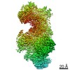
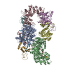
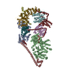
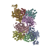
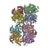
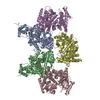
| Title | Structure of yeast cytoplasmic dynein with AAA3 Walker B mutation bound to Lis1 | ||||||
 Components Components |
| ||||||
 Keywords Keywords | MOTOR PROTEIN / motor / AAA | ||||||
| Function / homology |  Function and homology information Function and homology informationmicrotubule sliding / microtubule organizing center organization / nuclear migration along microtubule / dynein complex / microtubule plus-end binding / vesicle transport along microtubule / minus-end-directed microtubule motor activity / microtubule associated complex / microtubule-based movement / nuclear migration ...microtubule sliding / microtubule organizing center organization / nuclear migration along microtubule / dynein complex / microtubule plus-end binding / vesicle transport along microtubule / minus-end-directed microtubule motor activity / microtubule associated complex / microtubule-based movement / nuclear migration / Ubiquitin-Mediated Degradation of Phosphorylated Cdc25A / establishment of mitotic spindle orientation / Antigen processing: Ubiquitination & Proteasome degradation / dynein complex binding / cytoplasmic microtubule / kinetochore / spindle pole / nuclear envelope / microtubule / cell division / ATP binding / identical protein binding / nucleus / cytoplasm Similarity search - Function | ||||||
| Biological species |  | ||||||
| Method | ELECTRON MICROSCOPY / single particle reconstruction / cryo EM / Resolution: 3.1 Å | ||||||
 Authors Authors | Lahiri, I. / Reimer, J.M. / Leschziner, A.E. | ||||||
| Funding support |  United States, 1items United States, 1items
| ||||||
 Citation Citation | Journal: Elife / Year: 2022 Title: Structural basis for cytoplasmic dynein-1 regulation by Lis1. Authors: John P Gillies / Janice M Reimer / Eva P Karasmanis / Indrajit Lahiri / Zaw Min Htet / Andres E Leschziner / Samara L Reck-Peterson /  Abstract: The lissencephaly 1 gene, , is mutated in patients with the neurodevelopmental disease lissencephaly. The Lis1 protein is conserved from fungi to mammals and is a key regulator of cytoplasmic dynein- ...The lissencephaly 1 gene, , is mutated in patients with the neurodevelopmental disease lissencephaly. The Lis1 protein is conserved from fungi to mammals and is a key regulator of cytoplasmic dynein-1, the major minus-end-directed microtubule motor in many eukaryotes. Lis1 is the only dynein regulator known to bind directly to dynein's motor domain, and by doing so alters dynein's mechanochemistry. Lis1 is required for the formation of fully active dynein complexes, which also contain essential cofactors: dynactin and an activating adaptor. Here, we report the first high-resolution structure of the yeast dynein-Lis1 complex. Our 3.1 Å structure reveals, in molecular detail, the major contacts between dynein and Lis1 and between Lis1's ß-propellers. Structure-guided mutations in Lis1 and dynein show that these contacts are required for Lis1's ability to form fully active human dynein complexes and to regulate yeast dynein's mechanochemistry and in vivo function. | ||||||
| History |
|
- Structure visualization
| Movie |
Movie viewer |
|---|---|
| Structure viewer | Molecule: MolmilJmol/JSmol |
- Downloads & links
-Download
| PDBx/mmCIF format | 7mgm.cif.gz | 558.6 KB | Display | PDBx/mmCIF format |
|---|---|---|---|---|
| PDB format | pdb7mgm.ent.gz | 435.4 KB | Display | PDB format |
| PDBx/mmJSON format | 7mgm.json.gz | Tree view | PDBx/mmJSON format | |
| Others |  Other downloads Other downloads |
-Validation report
| Arichive directory | https://data.pdbj.org/pub/pdb/validation_reports/mg/7mgmftp://data.pdbj.org/pub/pdb/validation_reports/mg/7mgm | HTTPS FTP |
|---|
-Related structure data
| Related structure data |  23829MC M: map data used to model this data C: citing same article ( |
|---|---|
| Similar structure data |


-Links
 PDBj
PDBj


- Assembly
| Deposited unit | 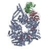
|
|---|---|
| 1 |
|
-Components
| #1: Protein | Mass: 331524.000 Da / Num. of mol.: 1 / Source method: isolated from a natural source / Source: (natural) | ||||||||
|---|---|---|---|---|---|---|---|---|---|
| #2: Protein | Mass: 57030.617 Da / Num. of mol.: 2 / Source method: isolated from a natural source / Source: (natural) #3: Chemical |   Mass: 507.181 Da / Num. of mol.: 3 / Source method: obtained synthetically / Formula: C10H16N5O13P3 / Comment: ATP, energy-carrying molecule*YM Mass: 507.181 Da / Num. of mol.: 3 / Source method: obtained synthetically / Formula: C10H16N5O13P3 / Comment: ATP, energy-carrying molecule*YM#4: Chemical | ChemComp-ADP / |   Mass: 427.201 Da / Num. of mol.: 1 / Source method: obtained synthetically / Formula: C10H15N5O10P2 / Comment: ADP, energy-carrying molecule*YM Mass: 427.201 Da / Num. of mol.: 1 / Source method: obtained synthetically / Formula: C10H15N5O10P2 / Comment: ADP, energy-carrying molecule*YM#5: Chemical |   Mass: 24.305 Da / Num. of mol.: 2 / Source method: obtained synthetically / Formula: Mg Mass: 24.305 Da / Num. of mol.: 2 / Source method: obtained synthetically / Formula: MgHas ligand of interest | N | |
-Experimental details
-Experiment
| Experiment | Method: ELECTRON MICROSCOPY |
|---|---|
| EM experiment | Aggregation state: PARTICLE / 3D reconstruction method: single particle reconstruction |
- Sample preparation
| Component | Name: Complex of yeast dynein bound by two Lis1s in the presence of ATP-Va Type: COMPLEX / Entity ID: #1-#2 / Source: NATURAL |
|---|---|
| Molecular weight | Experimental value: NO |
| Source (natural) | Organism: |
| Buffer solution | pH: 7.4 |
| Specimen | Embedding applied: NO / Shadowing applied: NO / Staining applied: NO / Vitrification applied: YES Details: The yeast dynein was biotinylated prior to complex formation. |
| Specimen support | Details: Quantifoil R2/2 grids with gold foil were used. A monolayer of streptavidin crystals was deposited prior to applying the sample. The streptavidin monolayer acted as an affinity surface for ...Details: Quantifoil R2/2 grids with gold foil were used. A monolayer of streptavidin crystals was deposited prior to applying the sample. The streptavidin monolayer acted as an affinity surface for the biotinylated sample. |
| Vitrification | Instrument: FEI VITROBOT MARK II / Cryogen name: ETHANE / Humidity: 100 % / Chamber temperature: 295 K |
- Electron microscopy imaging
| Experimental equipment |  Model: Titan Krios / Image courtesy: FEI Company |
|---|---|
| Microscopy | Model: FEI TITAN KRIOS |
| Electron gun | Electron source:  FIELD EMISSION GUN / Accelerating voltage: 300 kV / Illumination mode: FLOOD BEAM FIELD EMISSION GUN / Accelerating voltage: 300 kV / Illumination mode: FLOOD BEAM |
| Electron lens | Mode: BRIGHT FIELD / Nominal defocus max: 2700 nm / Nominal defocus min: 2000 nm / Cs: 2.7 mm / Alignment procedure: COMA FREE |
| Specimen holder | Cryogen: NITROGEN / Specimen holder model: FEI TITAN KRIOS AUTOGRID HOLDER / Temperature (max): 70 K / Temperature (min): 70 K |
| Image recording | Average exposure time: 10 sec. / Electron dose: 58.3 e/Å2 / Detector mode: SUPER-RESOLUTION / Film or detector model: GATAN K2 SUMMIT (4k x 4k) / Num. of grids imaged: 1 / Num. of real images: 2229 |
| Image scans | Movie frames/image: 50 / Used frames/image: 1-50 |
- Processing
| Software | Name: PHENIX / Version: 1.18.2_3874: / Classification: refinement | ||||||||||||||||||||||||
|---|---|---|---|---|---|---|---|---|---|---|---|---|---|---|---|---|---|---|---|---|---|---|---|---|---|
| CTF correction | Type: PHASE FLIPPING AND AMPLITUDE CORRECTION | ||||||||||||||||||||||||
| 3D reconstruction | Resolution: 3.1 Å / Resolution method: FSC 0.143 CUT-OFF / Num. of particles: 83975 / Symmetry type: POINT | ||||||||||||||||||||||||
| Refine LS restraints |
|
