 Movie
Movie Controller

[English] 日本語
 Yorodumi
- EMDB-23829: Structure of yeast cytoplasmic dynein with AAA3 Walker B mutation... -
+ Open data

- Basic information
| Entry | Database: EMDB / ID: EMD-23829 | |||||||||
|---|---|---|---|---|---|---|---|---|---|---|
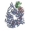
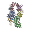
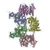
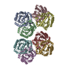
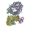
| Title | Structure of yeast cytoplasmic dynein with AAA3 Walker B mutation bound to Lis1 | |||||||||
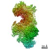
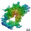
 Map data Map data | scDynein E2488Q bound to two Lis1s | |||||||||
 Sample Sample |
| |||||||||
 Keywords Keywords | motor / AAA / MOTOR PROTEIN | |||||||||
| Function / homology |  Function and homology information Function and homology informationmicrotubule sliding / microtubule organizing center organization / nuclear migration along microtubule / dynein complex / microtubule plus-end binding / vesicle transport along microtubule / minus-end-directed microtubule motor activity / microtubule associated complex / microtubule-based movement / nuclear migration ...microtubule sliding / microtubule organizing center organization / nuclear migration along microtubule / dynein complex / microtubule plus-end binding / vesicle transport along microtubule / minus-end-directed microtubule motor activity / microtubule associated complex / microtubule-based movement / nuclear migration / Ubiquitin-Mediated Degradation of Phosphorylated Cdc25A / establishment of mitotic spindle orientation / Antigen processing: Ubiquitination & Proteasome degradation / dynein complex binding / cytoplasmic microtubule / kinetochore / spindle pole / nuclear envelope / microtubule / cell division / ATP binding / identical protein binding / nucleus / cytoplasm Similarity search - Function | |||||||||
| Biological species |  | |||||||||
| Method | single particle reconstruction / cryo EM / Resolution: 3.1 Å | |||||||||
 Authors Authors | Lahiri I / Reimer JM | |||||||||
| Funding support |  United States, 1 items United States, 1 items
| |||||||||
 Citation Citation | Journal: Elife / Year: 2022 Title: Structural basis for cytoplasmic dynein-1 regulation by Lis1. Authors: John P Gillies / Janice M Reimer / Eva P Karasmanis / Indrajit Lahiri / Zaw Min Htet / Andres E Leschziner / Samara L Reck-Peterson /  Abstract: The lissencephaly 1 gene, , is mutated in patients with the neurodevelopmental disease lissencephaly. The Lis1 protein is conserved from fungi to mammals and is a key regulator of cytoplasmic dynein- ...The lissencephaly 1 gene, , is mutated in patients with the neurodevelopmental disease lissencephaly. The Lis1 protein is conserved from fungi to mammals and is a key regulator of cytoplasmic dynein-1, the major minus-end-directed microtubule motor in many eukaryotes. Lis1 is the only dynein regulator known to bind directly to dynein's motor domain, and by doing so alters dynein's mechanochemistry. Lis1 is required for the formation of fully active dynein complexes, which also contain essential cofactors: dynactin and an activating adaptor. Here, we report the first high-resolution structure of the yeast dynein-Lis1 complex. Our 3.1 Å structure reveals, in molecular detail, the major contacts between dynein and Lis1 and between Lis1's ß-propellers. Structure-guided mutations in Lis1 and dynein show that these contacts are required for Lis1's ability to form fully active human dynein complexes and to regulate yeast dynein's mechanochemistry and in vivo function. | |||||||||
| History |
|
- Structure visualization
| Movie |
Movie viewer |
|---|---|
| Structure viewer | EM map: SurfViewMolmilJmol/JSmol |
| Supplemental images |

- Downloads & links
-EMDB archive
| Map data | emd_23829.map.gz | 93.9 MB | EMDB map data format | |
|---|---|---|---|---|
| Header (meta data) | emd-23829-v30.xmlemd-23829.xml | 19.7 KB 19.7 KB | Display Display | EMDB header |
| FSC (resolution estimation) | emd_23829_fsc.xml | 10.6 KB | Display | FSC data file |
| Images | 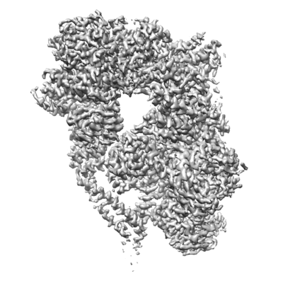 emd_23829.png emd_23829.png | 123.6 KB | ||
| Filedesc metadata | emd-23829.cif.gz | 7.7 KB | ||
| Others | emd_23829_half_map_1.map.gzemd_23829_half_map_2.map.gz | 80.4 MB 80.4 MB | ||
| Archive directory |  http://ftp.pdbj.org/pub/emdb/structures/EMD-23829ftp://ftp.pdbj.org/pub/emdb/structures/EMD-23829 http://ftp.pdbj.org/pub/emdb/structures/EMD-23829ftp://ftp.pdbj.org/pub/emdb/structures/EMD-23829 | HTTPS FTP |
-Related structure data
| Related structure data |  7mgmMC M: atomic model generated by this map C: citing same article ( |
|---|---|
| Similar structure data |


-Links
| EMDB pages | EMDB (EBI/PDBe) / EMDataResource |
|---|---|
| Related items in Molecule of the Month |


-Map
| File | Download / File: emd_23829.map.gz / Format: CCP4 / Size: 103 MB / Type: IMAGE STORED AS FLOATING POINT NUMBER (4 BYTES) | ||||||||||||||||||||||||||||||||||||||||||||||||||||||||||||
|---|---|---|---|---|---|---|---|---|---|---|---|---|---|---|---|---|---|---|---|---|---|---|---|---|---|---|---|---|---|---|---|---|---|---|---|---|---|---|---|---|---|---|---|---|---|---|---|---|---|---|---|---|---|---|---|---|---|---|---|---|---|
| Annotation | scDynein E2488Q bound to two Lis1s | ||||||||||||||||||||||||||||||||||||||||||||||||||||||||||||
| Projections & slices | Image control
Images are generated by Spider. | ||||||||||||||||||||||||||||||||||||||||||||||||||||||||||||
| Voxel size | X=Y=Z: 1.31 Å | ||||||||||||||||||||||||||||||||||||||||||||||||||||||||||||
| Density |
| ||||||||||||||||||||||||||||||||||||||||||||||||||||||||||||
| Symmetry | Space group: 1 | ||||||||||||||||||||||||||||||||||||||||||||||||||||||||||||
| Details | EMDB XML:
CCP4 map header:
| ||||||||||||||||||||||||||||||||||||||||||||||||||||||||||||
 Z (Sec.)
 Y (Row.)
 X (Col.)


-Supplemental data
-Half map: Half map 1
| File | emd_23829_half_map_1.map | ||||||||||||
|---|---|---|---|---|---|---|---|---|---|---|---|---|---|
| Annotation | Half map 1 | ||||||||||||
| Projections & Slices |
| ||||||||||||
| Density Histograms |


-Half map: Half map 2
| File | emd_23829_half_map_2.map | ||||||||||||
|---|---|---|---|---|---|---|---|---|---|---|---|---|---|
| Annotation | Half map 2 | ||||||||||||
| Projections & Slices |
| ||||||||||||
| Density Histograms |


- Sample components
-Entire : Complex of yeast dynein bound by two Lis1s in the presence of ATP-Va
| Entire | Name: Complex of yeast dynein bound by two Lis1s in the presence of ATP-Va |
|---|---|
| Components |
|
-Supramolecule #1: Complex of yeast dynein bound by two Lis1s in the presence of ATP-Va
| Supramolecule | Name: Complex of yeast dynein bound by two Lis1s in the presence of ATP-Va type: complex / ID: 1 / Parent: 0 / Macromolecule list: #1-#2 |
|---|---|
| Source (natural) | Organism: |
-Macromolecule #1: dynein AAA3-WalkerB mutant (E2488Q)
| Macromolecule | Name: dynein AAA3-WalkerB mutant (E2488Q) / type: protein_or_peptide / ID: 1 / Number of copies: 1 / Enantiomer: LEVO |
|---|---|
| Source (natural) | Organism: |
| Molecular weight | Theoretical: 331.524 KDa |
| Sequence | String: GDQLTHVVEE VKTYDLVWRS IKNLWEDVQR TFETPWCRVD VLLLQSDLAN FLRRADELPR AVKQFEMYKS LFSQVNMLTS VNKILVELK DGALKPRHWN MIFRDIGKRQ IQKNLLDKLE FSLKDVMVLN LTLNEILLTK IIERAQKEFV IEKSLNRIKK F WKEAQYEV ...String: GDQLTHVVEE VKTYDLVWRS IKNLWEDVQR TFETPWCRVD VLLLQSDLAN FLRRADELPR AVKQFEMYKS LFSQVNMLTS VNKILVELK DGALKPRHWN MIFRDIGKRQ IQKNLLDKLE FSLKDVMVLN LTLNEILLTK IIERAQKEFV IEKSLNRIKK F WKEAQYEV IEHSSGLKLV REWDVLEQAC KEDLEELVSM KASNYYKIFE QDCLDLESKL TKLSEIQVNW VEVQFYWLDL YG ILGENLD IQNFLPLETS KFKSLTSEYK MITTRAFQLD TTIEVIHIPN FDTTLKLTID SLKMIKSSLS TFLERQRRQF PRF YFLGND DLLKIIGSGK HHDQVSKFMK KMFGSIESII FFEDSITGVR SVEGEVLNLN EKIELKDSIQ AQEWLNILDT EIKL SVFTQ FRDCLGQLKD GTDIEVVVSK YIFQAILLSA QVMWTELVEK CLQTNEFSKY WKEVDMKIKG LLDKLNKSSD NVKKK IEAL LVEYLHFNNV IGQLKNCSTK EEARLLWAKV QKFYQKNDTL DDLNSVFISQ SGYLLQYKFE YIGIPERLIY TPLLLV GFA TLTDSLHQKY GGCFFGPAGT GKTETVKAFG QNLGRVVVVF NCDDSFDYQV LSRLLVGITQ IGAWGCFDEF NRLDEKV LS AVSANIQQIQ NGLQVGKSHI TLLEEETPLS PHTAVFITLN PGYNGRSELP ENLKKSFREF SMKSPQSGTI AEMILQIM G FEDSKSLASK IVHFLELLSS KCSSMNHYHF GLRTLKGVLR NCSPLVSEFG EGEKTVVESL KRVILPSLGD TDELVFKDE LSKIFDSAGT PLNSKAIVQC LKDAGQRSGF SMSEEFLKKC MQFYYMQKTQ QALILVGKAG CGKTATWKTV IDAMAIFDGH ANVVYVIDT KVLTKESLYG SMLKATLEWR DGLFTSILRR VNDDITGTFK NSRIWVVFDS DLDPEYVEAM NSVLDDNKIL T LPNGERLP IPPNFRILFE TDNLDHTTPA TITRCGLLWF STDVCSISSK IDHLLNKSYE ALDNKLSMFE LDKLKDLISD SF DMASLTN IFTCSNDLVH ILGVRTFNKL ETAVQLAVHL ISSYRQWFQN LDDKSLKDVI TLLIKRSLLY ALAGDSTGES QRA FIQTIN TYFGHDSQEL SDYSTIVIAN DKLSFSSFCS EIPSVSLEAH EVMRPDIVIP TIDTIKHEKI FYDLLNSKRG IILC GPPGS GKTMIMNNAL RNSSLYDVVG INFSKDTTTE HILSALHRHT NYVTTSKGLT LLPKSDIKNL VLFCDQINLP KLDKY GSQN VVLFLRQLME KQGFWKTPEN KWVTIERIHI VGACNPPTDP GRIPMSERFT RHAAILYLGY PSGKSLSQIY EIYYKA IFK LVPEFRSYTE PFARASVHLY NECKARYSTG LQSHYLFSPR ELTRLVRGVY TAINTGPRQT LRSLIRLWAY EAWRIFA DR LVGVKEKNSF EQLLYETVDK YLPNQDLGNI SSTSLLFSGL LSLDFKEVNK TDLVNFIEER FKTFCDEELE VPMVIHES M VDHILRIDRA LKQVQGHMML IGASRTGKTI LTRFVAWLNG LKIVQPKIHR HSNLSDFDMI LKKAISDCSL KESRTCLII DESNILETAF LERMNTLLAN ADIPDLFQGE EYDKLLNNLR NKTRSLGLLL DTEQELYDWF VGEIAKNLHV VFTICDPTNN KSSAMISSP ALFNRCIINW MGDWDTKTMS QVANNMVDVV PMEFTDFIVP EVNKELVFTE PIQTIRDAVV NILIHFDRNF Y QKMKVGVN PRSPGYFIDG LRALVKLVTA KYQDLQENQR FVNVGLEKLN ESVLKVNELN KTLSKKSTEL TEKEKEARST LD KMLMEQN ESERKQEATE EIKKILKVQE EDIRKRKEVV MKSIQDIEPT ILEAQRGVKN IKKQQLTEIR SMVNPPSGVK IVM EAVCAI LGYQFSNWRD IQQFIRKDDF IHNIVHYDTT LHMKPQIRKY MEEEFLSDPN FTYETINRAS KACGPLYQWV NAQI NFSKV LENVDPLRQE MKRIEFESLK TKANLLAAEE MTQDLEASIE VSKQKYSLLI RDVEAIKTEM SNVQANLDRS ISLVK SLTF EKERWLNTTK QFSKTSQELI GNCIISSIYE TYFGHLNERE RGDMLVILKR LLGKFAVKYD VNYRFIDYLV TLDEKM KWL ECGLDKNDYF LENMSIVMNS QDAVPFLLDP SSHMITVISN YYGNKTVLLS FLEEGFVKRL ENAVRFGSVV IIQDGEF FD PIISRLISRE FNHAGNRVTV EIGDHEVDVS GDFKLFIHSC DPSGDIPIFL RSRVRLVHFV TNKESIETRI FDITLTEE N AEMQRKREDL IKLNTEYRLK LKNLEKRLLE ELNNSQGNML ENDELMVTLN NLKKEAMNIE KKLSESEEFF PQFDNLVEE YSIIGKHSVK IFSMLEKFGQ FHWFYGISIG QFLSCFKRVF IKKSRETRAA RTRVDEILWL LYQEVYCQFS TALDKKFKMI MAMTMFCLY KFDIESEQYK EAVLTMIGVL SESSDGVPKL TVDTNDDLRY LWDYVTTKSY ISALNWFKNE FFVDEWNIAD V VANSENNY FTMASERDVD GTFKLIELAK ASKESLKIIP LGSIENLNYA QEEISKSKIE GGWILLQNIQ MSLSWVKTYL HK HVEETKA AEEHEKFKMF MTCHLTGDKL PAPLLQRTDR VVYEDIPGIL DTVKDLWGSQ FFTGKISGVW SVYCTFLLSW FHA LITART RLVPHGFSKK YYFNDCDFQF ASVYLENVLA TNSTNNIPWA QVRDHIATIV YGGKIDEEKD LEVVAKLCAH VFCG SDNLQ IVPGVRIPQP LLQQSEEEER ARLTAILSNT IEPADSLSSW LQLPRESILD YERLQAKEVA SSTEQLLQEM UniProtKB: Dynein heavy chain, cytoplasmic |
-Macromolecule #2: Nuclear distribution protein PAC1
| Macromolecule | Name: Nuclear distribution protein PAC1 / type: protein_or_peptide / ID: 2 / Number of copies: 2 / Enantiomer: LEVO |
|---|---|
| Source (natural) | Organism: |
| Molecular weight | Theoretical: 57.030617 KDa |
| Sequence | String: GMTNWQQQLP LTDTQKNELD KSVLRYLNWN YKQTVRHEHA QDYESVRHAI VTLSGFLLQE SVDRQEFISN NDTSNESMVD IDELLLPKK WNSIVRLQKK IIELEQNTET LVSQIKDLNT QVSELAQFKP TTSNGTSAHN VLKWIPRNLP SCLINVESSV T SVKLHPNL ...String: GMTNWQQQLP LTDTQKNELD KSVLRYLNWN YKQTVRHEHA QDYESVRHAI VTLSGFLLQE SVDRQEFISN NDTSNESMVD IDELLLPKK WNSIVRLQKK IIELEQNTET LVSQIKDLNT QVSELAQFKP TTSNGTSAHN VLKWIPRNLP SCLINVESSV T SVKLHPNL PIVFVATDHG KLYAFDLFNY TIPLASLQSH TKAITSMDVL FTNYTNSSKK NYLVIVTASK DLQIHVFKWV SE ECKFQQI RSLLGHEHIV SAVKIWQKNN DVHIASCSRD QTVKIWDFHN GWSLKTFQPH SQWVRSIDVL GDYIISGSHD TTL RLTHWP SGNGLSVGTG HEFPIEKVKF IHFIEDSPEI RFRTPSTDRY KNWGMQYCVS ASRDRTIKIW EIPLPTLMAH RAPI PNPTD SNFRCVLTLK GHLSWVRDIS IRGQYLFSCA DDKSVRCWDL NTGQCLHVWE KLHTGFVNCL DLDVDFDSNV TPRQM MVTG GLDCKSNVFM R UniProtKB: Nuclear distribution protein PAC1 |
-Macromolecule #3: ADENOSINE-5'-TRIPHOSPHATE
| Macromolecule | Name: ADENOSINE-5'-TRIPHOSPHATE / type: ligand / ID: 3 / Number of copies: 3 / Formula: ATP |
|---|---|
| Molecular weight | Theoretical: 507.181 Da |
| Chemical component information |  ChemComp-ATP: |
-Macromolecule #4: ADENOSINE-5'-DIPHOSPHATE
| Macromolecule | Name: ADENOSINE-5'-DIPHOSPHATE / type: ligand / ID: 4 / Number of copies: 1 / Formula: ADP |
|---|---|
| Molecular weight | Theoretical: 427.201 Da |
| Chemical component information |  ChemComp-ADP: |
-Macromolecule #5: MAGNESIUM ION
| Macromolecule | Name: MAGNESIUM ION / type: ligand / ID: 5 / Number of copies: 2 / Formula: MG |
|---|---|
| Molecular weight | Theoretical: 24.305 Da |
-Experimental details
-Structure determination
| Method | cryo EM |
|---|---|
 Processing Processing | single particle reconstruction |
| Aggregation state | particle |
-Sample preparation
| Buffer | pH: 7.4 |
|---|---|
| Grid | Details: Quantifoil R2/2 grids with gold foil were used. A monolayer of streptavidin crystals was deposited prior to applying the sample. The streptavidin monolayer acted as an affinity surface for ...Details: Quantifoil R2/2 grids with gold foil were used. A monolayer of streptavidin crystals was deposited prior to applying the sample. The streptavidin monolayer acted as an affinity surface for the biotinylated sample. |
| Vitrification | Cryogen name: ETHANE / Chamber humidity: 100 % / Chamber temperature: 295 K / Instrument: FEI VITROBOT MARK II |
| Details | The yeast dynein was biotinylated prior to complex formation. |
- Electron microscopy
| Microscope | FEI TITAN KRIOS |
|---|---|
| Temperature | Min: 70.0 K / Max: 70.0 K |
| Image recording | Film or detector model: GATAN K2 SUMMIT (4k x 4k) / Detector mode: SUPER-RESOLUTION / Digitization - Frames/image: 1-50 / Number grids imaged: 1 / Number real images: 2229 / Average exposure time: 10.0 sec. / Average electron dose: 58.3 e/Å2 |
| Electron beam | Acceleration voltage: 300 kV / Electron source:  FIELD EMISSION GUN FIELD EMISSION GUN |
| Electron optics | Illumination mode: FLOOD BEAM / Imaging mode: BRIGHT FIELD / Cs: 2.7 mm / Nominal defocus max: 2.7 µm / Nominal defocus min: 2.0 µm |
| Sample stage | Specimen holder model: FEI TITAN KRIOS AUTOGRID HOLDER / Cooling holder cryogen: NITROGEN |
| Experimental equipment |  Model: Titan Krios / Image courtesy: FEI Company |

