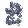
 Movie
Movie Controller

[English] 日本語
 Yorodumi
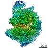
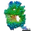
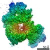
- PDB-7k1b: CryoEM structure of DNA-PK catalytic subunit complexed with DNA (... -
+ Open data

- Basic information
| Entry | Database: PDB / ID: 7k1b | ||||||
|---|---|---|---|---|---|---|---|
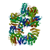
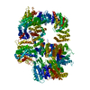
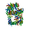
| Title | CryoEM structure of DNA-PK catalytic subunit complexed with DNA (Complex II) | ||||||
 Components Components |
| ||||||
 Keywords Keywords | DNA BINDING PROTEIN/DNA / NHEJ / V(D)J recombination / DNA repair / DNA BINDING PROTEIN / DNA BINDING PROTEIN-DNA complex | ||||||
| Function / homology |  Function and homology information Function and homology informationMHC class II antigen presentation / Neutrophil degranulation / entry into host cell by a symbiont-containing vacuole / Hydrolases; Acting on peptide bonds (peptidases); Aspartic endopeptidases / protein autoprocessing / transport vesicle / protein processing / aspartic-type endopeptidase activity / proteolysis Similarity search - Function | ||||||
| Biological species |  Homo sapiens (human) Homo sapiens (human)synthetic construct (others) | ||||||
| Method | ELECTRON MICROSCOPY / single particle reconstruction / cryo EM / Resolution: 4.3 Å | ||||||
 Authors Authors | Chen, X. / Gellert, M. / Yang, W. | ||||||
| Funding support |  United States, 1items United States, 1items
| ||||||
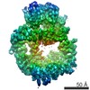
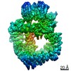
 Citation Citation | Journal: Mol Cell / Year: 2021 Title: Structure of an activated DNA-PK and its implications for NHEJ. Authors: Xuemin Chen / Xiang Xu / Yun Chen / Joyce C Cheung / Huaibin Wang / Jiansen Jiang / Natalia de Val / Tara Fox / Martin Gellert / Wei Yang / Abstract: DNA-dependent protein kinase (DNA-PK), like all phosphatidylinositol 3-kinase-related kinases (PIKKs), is composed of conserved FAT and kinase domains (FATKINs) along with solenoid structures made of ...DNA-dependent protein kinase (DNA-PK), like all phosphatidylinositol 3-kinase-related kinases (PIKKs), is composed of conserved FAT and kinase domains (FATKINs) along with solenoid structures made of HEAT repeats. These kinases are activated in response to cellular stress signals, but the mechanisms governing activation and regulation remain unresolved. For DNA-PK, all existing structures represent inactive states with resolution limited to 4.3 Å at best. Here, we report the cryoelectron microscopy (cryo-EM) structures of DNA-PKcs (DNA-PK catalytic subunit) bound to a DNA end or complexed with Ku70/80 and DNA in both inactive and activated forms at resolutions of 3.7 Å overall and 3.2 Å for FATKINs. These structures reveal the sequential transition of DNA-PK from inactive to activated forms. Most notably, activation of the kinase involves previously unknown stretching and twisting within individual solenoid segments and loosens DNA-end binding. This unprecedented structural plasticity of helical repeats may be a general regulatory mechanism of HEAT-repeat proteins. | ||||||
| History |
|
- Structure visualization
| Movie |
Movie viewer |
|---|---|
| Structure viewer | Molecule: MolmilJmol/JSmol |
- Downloads & links
-Download
| PDBx/mmCIF format | 7k1b.cif.gz | 665.7 KB | Display | PDBx/mmCIF format |
|---|---|---|---|---|
| PDB format | pdb7k1b.ent.gz | 528 KB | Display | PDB format |
| PDBx/mmJSON format | 7k1b.json.gz | Tree view | PDBx/mmJSON format | |
| Others |  Other downloads Other downloads |
-Validation report
| Summary document | 7k1b_validation.pdf.gz | 1001.1 KB | Display | wwPDB validaton report |
|---|---|---|---|---|
| Full document | 7k1b_full_validation.pdf.gz | 1 MB | Display | |
| Data in XML | 7k1b_validation.xml.gz | 92.9 KB | Display | |
| Data in CIF | 7k1b_validation.cif.gz | 142.7 KB | Display | |
| Arichive directory | https://data.pdbj.org/pub/pdb/validation_reports/k1/7k1bftp://data.pdbj.org/pub/pdb/validation_reports/k1/7k1b | HTTPS FTP |
-Related structure data
| Related structure data | 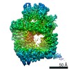 22623MC  7k0yC 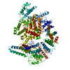 7k10C  7k11C  7k17C  7k19C  7k1jC 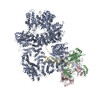 7k1kC  7k1nC C: citing same article ( M: map data used to model this data |
|---|---|
| Similar structure data |


-Links
 PDBj
PDBj


- Assembly
| Deposited unit | 
|
|---|---|
| 1 |
|
-Components
| #1: Protein | Mass: 469673.219 Da / Num. of mol.: 1 / Source method: isolated from a natural source / Source: (natural) Homo sapiens (human)References: UniProt: P78527, non-specific serine/threonine protein kinase | ||
|---|---|---|---|
| #2: DNA chain | Mass: 7311.714 Da / Num. of mol.: 2 / Source method: obtained synthetically / Source: (synth.) synthetic construct (others) #3: DNA chain | | Mass: 4956.243 Da / Num. of mol.: 1 / Source method: obtained synthetically / Source: (synth.) synthetic construct (others) |
-Experimental details
-Experiment
| Experiment | Method: ELECTRON MICROSCOPY |
|---|---|
| EM experiment | Aggregation state: PARTICLE / 3D reconstruction method: single particle reconstruction |
- Sample preparation
| Component | Name: complex II / Type: COMPLEX / Entity ID: all / Source: MULTIPLE SOURCES | ||||||||||||
|---|---|---|---|---|---|---|---|---|---|---|---|---|---|
| Source (natural) |
| ||||||||||||
| Buffer solution | pH: 7.9 | ||||||||||||
| Specimen | Embedding applied: NO / Shadowing applied: NO / Staining applied: NO / Vitrification applied: YES | ||||||||||||
| Vitrification | Cryogen name: ETHANE |
- Electron microscopy imaging
| Experimental equipment |  Model: Titan Krios / Image courtesy: FEI Company |
|---|---|
| Microscopy | Model: FEI TITAN KRIOS |
| Electron gun | Electron source:  FIELD EMISSION GUN / Accelerating voltage: 300 kV / Illumination mode: FLOOD BEAM FIELD EMISSION GUN / Accelerating voltage: 300 kV / Illumination mode: FLOOD BEAM |
| Electron lens | Mode: BRIGHT FIELD |
| Image recording | Electron dose: 45 e/Å2 / Film or detector model: GATAN K2 SUMMIT (4k x 4k) |
- Processing
| Software | Name: PHENIX / Version: 1.14_3260: / Classification: refinement | ||||||||||||||||||||||||
|---|---|---|---|---|---|---|---|---|---|---|---|---|---|---|---|---|---|---|---|---|---|---|---|---|---|
| CTF correction | Type: PHASE FLIPPING AND AMPLITUDE CORRECTION | ||||||||||||||||||||||||
| 3D reconstruction | Resolution: 4.3 Å / Resolution method: FSC 0.143 CUT-OFF / Num. of particles: 46407 / Symmetry type: POINT | ||||||||||||||||||||||||
| Refine LS restraints |
|
