National Institutes of Health/National Institute Of Allergy and Infectious Diseases (NIH/NIAID)
AI125535
United States
Citation
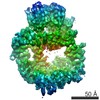
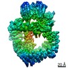
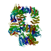
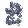
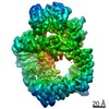
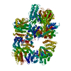
Journal: Proc Natl Acad Sci U S A / Year: 2017 Title: Cryo-EM structure of the DNA-PK holoenzyme. Authors: Humayun Sharif / Yang Li / Yuanchen Dong / Liyi Dong / Wei Li Wang / Youdong Mao / Hao Wu / Abstract: DNA-dependent protein kinase (DNA-PK) is a large protein complex central to the nonhomologous end joining (NHEJ) DNA-repair pathway. It comprises the DNA-PK catalytic subunit (DNA-PKcs) and the ...DNA-dependent protein kinase (DNA-PK) is a large protein complex central to the nonhomologous end joining (NHEJ) DNA-repair pathway. It comprises the DNA-PK catalytic subunit (DNA-PKcs) and the heterodimer of DNA-binding proteins Ku70 and Ku80. Here, we report the cryo-electron microscopy (cryo-EM) structures of human DNA-PKcs at 4.4-Å resolution and the DNA-PK holoenzyme at 5.8-Å resolution. The DNA-PKcs structure contains three distinct segments: the N-terminal region with an arm and a bridge, the circular cradle, and the head that includes the kinase domain. Two perpendicular apertures exist in the structure, which are sufficiently large for the passage of dsDNA. The DNA-PK holoenzyme cryo-EM map reveals density for the C-terminal globular domain of Ku80 that interacts with the arm of DNA-PKcs. The Ku80-binding site is adjacent to the previously identified density for the DNA-binding region of the Ku70/Ku80 complex, suggesting concerted DNA interaction by DNA-PKcs and the Ku complex.
History
Deposition
Jun 4, 2017
Deposition site: RCSB / Processing site: RCSB
Revision 1.0
Jul 19, 2017
Provider: repository / Type: Initial release
Revision 1.0
Jul 19, 2017
Data content type: EM metadata / Data content type: EM metadata / Provider: repository / Type: Initial release
Revision 1.0
Jul 19, 2017
Data content type: Image / Data content type: Image / Provider: repository / Type: Initial release
Revision 1.0
Jul 19, 2017
Data content type: Primary map / Data content type: Primary map / Provider: repository / Type: Initial release
Revision 1.0
Jul 19, 2017
Data content type: Image / Data content type: Image / Provider: repository / Type: Initial release
Revision 1.0
Jul 19, 2017
Data content type: Primary map / Data content type: Primary map / Provider: repository / Type: Initial release
Revision 1.0
Jul 19, 2017
Data content type: Image / Data content type: Image / Provider: repository / Type: Initial release
Revision 1.0
Jul 19, 2017
Data content type: Primary map / Data content type: Primary map / Provider: repository / Type: Initial release
Revision 1.0
Jul 19, 2017
Data content type: Image / Data content type: Image / Provider: repository / Type: Initial release
Revision 1.0
Jul 19, 2017
Data content type: Primary map / Data content type: Primary map / Provider: repository / Type: Initial release
Revision 1.0
Jul 19, 2017
Data content type: Image / Data content type: Image / Provider: repository / Type: Initial release
Revision 1.0
Jul 19, 2017
Data content type: Primary map / Data content type: Primary map / Provider: repository / Type: Initial release
Data content type: EM metadata / Data content type: EM metadata / EM metadata / Group: Data processing / Experimental summary / Data content type: EM metadata / EM metadata / Category: em_admin / em_software / Data content type: EM metadata / EM metadata / Item: _em_admin.last_update / _em_software.name
Data content type: EM metadata / Data content type: EM metadata / EM metadata / Group: Data processing / Experimental summary / Data content type: EM metadata / EM metadata / Category: em_admin / em_software / Data content type: EM metadata / EM metadata / Item: _em_admin.last_update / _em_software.name
In the structure databanks used in Yorodumi, some data are registered as the other names, "COVID-19 virus" and "2019-nCoV". Here are the details of the virus and the list of structure data.
Jan 31, 2019. EMDB accession codes are about to change! (news from PDBe EMDB page)
EMDB accession codes are about to change! (news from PDBe EMDB page)
The allocation of 4 digits for EMDB accession codes will soon come to an end. Whilst these codes will remain in use, new EMDB accession codes will include an additional digit and will expand incrementally as the available range of codes is exhausted. The current 4-digit format prefixed with “EMD-” (i.e. EMD-XXXX) will advance to a 5-digit format (i.e. EMD-XXXXX), and so on. It is currently estimated that the 4-digit codes will be depleted around Spring 2019, at which point the 5-digit format will come into force.
The EM Navigator/Yorodumi systems omit the EMD- prefix.
Related info.:Q: What is EMD? / ID/Accession-code notation in Yorodumi/EM Navigator
Yorodumi is a browser for structure data from EMDB, PDB, SASBDB, etc.
This page is also the successor to EM Navigator detail page, and also detail information page/front-end page for Omokage search.
The word "yorodu" (or yorozu) is an old Japanese word meaning "ten thousand". "mi" (miru) is to see.
Related info.:EMDB / PDB / SASBDB / Comparison of 3 databanks / Yorodumi Search / Aug 31, 2016. New EM Navigator & Yorodumi / Yorodumi Papers / Jmol/JSmol / Function and homology information / Changes in new EM Navigator and Yorodumi
 Movie
Movie Controller


 Open data
 Basic information
 Components
 Keywords
 Function and homology information
 Homo sapiens (human)
 Authors
 United States, 1items
  Citation
 Structure visualization
 Downloads & links
 Other downloads


 PDBj
PDBj


 Assembly
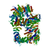
 Sample preparation
 Electron microscopy imaging
 FIELD EMISSION GUN / Accelerating voltage: 200 kV / Illumination mode: FLOOD BEAM
 Processing