 Movie
Movie Controller

[English] 日本語
 Yorodumi
- PDB-7ado: Cryo-EM structure of human ER membrane protein complex in lipid n... -
+ Open data

- Basic information
| Entry | Database: PDB / ID: 7ado | |||||||||
|---|---|---|---|---|---|---|---|---|---|---|
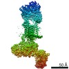
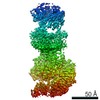
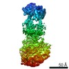
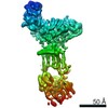
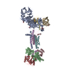
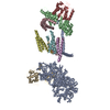
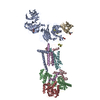
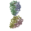
| Title | Cryo-EM structure of human ER membrane protein complex in lipid nanodiscs | |||||||||
 Components Components |
| |||||||||
 Keywords Keywords | MEMBRANE PROTEIN / ER membrane protein / EMC / Membrane protein biogenesis | |||||||||
| Function / homology |  Function and homology information Function and homology informationextrinsic component of endoplasmic reticulum membrane / EMC complex / : / omegasome membrane / protein insertion into ER membrane by stop-transfer membrane-anchor sequence / magnesium ion transport / tail-anchored membrane protein insertion into ER membrane / Miscellaneous transport and binding events / cobalt ion transmembrane transporter activity / ferrous iron transmembrane transporter activity ...extrinsic component of endoplasmic reticulum membrane / EMC complex / : / omegasome membrane / protein insertion into ER membrane by stop-transfer membrane-anchor sequence / magnesium ion transport / tail-anchored membrane protein insertion into ER membrane / Miscellaneous transport and binding events / cobalt ion transmembrane transporter activity / ferrous iron transmembrane transporter activity / copper ion transport / magnesium ion transmembrane transporter activity / RHOA GTPase cycle / autophagosome assembly / positive regulation of endothelial cell proliferation / positive regulation of endothelial cell migration / positive regulation of angiogenesis / carbohydrate binding / angiogenesis / early endosome membrane / early endosome / Golgi membrane / apoptotic process / endoplasmic reticulum membrane / endoplasmic reticulum / Golgi apparatus / protein-containing complex / extracellular region / membrane / plasma membrane / cytoplasm Similarity search - Function | |||||||||
| Biological species |  Homo sapiens (human) Homo sapiens (human) | |||||||||
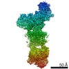
| Method | ELECTRON MICROSCOPY / single particle reconstruction / cryo EM / Resolution: 3.39 Å | |||||||||
 Authors Authors | Braeuning, B. / Prabu, J.R. / Miller-Vedam, L.E. / Weissman, J.S. / Frost, A. / Schulman, B.A. | |||||||||
| Funding support |  Germany, 2items Germany, 2items
| |||||||||
 Citation Citation | Journal: Elife / Year: 2020 Title: Structural and mechanistic basis of the EMC-dependent biogenesis of distinct transmembrane clients. Authors: Lakshmi E Miller-Vedam / Bastian Bräuning / Katerina D Popova / Nicole T Schirle Oakdale / Jessica L Bonnar / Jesuraj R Prabu / Elizabeth A Boydston / Natalia Sevillano / Matthew J ...Authors: Lakshmi E Miller-Vedam / Bastian Bräuning / Katerina D Popova / Nicole T Schirle Oakdale / Jessica L Bonnar / Jesuraj R Prabu / Elizabeth A Boydston / Natalia Sevillano / Matthew J Shurtleff / Robert M Stroud / Charles S Craik / Brenda A Schulman / Adam Frost / Jonathan S Weissman /  Abstract: Membrane protein biogenesis in the endoplasmic reticulum (ER) is complex and failure-prone. The ER membrane protein complex (EMC), comprising eight conserved subunits, has emerged as a central player ...Membrane protein biogenesis in the endoplasmic reticulum (ER) is complex and failure-prone. The ER membrane protein complex (EMC), comprising eight conserved subunits, has emerged as a central player in this process. Yet, we have limited understanding of how EMC enables insertion and integrity of diverse clients, from tail-anchored to polytopic transmembrane proteins. Here, yeast and human EMC cryo-EM structures reveal conserved intricate assemblies and human-specific features associated with pathologies. Structure-based functional studies distinguish between two separable EMC activities, as an insertase regulating tail-anchored protein levels and a broader role in polytopic membrane protein biogenesis. These depend on mechanistically coupled yet spatially distinct regions including two lipid-accessible membrane cavities which confer client-specific regulation, and a non-insertase EMC function mediated by the EMC lumenal domain. Our studies illuminate the structural and mechanistic basis of EMC's multifunctionality and point to its role in differentially regulating the biogenesis of distinct client protein classes. | |||||||||
| History |
|
- Structure visualization
| Movie |
Movie viewer |
|---|---|
| Structure viewer | Molecule: MolmilJmol/JSmol |
- Downloads & links
-Download
| PDBx/mmCIF format | 7ado.cif.gz | 397.7 KB | Display | PDBx/mmCIF format |
|---|---|---|---|---|
| PDB format | pdb7ado.ent.gz | 312.6 KB | Display | PDB format |
| PDBx/mmJSON format | 7ado.json.gz | Tree view | PDBx/mmJSON format | |
| Others |  Other downloads Other downloads |
-Validation report
| Arichive directory | https://data.pdbj.org/pub/pdb/validation_reports/ad/7adoftp://data.pdbj.org/pub/pdb/validation_reports/ad/7ado | HTTPS FTP |
|---|
-Related structure data
| Related structure data | 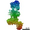 11732MC 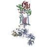 7adpC 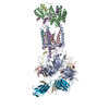 7kraC 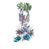 7ktxC M: map data used to model this data C: citing same article ( |
|---|---|
| Similar structure data |
-Links
 PDBj
PDBj


- Assembly
| Deposited unit | 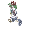
|
|---|---|
| 1 |
|
-Components
-ER membrane protein complex subunit ... , 8 types, 8 molecules ABCDFGHI
| #1: Protein | Mass: 111886.141 Da / Num. of mol.: 1 Source method: isolated from a genetically manipulated source Source: (gene. exp.) Homo sapiens (human) / Gene: EMC1, KIAA0090, PSEC0263 / Plasmid: pEG / Cell line (production host): HEK293 GnTI- / Production host: Homo sapiens (human) / References: UniProt: Q8N766 |
|---|---|
| #2: Protein | Mass: 34882.531 Da / Num. of mol.: 1 Source method: isolated from a genetically manipulated source Source: (gene. exp.) Homo sapiens (human) / Gene: EMC2, KIAA0103, TTC35 / Plasmid: pEG / Cell line (production host): HEK293 GnTI- / Production host: Homo sapiens (human) / References: UniProt: Q15006 |
| #3: Protein | Mass: 29981.924 Da / Num. of mol.: 1 Source method: isolated from a genetically manipulated source Source: (gene. exp.) Homo sapiens (human) / Gene: EMC3, TMEM111 / Plasmid: pEG / Cell line (production host): HEK293 GnTI- / Production host: Homo sapiens (human) / References: UniProt: Q9P0I2 |
| #4: Protein | Mass: 20104.572 Da / Num. of mol.: 1 Source method: isolated from a genetically manipulated source Details: There is no mutations but sequence from model and input are not aligning well. Source: (gene. exp.) Homo sapiens (human) / Gene: EMC4, TMEM85, HSPC184, PIG17 / Plasmid: pEG / Cell line (production host): HEK293 GnTI- / Production host: Homo sapiens (human) / References: UniProt: Q5J8M3 |
| #6: Protein | Mass: 12029.248 Da / Num. of mol.: 1 Source method: isolated from a genetically manipulated source Source: (gene. exp.) Homo sapiens (human) / Gene: EMC6, TMEM93 / Plasmid: pEG / Cell line (production host): HEK293 GnTI- / Production host: Homo sapiens (human) / References: UniProt: Q9BV81 |
| #7: Protein | Mass: 26501.586 Da / Num. of mol.: 1 Source method: isolated from a genetically manipulated source Source: (gene. exp.) Homo sapiens (human) / Gene: EMC7, C11orf3, C15orf24, HT022, UNQ905/PRO1926 / Plasmid: pEG / Cell line (production host): HEK293 GnTI- / Production host: Homo sapiens (human) / References: UniProt: Q9NPA0 |
| #8: Protein | Mass: 23807.076 Da / Num. of mol.: 1 Source method: isolated from a genetically manipulated source Details: Map modelled as EMC8; both EMC8 and its paralog EMC9 (44% identity) were co-expressed and turns out they can both occupy same position on complex in mutually exclusive manner. We chose to ...Details: Map modelled as EMC8; both EMC8 and its paralog EMC9 (44% identity) were co-expressed and turns out they can both occupy same position on complex in mutually exclusive manner. We chose to model as EMC8 but note that map is likely superposition of both. Source: (gene. exp.) Homo sapiens (human) / Gene: EMC8, C16orf2, C16orf4, COX4AL, COX4NB, FAM158B, NOC4 / Plasmid: pEG / Cell line (production host): HEK293 GnTI- / Production host: Homo sapiens (human) / References: UniProt: O43402 |
| #9: Protein | Mass: 27446.875 Da / Num. of mol.: 1 Source method: isolated from a genetically manipulated source Source: (gene. exp.) Homo sapiens (human) / Gene: EMC10, C19orf63, HSM1, INM02, UNQ764/PRO1556 / Plasmid: pEG / Cell line (production host): HEK293 GnTI- / Production host: Homo sapiens (human) / References: UniProt: Q5UCC4 |
-Protein / Protein/peptide / Sugars / Non-polymers , 4 types, 11 molecules EK

| #10: Protein/peptide | Mass: 1379.692 Da / Num. of mol.: 1 Source method: isolated from a genetically manipulated source Details: This is transmembrane alpha-helix which was modeled as poly-alanine. Source: (gene. exp.) Homo sapiens (human) / Plasmid: pEG / Production host: Homo sapiens (human) | ||||
|---|---|---|---|---|---|
| #11: Sugar | ChemComp-NAG /  Type: D-saccharide, beta linking / Mass: 221.208 Da / Num. of mol.: 4 / Source method: obtained synthetically / Formula: C8H15NO6 Type: D-saccharide, beta linking / Mass: 221.208 Da / Num. of mol.: 4 / Source method: obtained synthetically / Formula: C8H15NO6#12: Chemical | ChemComp-PCW /  Mass: 787.121 Da / Num. of mol.: 5 / Source method: obtained synthetically / Formula: C44H85NO8P / Comment: DOPC, phospholipid*YM Mass: 787.121 Da / Num. of mol.: 5 / Source method: obtained synthetically / Formula: C44H85NO8P / Comment: DOPC, phospholipid*YM#5: Protein | | Mass: 15703.762 Da / Num. of mol.: 1 Source method: isolated from a genetically manipulated source Source: (gene. exp.) Homo sapiens (human) / Gene: MMGT1, EMC5, TMEM32 / Plasmid: pEG / Cell line (production host): HEK293 GnTI- / Production host: Homo sapiens (human) / References: UniProt: Q8N4V1 |
-Details
| Has ligand of interest | N |
|---|---|
| Has protein modification | Y |
-Experimental details
-Experiment
| Experiment | Method: ELECTRON MICROSCOPY |
|---|---|
| EM experiment | Aggregation state: PARTICLE / 3D reconstruction method: single particle reconstruction |
- Sample preparation
| Component | Name: Human endoplasmic reticulum membrane protein complex (EMC) in POPC nanodiscs Type: COMPLEX / Entity ID: #1-#10 / Source: RECOMBINANT |
|---|---|
| Molecular weight | Value: 0.3 MDa |
| Source (natural) | Organism: Homo sapiens (human) |
| Source (recombinant) | Organism: Homo sapiens (human) / Strain: HEK293 GnTI- / Plasmid: pEG |
| Buffer solution | pH: 6 Details: 10 mM ammonium citrate pH 6.0, 100 mM sodium chloride, 0.25 mM TCEP |
| Specimen | Conc.: 2 mg/ml / Embedding applied: NO / Shadowing applied: NO / Staining applied: NO / Vitrification applied: YES |
| Specimen support | Grid material: COPPER / Grid mesh size: 300 divisions/in. / Grid type: Quantifoil R1.2/1.3 |
| Vitrification | Instrument: FEI VITROBOT MARK III / Cryogen name: ETHANE / Humidity: 100 % / Chamber temperature: 277 K |
- Electron microscopy imaging
| Experimental equipment |  Model: Titan Krios / Image courtesy: FEI Company |
|---|---|
| Microscopy | Model: FEI TITAN KRIOS / Date: Dec 23, 2019 |
| Electron gun | Electron source:  FIELD EMISSION GUN / Accelerating voltage: 300 kV / Illumination mode: FLOOD BEAM FIELD EMISSION GUN / Accelerating voltage: 300 kV / Illumination mode: FLOOD BEAM |
| Electron lens | Mode: BRIGHT FIELD |
| Image recording | Average exposure time: 3 sec. / Electron dose: 72 e/Å2 / Film or detector model: GATAN K3 BIOQUANTUM (6k x 4k) |
- Processing
| EM software |
| |||||||||||||||
|---|---|---|---|---|---|---|---|---|---|---|---|---|---|---|---|---|
| CTF correction | Type: PHASE FLIPPING AND AMPLITUDE CORRECTION | |||||||||||||||
| Symmetry | Point symmetry: C1 (asymmetric) | |||||||||||||||
| 3D reconstruction | Resolution: 3.39 Å / Resolution method: FSC 0.143 CUT-OFF / Num. of particles: 177560 / Symmetry type: POINT | |||||||||||||||
| Atomic model building | Protocol: AB INITIO MODEL |
