 Movie
Movie Controller

[English] 日本語
 Yorodumi
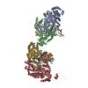
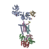
- PDB-6ww7: Structure of the human ER membrane protein complex in a lipid nanodisc -
+ Open data

- Basic information
| Entry | Database: PDB / ID: 6ww7 | |||||||||
|---|---|---|---|---|---|---|---|---|---|---|
| Title | Structure of the human ER membrane protein complex in a lipid nanodisc | |||||||||
 Components Components |
| |||||||||
 Keywords Keywords | MEMBRANE PROTEIN / Insertase / endoplasmic reticulum / transmembrane chaperone | |||||||||
| Function / homology |  Function and homology information Function and homology informationextrinsic component of endoplasmic reticulum membrane / : / EMC complex / omegasome membrane / protein insertion into ER membrane by stop-transfer membrane-anchor sequence / magnesium ion transport / tail-anchored membrane protein insertion into ER membrane / Miscellaneous transport and binding events / cobalt ion transmembrane transporter activity / ferrous iron transmembrane transporter activity ...extrinsic component of endoplasmic reticulum membrane / : / EMC complex / omegasome membrane / protein insertion into ER membrane by stop-transfer membrane-anchor sequence / magnesium ion transport / tail-anchored membrane protein insertion into ER membrane / Miscellaneous transport and binding events / cobalt ion transmembrane transporter activity / ferrous iron transmembrane transporter activity / copper ion transport / magnesium ion transmembrane transporter activity / RHOA GTPase cycle / autophagosome assembly / positive regulation of endothelial cell proliferation / positive regulation of endothelial cell migration / positive regulation of angiogenesis / carbohydrate binding / angiogenesis / early endosome membrane / early endosome / Golgi membrane / endoplasmic reticulum membrane / Golgi apparatus / endoplasmic reticulum / protein-containing complex / extracellular region / membrane / plasma membrane / cytoplasm Similarity search - Function | |||||||||
| Biological species |  Homo sapiens (human) Homo sapiens (human) | |||||||||
| Method | ELECTRON MICROSCOPY / single particle reconstruction / cryo EM / Resolution: 3.4 Å | |||||||||
 Authors Authors | Tomaleri, G.P. / Januszyk, K. / Pleiner, T. / Inglis, A.J. / Voorhees, R.M. | |||||||||
| Funding support |  United States, 1items United States, 1items
| |||||||||
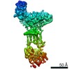
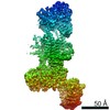
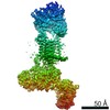
 Citation Citation | Journal: Science / Year: 2020 Title: Structural basis for membrane insertion by the human ER membrane protein complex. Authors: Tino Pleiner / Giovani Pinton Tomaleri / Kurt Januszyk / Alison J Inglis / Masami Hazu / Rebecca M Voorhees / Abstract: A defining step in the biogenesis of a membrane protein is the insertion of its hydrophobic transmembrane helices into the lipid bilayer. The nine-subunit endoplasmic reticulum (ER) membrane protein ...A defining step in the biogenesis of a membrane protein is the insertion of its hydrophobic transmembrane helices into the lipid bilayer. The nine-subunit endoplasmic reticulum (ER) membrane protein complex (EMC) is a conserved co- and posttranslational insertase at the ER. We determined the structure of the human EMC in a lipid nanodisc to an overall resolution of 3.4 angstroms by cryo-electron microscopy, permitting building of a nearly complete atomic model. We used structure-guided mutagenesis to demonstrate that substrate insertion requires a methionine-rich cytosolic loop and occurs via an enclosed hydrophilic vestibule within the membrane formed by the subunits EMC3 and EMC6. We propose that the EMC uses local membrane thinning and a positively charged patch to decrease the energetic barrier for insertion into the bilayer. | |||||||||
| History |
|
- Structure visualization
| Movie |
Movie viewer |
|---|---|
| Structure viewer | Molecule: MolmilJmol/JSmol |
- Downloads & links
-Download
| PDBx/mmCIF format | 6ww7.cif.gz | 385.4 KB | Display | PDBx/mmCIF format |
|---|---|---|---|---|
| PDB format | pdb6ww7.ent.gz | 303.5 KB | Display | PDB format |
| PDBx/mmJSON format | 6ww7.json.gz | Tree view | PDBx/mmJSON format | |
| Others |  Other downloads Other downloads |
-Validation report
| Arichive directory | https://data.pdbj.org/pub/pdb/validation_reports/ww/6ww7ftp://data.pdbj.org/pub/pdb/validation_reports/ww/6ww7 | HTTPS FTP |
|---|
-Related structure data
| Related structure data | 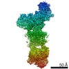 21929MC 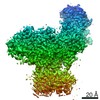 21930MC 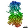 21931MC M: map data used to model this data C: citing same article ( |
|---|---|
| Similar structure data |
-Links
 PDBj
PDBj


- Assembly
| Deposited unit | 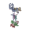
|
|---|---|
| 1 |
|
-Components
-ER membrane protein complex subunit ... , 8 types, 8 molecules ABCDFGHI
| #1: Protein | Mass: 111886.141 Da / Num. of mol.: 1 / Source method: isolated from a natural source / Source: (natural) Homo sapiens (human) / Cell line: HEK293 / References: UniProt: Q8N766 |
|---|---|
| #2: Protein | Mass: 34882.531 Da / Num. of mol.: 1 / Source method: isolated from a natural source / Source: (natural) Homo sapiens (human) / Cell line: HEK293 / References: UniProt: Q15006 |
| #3: Protein | Mass: 29981.924 Da / Num. of mol.: 1 / Source method: isolated from a natural source / Source: (natural) Homo sapiens (human) / Cell line: HEK293 / References: UniProt: Q9P0I2 |
| #4: Protein/peptide | Mass: 1209.482 Da / Num. of mol.: 1 / Source method: isolated from a natural source / Source: (natural) Homo sapiens (human) / Cell line: HEK293 |
| #6: Protein | Mass: 12029.248 Da / Num. of mol.: 1 / Source method: isolated from a natural source / Source: (natural) Homo sapiens (human) / Cell line: HEK293 / References: UniProt: Q9BV81 |
| #7: Protein | Mass: 26501.586 Da / Num. of mol.: 1 / Source method: isolated from a natural source / Source: (natural) Homo sapiens (human) / Cell line: HEK293 / References: UniProt: Q9NPA0 |
| #8: Protein | Mass: 23807.076 Da / Num. of mol.: 1 / Source method: isolated from a natural source / Source: (natural) Homo sapiens (human) / Cell line: HEK293 / References: UniProt: O43402 |
| #9: Protein | Mass: 27375.797 Da / Num. of mol.: 1 / Source method: isolated from a natural source / Source: (natural) Homo sapiens (human) / Cell line: HEK293 / References: UniProt: Q5UCC4 |
-Protein , 1 types, 1 molecules E
| #5: Protein | Mass: 14706.786 Da / Num. of mol.: 1 / Source method: isolated from a natural source / Source: (natural) Homo sapiens (human) / Cell line: HEK293 / References: UniProt: Q8N4V1 |
|---|
-Sugars , 2 types, 4 molecules 
| #10: Polysaccharide | Source method: isolated from a genetically manipulated source #11: Sugar |  Type: D-saccharide, beta linking / Mass: 221.208 Da / Num. of mol.: 2 Type: D-saccharide, beta linking / Mass: 221.208 Da / Num. of mol.: 2Source method: isolated from a genetically manipulated source Formula: C8H15NO6 |
|---|
-Details
| Has ligand of interest | Y |
|---|---|
| Has protein modification | Y |
-Experimental details
-Experiment
| Experiment | Method: ELECTRON MICROSCOPY |
|---|---|
| EM experiment | Aggregation state: PARTICLE / 3D reconstruction method: single particle reconstruction |
- Sample preparation
| Component | Name: Human ER Membrane Protein Complex / Type: COMPLEX / Entity ID: #1-#8 / Source: NATURAL | ||||||||||||||||||||||||
|---|---|---|---|---|---|---|---|---|---|---|---|---|---|---|---|---|---|---|---|---|---|---|---|---|---|
| Molecular weight | Experimental value: NO | ||||||||||||||||||||||||
| Source (natural) | Organism: Homo sapiens (human) / Cell: HEK293 | ||||||||||||||||||||||||
| Buffer solution | pH: 7.5 | ||||||||||||||||||||||||
| Buffer component |
| ||||||||||||||||||||||||
| Specimen | Conc.: 0.2 mg/ml / Embedding applied: NO / Shadowing applied: NO / Staining applied: NO / Vitrification applied: YES Details: Sample solubilized and purified in DDM, then reconstituted into lipid nanodisc. | ||||||||||||||||||||||||
| Specimen support | Grid material: GOLD / Grid mesh size: 300 divisions/in. / Grid type: Quantifoil R1.2/1.3 | ||||||||||||||||||||||||
| Vitrification | Cryogen name: ETHANE / Humidity: 95 % / Chamber temperature: 279 K |
- Electron microscopy imaging
| Experimental equipment |  Model: Titan Krios / Image courtesy: FEI Company |
|---|---|
| Microscopy | Model: FEI TITAN KRIOS |
| Electron gun | Electron source:  FIELD EMISSION GUN / Accelerating voltage: 300 kV / Illumination mode: SPOT SCAN FIELD EMISSION GUN / Accelerating voltage: 300 kV / Illumination mode: SPOT SCAN |
| Electron lens | Mode: DARK FIELD / Nominal magnification: 130000 X / Calibrated magnification: 59808 X / Cs: 2.7 mm / Alignment procedure: COMA FREE |
| Specimen holder | Cryogen: NITROGEN / Specimen holder model: FEI TITAN KRIOS AUTOGRID HOLDER |
| Image recording | Average exposure time: 2 sec. / Electron dose: 59.2 e/Å2 / Film or detector model: GATAN K3 (6k x 4k) / Num. of grids imaged: 10 / Num. of real images: 6345 |
| EM imaging optics | Energyfilter name: GIF Quantum LS / Energyfilter slit width: 20 eV |
- Processing
| EM software |
| |||||||||||||||||||||||||||||||||||
|---|---|---|---|---|---|---|---|---|---|---|---|---|---|---|---|---|---|---|---|---|---|---|---|---|---|---|---|---|---|---|---|---|---|---|---|---|
| CTF correction | Type: PHASE FLIPPING AND AMPLITUDE CORRECTION | |||||||||||||||||||||||||||||||||||
| Particle selection | Num. of particles selected: 1034250 | |||||||||||||||||||||||||||||||||||
| Symmetry | Point symmetry: C1 (asymmetric) | |||||||||||||||||||||||||||||||||||
| 3D reconstruction | Resolution: 3.4 Å / Resolution method: FSC 0.143 CUT-OFF / Num. of particles: 188746 / Symmetry type: POINT | |||||||||||||||||||||||||||||||||||
| Atomic model building | Space: REAL | |||||||||||||||||||||||||||||||||||
| Refinement | Stereochemistry target values: GeoStd + Monomer Library + CDL v1.2 | |||||||||||||||||||||||||||||||||||
| Displacement parameters | Biso mean: 64.57 Å2 | |||||||||||||||||||||||||||||||||||
| Refine LS restraints |
|
