- EMDB-21929: Human ER membrane protein complex in a lipid nanodisc, overall map -
+
Open data
ID or keywords:
Loading...
-
Basic information
Entry
Database: EMDB / ID: EMD-21929
Title
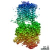
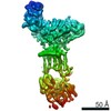
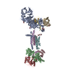
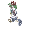
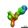
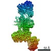
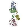
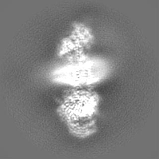
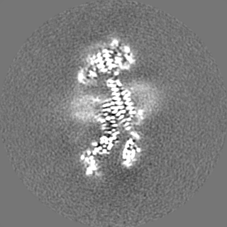
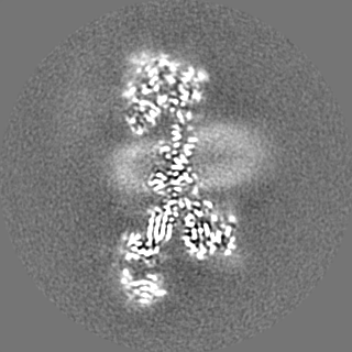
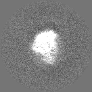
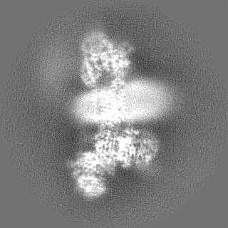
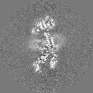
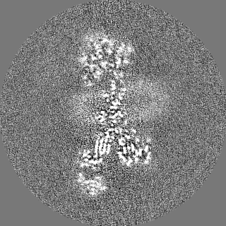
Human ER membrane protein complex in a lipid nanodisc, overall map
Map data
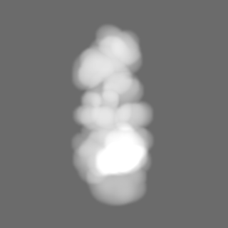
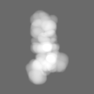
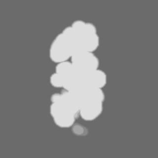
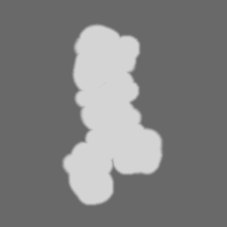
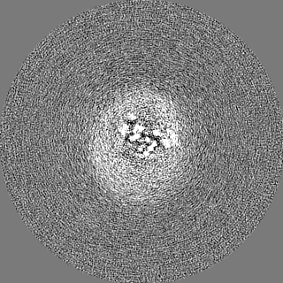
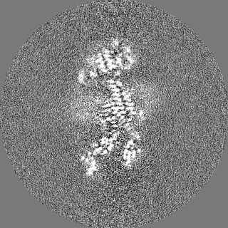
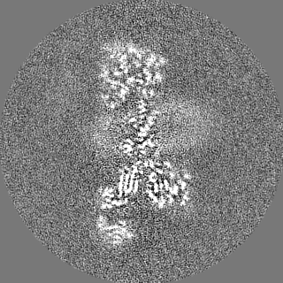
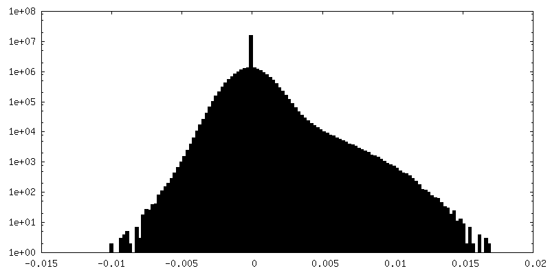
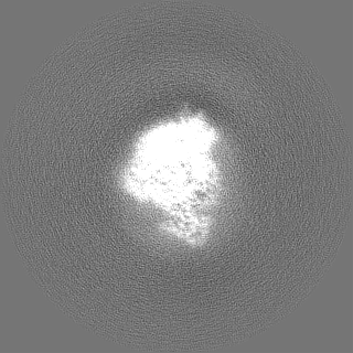
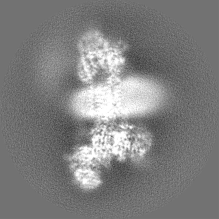
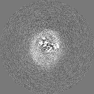
'Overall' human EMC post-processed, sharpened, and masked map.
Sample
Complex: Human ER Membrane Protein Complex
Protein or peptide: ER membrane protein complex subunit 1
Protein or peptide: ER membrane protein complex subunit 2
Protein or peptide: ER membrane protein complex subunit 3
Protein or peptide: ER Membrane Protein Complex Subunit 4
Protein or peptide: Membrane magnesium transporter 1
Protein or peptide: ER membrane protein complex subunit 6
Protein or peptide: ER membrane protein complex subunit 7
Protein or peptide: ER membrane protein complex subunit 8
Protein or peptide: ER membrane protein complex subunit 10
Ligand: 2-acetamido-2-deoxy-beta-D-glucopyranose
Keywords
Insertase / endoplasmic reticulum / transmembrane chaperone / MEMBRANE PROTEIN
Function / homology
Function and homology information
extrinsic component of endoplasmic reticulum membrane / EMC complex / : / omegasome membrane / protein insertion into ER membrane by stop-transfer membrane-anchor sequence / magnesium ion transport / tail-anchored membrane protein insertion into ER membrane / Miscellaneous transport and binding events / cobalt ion transmembrane transporter activity / ferrous iron transmembrane transporter activity ...extrinsic component of endoplasmic reticulum membrane / EMC complex / : / omegasome membrane / protein insertion into ER membrane by stop-transfer membrane-anchor sequence / magnesium ion transport / tail-anchored membrane protein insertion into ER membrane / Miscellaneous transport and binding events / cobalt ion transmembrane transporter activity / ferrous iron transmembrane transporter activity / copper ion transport / magnesium ion transmembrane transporter activity / RHOA GTPase cycle / autophagosome assembly / positive regulation of endothelial cell proliferation / positive regulation of endothelial cell migration / positive regulation of angiogenesis / carbohydrate binding / angiogenesis / early endosome membrane / early endosome / Golgi membrane / endoplasmic reticulum membrane / endoplasmic reticulum / Golgi apparatus / protein-containing complex / extracellular region / membrane / plasma membrane / cytoplasm Similarity search - Function
: / EMC1 N-terminal beta-propeller domain / ER membrane protein complex subunit 8/9 / : / Uncharacterised protein family (UPF0172) / EMC2 TPR-like repeat domain / ER membrane protein complex subunit 7, beta-sandwich domain / ER membrane protein complex subunit 7 / ER membrane protein complex subunit 7, beta-sandwich domain / ER membrane protein complex subunit 6 ...: / EMC1 N-terminal beta-propeller domain / ER membrane protein complex subunit 8/9 / : / Uncharacterised protein family (UPF0172) / EMC2 TPR-like repeat domain / ER membrane protein complex subunit 7, beta-sandwich domain / ER membrane protein complex subunit 7 / ER membrane protein complex subunit 7, beta-sandwich domain / ER membrane protein complex subunit 6 / ER membrane protein complex subunit 3 / ER membrane protein complex subunit 1, C-terminal / Membrane magnesium transporter / ER membrane protein complex subunit 1 / ER membrane protein complex subunit 6-like / EMC6 / ER membrane protein complex subunit 1, second beta-propeller / Membrane magnesium transporter / ER membrane protein complex subunit 10 / ER membrane protein complex subunit 2-like / Integral membrane protein EMC3/TMCO1-like / Integral membrane protein EMC3/TMCO1-like / Integral membrane protein DUF106 / Carbohydrate-binding-like fold / Quinoprotein alcohol dehydrogenase-like superfamily / TPR repeat region circular profile. / TPR repeat profile. / MPN domain / MPN domain profile. / Tetratricopeptide repeats / Tetratricopeptide repeat / Tetratricopeptide-like helical domain superfamily / WD40/YVTN repeat-like-containing domain superfamily Similarity search - Domain/homology
ER membrane protein complex subunit 8 / ER membrane protein complex subunit 2 / ER membrane protein complex subunit 10 / ER membrane protein complex subunit 5 / ER membrane protein complex subunit 1 / ER membrane protein complex subunit 6 / Endoplasmic reticulum membrane protein complex subunit 7 / ER membrane protein complex subunit 3 Similarity search - Component
Biological species
Homo sapiens (human)
Method
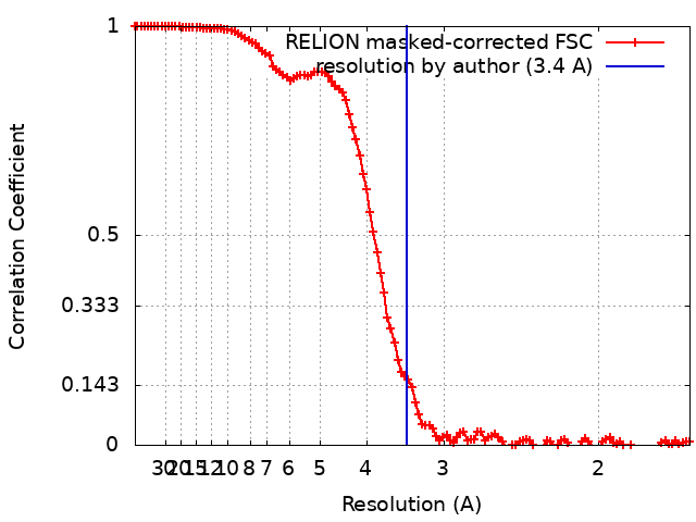
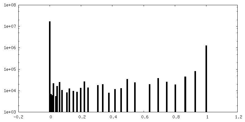
single particle reconstruction / cryo EM / Resolution: 3.4 Å
National Institutes of Health/National Institute of General Medical Sciences (NIH/NIGMS)
DP2GM137412
United States
Citation
Journal: Science / Year: 2020 Title: Structural basis for membrane insertion by the human ER membrane protein complex. Authors: Tino Pleiner / Giovani Pinton Tomaleri / Kurt Januszyk / Alison J Inglis / Masami Hazu / Rebecca M Voorhees / Abstract: A defining step in the biogenesis of a membrane protein is the insertion of its hydrophobic transmembrane helices into the lipid bilayer. The nine-subunit endoplasmic reticulum (ER) membrane protein ...A defining step in the biogenesis of a membrane protein is the insertion of its hydrophobic transmembrane helices into the lipid bilayer. The nine-subunit endoplasmic reticulum (ER) membrane protein complex (EMC) is a conserved co- and posttranslational insertase at the ER. We determined the structure of the human EMC in a lipid nanodisc to an overall resolution of 3.4 angstroms by cryo-electron microscopy, permitting building of a nearly complete atomic model. We used structure-guided mutagenesis to demonstrate that substrate insertion requires a methionine-rich cytosolic loop and occurs via an enclosed hydrophilic vestibule within the membrane formed by the subunits EMC3 and EMC6. We propose that the EMC uses local membrane thinning and a positively charged patch to decrease the energetic barrier for insertion into the bilayer.
History
Deposition
May 8, 2020
-
Header (metadata) release
May 27, 2020
-
Map release
May 27, 2020
-
Update
Nov 6, 2024
-
Current status
Nov 6, 2024
Processing site: RCSB / Status: Released
-
Structure visualization
Movie
Surface view with section colored by density value
Sample solubilized and purified in DDM, then reconstituted into lipid nanodisc.
-
Electron microscopy
Microscope
FEI TITAN KRIOS
Specialist optics
Energy filter - Name: GIF Quantum LS / Energy filter - Slit width: 20 eV
Image recording
Film or detector model: GATAN K3 (6k x 4k) / Number grids imaged: 10 / Number real images: 6345 / Average exposure time: 2.0 sec. / Average electron dose: 59.2 e/Å2
Electron beam
Acceleration voltage: 300 kV / Electron source: FIELD EMISSION GUN
Electron optics
Calibrated magnification: 59808 / Illumination mode: SPOT SCAN / Imaging mode: DARK FIELD / Cs: 2.7 mm / Nominal magnification: 130000
In the structure databanks used in Yorodumi, some data are registered as the other names, "COVID-19 virus" and "2019-nCoV". Here are the details of the virus and the list of structure data.
Jan 31, 2019. EMDB accession codes are about to change! (news from PDBe EMDB page)
EMDB accession codes are about to change! (news from PDBe EMDB page)
The allocation of 4 digits for EMDB accession codes will soon come to an end. Whilst these codes will remain in use, new EMDB accession codes will include an additional digit and will expand incrementally as the available range of codes is exhausted. The current 4-digit format prefixed with “EMD-” (i.e. EMD-XXXX) will advance to a 5-digit format (i.e. EMD-XXXXX), and so on. It is currently estimated that the 4-digit codes will be depleted around Spring 2019, at which point the 5-digit format will come into force.
The EM Navigator/Yorodumi systems omit the EMD- prefix.
Related info.:Q: What is EMD? / ID/Accession-code notation in Yorodumi/EM Navigator
Yorodumi is a browser for structure data from EMDB, PDB, SASBDB, etc.
This page is also the successor to EM Navigator detail page, and also detail information page/front-end page for Omokage search.
The word "yorodu" (or yorozu) is an old Japanese word meaning "ten thousand". "mi" (miru) is to see.
Related info.:EMDB / PDB / SASBDB / Comparison of 3 databanks / Yorodumi Search / Aug 31, 2016. New EM Navigator & Yorodumi / Yorodumi Papers / Jmol/JSmol / Function and homology information / Changes in new EM Navigator and Yorodumi
 Movie
Movie Controller

 Yorodumi
 Open data
 Basic information
 Map data
 Sample
 Keywords
 Function and homology information
 Homo sapiens (human)
 Authors
 United States, 1 items
  Citation
 Structure visualization
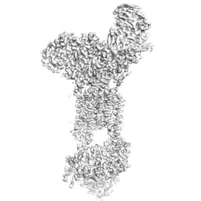
 Downloads & links
 emd_21929.png
 http://ftp.pdbj.org/pub/emdb/structures/EMD-21929


 Z (Sec.)
 Y (Row.)
 X (Col.)


 Sample components
 Processing
 Electron microscopy
 FIELD EMISSION GUN