- EMDB-23033: Cryo-EM structure of Saccharomyces cerevisiae ER membrane protein... -
+
Open data
ID or keywords:
Loading...
-
Basic information
Entry
Database: EMDB / ID: EMD-23033
Title
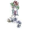
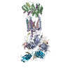
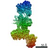
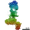
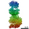
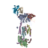
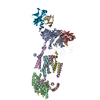
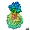
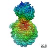
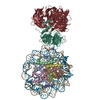
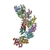
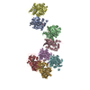
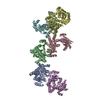
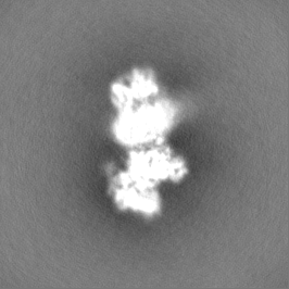
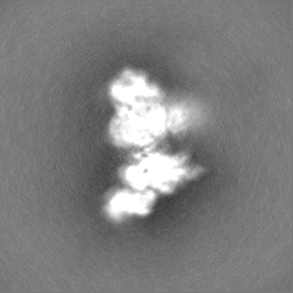
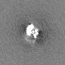
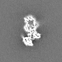
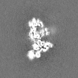
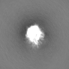
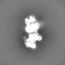
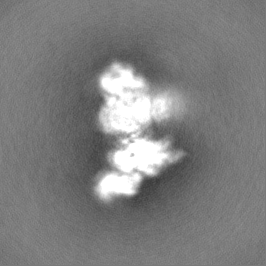
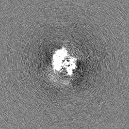
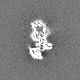
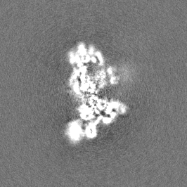
Cryo-EM structure of Saccharomyces cerevisiae ER membrane protein complex bound to a Fab in DDM detergent
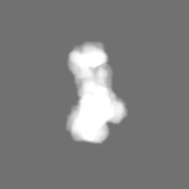
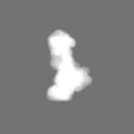
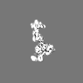
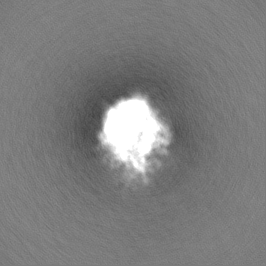
Map data
sharpened map
Sample
Complex: S. cerevisiae endoplasmic reticulum membrane protein complex (EMC) bound to Fab in DDM detergent
Complex: endoplasmic reticulum membrane protein complex
Protein or peptide: x 9 types
Complex: Fab DH4
Protein or peptide: x 2 types
Ligand: x 1 types
Keywords
ER membrane protein complex / EMC / membrane protein biogenesis / insertase / chaperone / endoplasmic / reticulum / MEMBRANE PROTEIN-IMMUNE SYSTEM complex
Function / homology
Function and homology information
EMC complex / protein insertion into ER membrane by stop-transfer membrane-anchor sequence / protein folding in endoplasmic reticulum / phospholipid transport / endoplasmic reticulum to Golgi vesicle-mediated transport / autophagosome assembly / phospholipid metabolic process / protein transport / protein-folding chaperone binding / endoplasmic reticulum membrane ...EMC complex / protein insertion into ER membrane by stop-transfer membrane-anchor sequence / protein folding in endoplasmic reticulum / phospholipid transport / endoplasmic reticulum to Golgi vesicle-mediated transport / autophagosome assembly / phospholipid metabolic process / protein transport / protein-folding chaperone binding / endoplasmic reticulum membrane / endoplasmic reticulum / membrane / nucleus Similarity search - Function
Protein Sop4 / : / Suppressor of PMA 1-7 protein / TMEM85/ER membrane protein complex subunit 4 / ER membrane protein complex subunit 4 / ER membrane protein complex subunit 6 / ER membrane protein complex subunit 3 / ER membrane protein complex subunit 1, C-terminal / Membrane magnesium transporter / ER membrane protein complex subunit 1 ...Protein Sop4 / : / Suppressor of PMA 1-7 protein / TMEM85/ER membrane protein complex subunit 4 / ER membrane protein complex subunit 4 / ER membrane protein complex subunit 6 / ER membrane protein complex subunit 3 / ER membrane protein complex subunit 1, C-terminal / Membrane magnesium transporter / ER membrane protein complex subunit 1 / ER membrane protein complex subunit 6-like / EMC6 / ER membrane protein complex subunit 1, second beta-propeller / Membrane magnesium transporter / ER membrane protein complex subunit 10 / ER membrane protein complex subunit 2-like / Integral membrane protein EMC3/TMCO1-like / Integral membrane protein EMC3/TMCO1-like / Integral membrane protein DUF106 / TPR repeat profile. / Tetratricopeptide repeat / Tetratricopeptide-like helical domain superfamily Similarity search - Domain/homology
ER membrane protein complex subunit 1 / ER membrane protein complex subunit 3 / Protein SOP4 / ER membrane protein complex subunit 5 / ER membrane protein complex subunit 2 / ER membrane protein complex subunit 4 / Endoplasmic reticulum membrane protein complex subunit 10 / ER membrane protein complex subunit 6 Similarity search - Component
Biological species
Saccharomyces cerevisiae BY4743 (yeast) / Homo sapiens (human)
Method
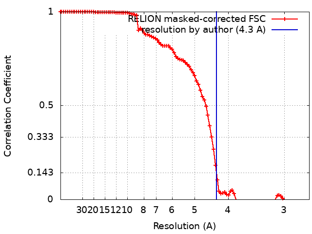
single particle reconstruction / cryo EM / Resolution: 4.3 Å
National Institutes of Health/Office of the Director
1DP2OD017690-01
United States
Citation
Journal: Elife / Year: 2020 Title: Structural and mechanistic basis of the EMC-dependent biogenesis of distinct transmembrane clients. Authors: Lakshmi E Miller-Vedam / Bastian Bräuning / Katerina D Popova / Nicole T Schirle Oakdale / Jessica L Bonnar / Jesuraj R Prabu / Elizabeth A Boydston / Natalia Sevillano / Matthew J ...Authors: Lakshmi E Miller-Vedam / Bastian Bräuning / Katerina D Popova / Nicole T Schirle Oakdale / Jessica L Bonnar / Jesuraj R Prabu / Elizabeth A Boydston / Natalia Sevillano / Matthew J Shurtleff / Robert M Stroud / Charles S Craik / Brenda A Schulman / Adam Frost / Jonathan S Weissman / Abstract: Membrane protein biogenesis in the endoplasmic reticulum (ER) is complex and failure-prone. The ER membrane protein complex (EMC), comprising eight conserved subunits, has emerged as a central player ...Membrane protein biogenesis in the endoplasmic reticulum (ER) is complex and failure-prone. The ER membrane protein complex (EMC), comprising eight conserved subunits, has emerged as a central player in this process. Yet, we have limited understanding of how EMC enables insertion and integrity of diverse clients, from tail-anchored to polytopic transmembrane proteins. Here, yeast and human EMC cryo-EM structures reveal conserved intricate assemblies and human-specific features associated with pathologies. Structure-based functional studies distinguish between two separable EMC activities, as an insertase regulating tail-anchored protein levels and a broader role in polytopic membrane protein biogenesis. These depend on mechanistically coupled yet spatially distinct regions including two lipid-accessible membrane cavities which confer client-specific regulation, and a non-insertase EMC function mediated by the EMC lumenal domain. Our studies illuminate the structural and mechanistic basis of EMC's multifunctionality and point to its role in differentially regulating the biogenesis of distinct client protein classes.
History
Deposition
Nov 24, 2020
-
Header (metadata) release
Dec 2, 2020
-
Map release
Dec 2, 2020
-
Update
Oct 30, 2024
-
Current status
Oct 30, 2024
Processing site: RCSB / Status: Released
-
Structure visualization
Movie
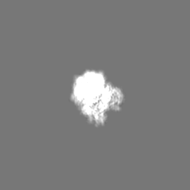
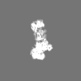
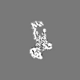
Surface view with section colored by density value
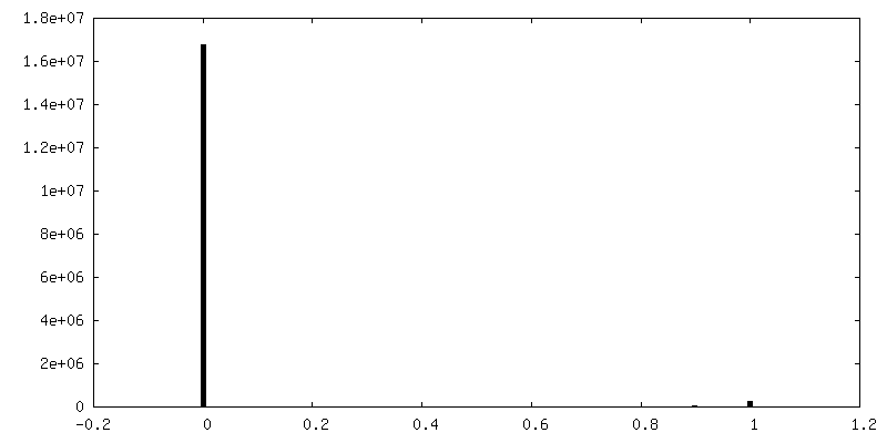
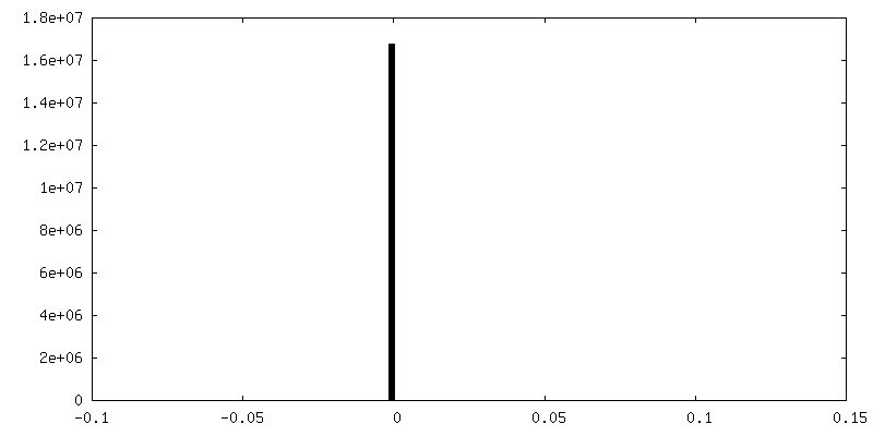
Resolution.type: BY AUTHOR / Resolution: 4.3 Å / Resolution method: FSC 0.143 CUT-OFF Details: These two datasets were processed independently initially. Upon selecting the best subset of data and then re-extracted and scaled to the same pixel size and combined to get to the final 4.3 ...Details: These two datasets were processed independently initially. Upon selecting the best subset of data and then re-extracted and scaled to the same pixel size and combined to get to the final 4.3 A resolution maps and model. Number images used: 83599
Initial angle assignment
Type: MAXIMUM LIKELIHOOD / Software - Name: RELION
Final angle assignment
Type: MAXIMUM LIKELIHOOD
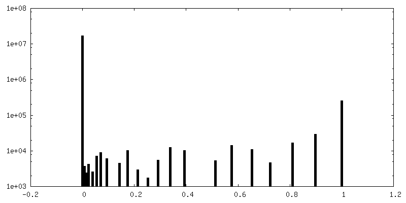
FSC plot (resolution estimation)
+
Image processing #2
Image processing ID
2
Image recording ID
2
Startup model
Type of model: INSILICO MODEL
Final reconstruction
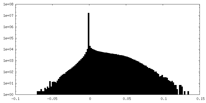
Resolution.type: BY AUTHOR / Resolution: 4.3 Å / Resolution method: FSC 0.143 CUT-OFF / Number images used: 170186
Initial angle assignment
Type: MAXIMUM LIKELIHOOD / Software - Name: RELION
Final angle assignment
Type: MAXIMUM LIKELIHOOD
FSC plot (resolution estimation)
-
Atomic model buiding 1
Refinement
Protocol: AB INITIO MODEL
Output model
PDB-7ktx: Cryo-EM structure of Saccharomyces cerevisiae ER membrane protein complex bound to a Fab in DDM detergent
+
About Yorodumi
-
News
-
Feb 9, 2022. New format data for meta-information of EMDB entries
New format data for meta-information of EMDB entries
Version 3 of the EMDB header file is now the official format.
The previous official version 1.9 will be removed from the archive.
In the structure databanks used in Yorodumi, some data are registered as the other names, "COVID-19 virus" and "2019-nCoV". Here are the details of the virus and the list of structure data.
Jan 31, 2019. EMDB accession codes are about to change! (news from PDBe EMDB page)
EMDB accession codes are about to change! (news from PDBe EMDB page)
The allocation of 4 digits for EMDB accession codes will soon come to an end. Whilst these codes will remain in use, new EMDB accession codes will include an additional digit and will expand incrementally as the available range of codes is exhausted. The current 4-digit format prefixed with “EMD-” (i.e. EMD-XXXX) will advance to a 5-digit format (i.e. EMD-XXXXX), and so on. It is currently estimated that the 4-digit codes will be depleted around Spring 2019, at which point the 5-digit format will come into force.
The EM Navigator/Yorodumi systems omit the EMD- prefix.
Related info.:Q: What is EMD? / ID/Accession-code notation in Yorodumi/EM Navigator
Yorodumi is a browser for structure data from EMDB, PDB, SASBDB, etc.
This page is also the successor to EM Navigator detail page, and also detail information page/front-end page for Omokage search.
The word "yorodu" (or yorozu) is an old Japanese word meaning "ten thousand". "mi" (miru) is to see.
Related info.:EMDB / PDB / SASBDB / Comparison of 3 databanks / Yorodumi Search / Aug 31, 2016. New EM Navigator & Yorodumi / Yorodumi Papers / Jmol/JSmol / Function and homology information / Changes in new EM Navigator and Yorodumi
 Movie
Movie Controller

 Yorodumi
 Open data
 Basic information
 Map data
 Sample
 Keywords
 Function and homology information
 Homo sapiens (human)
 Authors
 United States, 2 items
  Citation
 Structure visualization
 Downloads & links
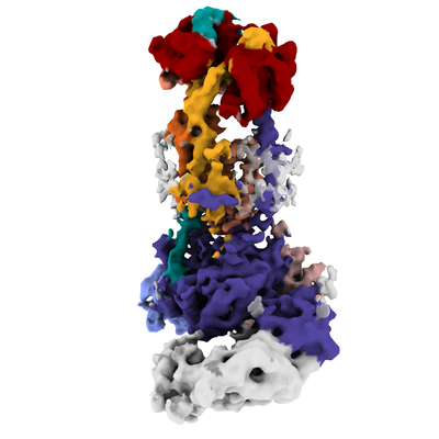 emd_23033.png
 http://ftp.pdbj.org/pub/emdb/structures/EMD-23033


 Z (Sec.)
 Y (Row.)
 X (Col.)


 Sample components

 Processing
 Electron microscopy #1
 FIELD EMISSION GUN