 Movie
Movie Controller


+ Open data

- Basic information
| Entry | Database: PDB / ID: 6znl | |||||||||
|---|---|---|---|---|---|---|---|---|---|---|
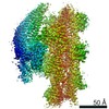
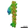
| Title | Cryo-EM structure of the dynactin complex | |||||||||
 Components Components |
| |||||||||
 Keywords Keywords | STRUCTURAL PROTEIN / Dynactin / Complex / Scaffold / Cytoskeleton | |||||||||
| Function / homology |  Function and homology information Function and homology informationAdvanced glycosylation endproduct receptor signaling / retrograde axonal transport of mitochondrion / Regulation of actin dynamics for phagocytic cup formation / EPHB-mediated forward signaling / Adherens junctions interactions / VEGFA-VEGFR2 Pathway / Cell-extracellular matrix interactions / RHO GTPases Activate WASPs and WAVEs / MAP2K and MAPK activation / RHOF GTPase cycle ...Advanced glycosylation endproduct receptor signaling / retrograde axonal transport of mitochondrion / Regulation of actin dynamics for phagocytic cup formation / EPHB-mediated forward signaling / Adherens junctions interactions / VEGFA-VEGFR2 Pathway / Cell-extracellular matrix interactions / RHO GTPases Activate WASPs and WAVEs / MAP2K and MAPK activation / RHOF GTPase cycle / Formation of the canonical BAF (cBAF) complex / Formation of the polybromo-BAF (pBAF) complex / Formation of the embryonic stem cell BAF (esBAF) complex / Formation of the non-canonical BAF (ncBAF) complex / GBP-mediated host defense / sterol sensor activity / Platelet degranulation / Gap junction degradation / Formation of annular gap junctions / UCH proteinases / dynactin complex / centriolar subdistal appendage / Clathrin-mediated endocytosis / centriole-centriole cohesion / positive regulation of neuromuscular junction development / Regulation of PLK1 Activity at G2/M Transition / Loss of Nlp from mitotic centrosomes / Loss of proteins required for interphase microtubule organization from the centrosome / Anchoring of the basal body to the plasma membrane / AURKA Activation by TPX2 / Recruitment of mitotic centrosome proteins and complexes / microtubule anchoring at centrosome / F-actin capping protein complex / WASH complex / Regulation of CDH1 Function / Formation of the dystrophin-glycoprotein complex (DGC) / lysosome to ER cholesterol transport / ventral spinal cord development / retromer complex / cytoskeleton-dependent cytokinesis / dynein complex / microtubule plus-end / mitotic nuclear membrane disassembly / positive regulation of microtubule nucleation / cellular response to cytochalasin B / regulation of transepithelial transport / morphogenesis of a polarized epithelium / structural constituent of postsynaptic actin cytoskeleton / protein localization to adherens junction / non-motile cilium assembly / barbed-end actin filament capping / dense body / Neutrophil degranulation / Tat protein binding / motor behavior / postsynaptic actin cytoskeleton / apical protein localization / neuron cellular homeostasis / retrograde transport, endosome to Golgi / adherens junction assembly / neuromuscular process / RHO GTPases activate IQGAPs / RHO GTPases Activate Formins / microtubule associated complex / HSP90 chaperone cycle for steroid hormone receptors (SHR) in the presence of ligand / MHC class II antigen presentation / nuclear migration / Recruitment of NuMA to mitotic centrosomes / tight junction / COPI-mediated anterograde transport / apical junction complex / intercellular bridge / establishment of mitotic spindle orientation / NuA4 histone acetyltransferase complex / regulation of norepinephrine uptake / transporter regulator activity / cell leading edge / cortical cytoskeleton / cleavage furrow / establishment or maintenance of cell polarity / neuromuscular junction development / nitric-oxide synthase binding / dynein complex binding / brush border / regulation of synaptic vesicle endocytosis / kinesin binding / regulation of protein localization to plasma membrane / microtubule-based process / positive regulation of double-strand break repair via homologous recombination / axonogenesis / stress fiber / cholesterol homeostasis / axon cytoplasm / neuron projection maintenance / centriole / calyx of Held / cell motility / nitric-oxide synthase regulator activity / mitotic spindle organization / regulation of mitotic spindle organization Similarity search - Function | |||||||||
| Biological species |   Sus scrofa (pig) Sus scrofa (pig) | |||||||||
| Method | ELECTRON MICROSCOPY / single particle reconstruction / cryo EM / Resolution: 3.8 Å | |||||||||
 Authors Authors | Lau, C.K. / Lacey, S.E. / Carter, A.P. | |||||||||
| Funding support |  United Kingdom, 2items United Kingdom, 2items
| |||||||||
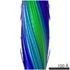
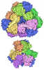
 Citation Citation | Journal: EMBO J / Year: 2021 Title: Cryo-EM reveals the complex architecture of dynactin's shoulder region and pointed end. Authors: Clinton K Lau / Francis J O'Reilly / Balaji Santhanam / Samuel E Lacey / Juri Rappsilber / Andrew P Carter /  Abstract: Dynactin is a 1.1 MDa complex that activates the molecular motor dynein for ultra-processive transport along microtubules. In order to do this, it forms a tripartite complex with dynein and a coiled- ...Dynactin is a 1.1 MDa complex that activates the molecular motor dynein for ultra-processive transport along microtubules. In order to do this, it forms a tripartite complex with dynein and a coiled-coil adaptor. Dynactin consists of an actin-related filament whose length is defined by its flexible shoulder domain. Despite previous cryo-EM structures, the molecular architecture of the shoulder and pointed end of the filament is still poorly understood due to the lack of high-resolution information in these regions. Here we combine multiple cryo-EM datasets and define precise masking strategies for particle signal subtraction and 3D classification. This overcomes domain flexibility and results in high-resolution maps into which we can build the shoulder and pointed end. The unique architecture of the shoulder securely houses the p150 subunit and positions the four identical p50 subunits in different conformations to bind dynactin's filament. The pointed end map allows us to build the first structure of p62 and reveals the molecular basis for cargo adaptor binding to different sites at the pointed end. | |||||||||
| History |
|
- Structure visualization
| Movie |
Movie viewer |
|---|---|
| Structure viewer | Molecule: MolmilJmol/JSmol |
- Downloads & links
-Download
| PDBx/mmCIF format | 6znl.cif.gz | 1.2 MB | Display | PDBx/mmCIF format |
|---|---|---|---|---|
| PDB format | pdb6znl.ent.gz | 974.9 KB | Display | PDB format |
| PDBx/mmJSON format | 6znl.json.gz | Tree view | PDBx/mmJSON format | |
| Others |  Other downloads Other downloads |
-Validation report
| Arichive directory | https://data.pdbj.org/pub/pdb/validation_reports/zn/6znlftp://data.pdbj.org/pub/pdb/validation_reports/zn/6znl | HTTPS FTP |
|---|
-Related structure data
| Related structure data | 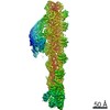 11313MC 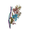 6znmC  6znnC  6znoC 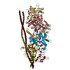 6zo4C C: citing same article ( M: map data used to model this data |
|---|---|
| Similar structure data |


-Links
 PDBj
PDBj



- Assembly
| Deposited unit | 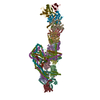
|
|---|---|
| 1 |
|
-Components
-Protein , 6 types, 13 molecules ABCDEFGIHJKLU
| #1: Protein | Mass: 42670.688 Da / Num. of mol.: 8 / Source method: isolated from a natural source / Source: (natural) Sus scrofa (pig) / References: UniProt: F2Z5G5#2: Protein | | Mass: 41782.660 Da / Num. of mol.: 1 / Source method: isolated from a natural source / Source: (natural) Sus scrofa (pig) / References: UniProt: Q6QAQ1#3: Protein | | Mass: 46250.785 Da / Num. of mol.: 1 / Source method: isolated from a natural source / Source: (natural) Sus scrofa (pig) / References: UniProt: I3LHK5#4: Protein | | Mass: 33059.848 Da / Num. of mol.: 1 / Source method: isolated from a natural source / Source: (natural) Sus scrofa (pig) / References: UniProt: A0PFK5#5: Protein | | Mass: 30669.768 Da / Num. of mol.: 1 / Source method: isolated from a natural source / Source: (natural) Sus scrofa (pig) / References: UniProt: A9XFX6#8: Protein | | Mass: 20703.910 Da / Num. of mol.: 1 / Source method: isolated from a natural source / Source: (natural) Sus scrofa (pig) / References: UniProt: D0G6S1 |
|---|
-Dynactin subunit ... , 5 types, 10 molecules MNmnOoVYZz
| #6: Protein | Mass: 44704.414 Da / Num. of mol.: 4 / Source method: isolated from a natural source / Source: (natural) Sus scrofa (pig) / References: UniProt: A0A5G2QD80#7: Protein | Mass: 21192.477 Da / Num. of mol.: 2 / Source method: isolated from a natural source / Source: (natural) Sus scrofa (pig) / References: UniProt: F1SEC0#9: Protein | | Mass: 20150.533 Da / Num. of mol.: 1 / Source method: isolated from a natural source / Source: (natural) Sus scrofa (pig) / References: UniProt: A0A286ZK88#10: Protein | | Mass: 52920.434 Da / Num. of mol.: 1 / Source method: isolated from a natural source / Source: (natural) Sus scrofa (pig) / References: UniProt: A0A4X1TB62#11: Protein | Mass: 142547.156 Da / Num. of mol.: 2 / Source method: isolated from a natural source / Source: (natural) Sus scrofa (pig) / References: UniProt: A0A287B8J2*PLUS |
|---|
-Non-polymers , 3 types, 13 molecules 

| #12: Chemical | ChemComp-ADP / 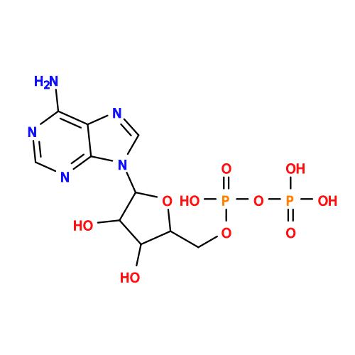 Mass: 427.201 Da / Num. of mol.: 9 / Source method: obtained synthetically / Formula: C10H15N5O10P2 / Comment: ADP, energy-carrying molecule*YM Mass: 427.201 Da / Num. of mol.: 9 / Source method: obtained synthetically / Formula: C10H15N5O10P2 / Comment: ADP, energy-carrying molecule*YM#13: Chemical | ChemComp-ATP / | 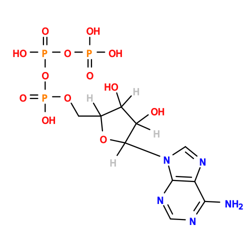 Mass: 507.181 Da / Num. of mol.: 1 / Source method: obtained synthetically / Formula: C10H16N5O13P3 / Comment: ATP, energy-carrying molecule*YM Mass: 507.181 Da / Num. of mol.: 1 / Source method: obtained synthetically / Formula: C10H16N5O13P3 / Comment: ATP, energy-carrying molecule*YM#14: Chemical |  Mass: 65.409 Da / Num. of mol.: 3 / Source method: obtained synthetically / Formula: Zn Mass: 65.409 Da / Num. of mol.: 3 / Source method: obtained synthetically / Formula: Zn |
|---|
-Details
| Has ligand of interest | N |
|---|
-Experimental details
-Experiment
| Experiment | Method: ELECTRON MICROSCOPY |
|---|---|
| EM experiment | Aggregation state: PARTICLE / 3D reconstruction method: single particle reconstruction |
- Sample preparation
| Component | Name: Dynactin complex / Type: COMPLEX / Entity ID: #1-#11 / Source: NATURAL |
|---|---|
| Molecular weight | Value: 1 MDa / Experimental value: NO |
| Source (natural) | Organism: Sus scrofa (pig) |
| Buffer solution | pH: 7.4 |
| Specimen | Embedding applied: NO / Shadowing applied: NO / Staining applied: NO / Vitrification applied: YES |
| Vitrification | Cryogen name: ETHANE |
- Electron microscopy imaging
| Experimental equipment |  Model: Titan Krios / Image courtesy: FEI Company |
|---|---|
| Microscopy | Model: FEI TITAN KRIOS |
| Electron gun | Electron source:  FIELD EMISSION GUN / Accelerating voltage: 300 kV / Illumination mode: FLOOD BEAM FIELD EMISSION GUN / Accelerating voltage: 300 kV / Illumination mode: FLOOD BEAM |
| Electron lens | Mode: BRIGHT FIELD |
| Image recording | Electron dose: 52 e/Å2 / Film or detector model: FEI FALCON II (4k x 4k) |
- Processing
| EM software |
| |||||||||
|---|---|---|---|---|---|---|---|---|---|---|
| Image processing | Details: All 4 image detectors used for reconstruction | |||||||||
| CTF correction | Type: PHASE FLIPPING AND AMPLITUDE CORRECTION | |||||||||
| 3D reconstruction | Resolution: 3.8 Å / Resolution method: FSC 0.143 CUT-OFF / Num. of particles: 336972 / Symmetry type: POINT | |||||||||
| Atomic model building | Protocol: AB INITIO MODEL |
