 Movie
Movie Controller

[English] 日本語
 Yorodumi
- EMDB-2237: Electron cyro-microscopy helical reconstruction of Par-3 N-termin... -
+ Open data

- Basic information
| Entry | Database: EMDB / ID: EMD-2237 | |||||||||
|---|---|---|---|---|---|---|---|---|---|---|
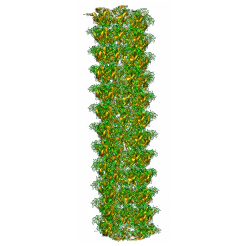
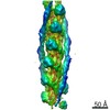
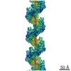
| Title | Electron cyro-microscopy helical reconstruction of Par-3 N-terminal domain | |||||||||
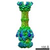
 Map data Map data | Reconstruction of Par3NTD | |||||||||
 Sample Sample |
| |||||||||
 Keywords Keywords | cell polarity / DUF3534 domain / self-association | |||||||||
| Function / homology |  Function and homology information Function and homology informationTight junction interactions / regulation of actin filament-based process / TGF-beta receptor signaling in EMT (epithelial to mesenchymal transition) / internode region of axon / regulation of cellular localization / apical constriction / PAR polarity complex / establishment of centrosome localization / establishment of epithelial cell polarity / lateral loop ...Tight junction interactions / regulation of actin filament-based process / TGF-beta receptor signaling in EMT (epithelial to mesenchymal transition) / internode region of axon / regulation of cellular localization / apical constriction / PAR polarity complex / establishment of centrosome localization / establishment of epithelial cell polarity / lateral loop / positive regulation of myelination / bicellular tight junction assembly / negative regulation of peptidyl-threonine phosphorylation / Schmidt-Lanterman incisure / establishment or maintenance of epithelial cell apical/basal polarity / myelination in peripheral nervous system / phosphatidylinositol-3-phosphate binding / wound healing, spreading of cells / centrosome localization / protein targeting to membrane / apical junction complex / establishment of cell polarity / cell leading edge / phosphatidylinositol-3,4,5-trisphosphate binding / positive regulation of receptor internalization / bicellular tight junction / axonal growth cone / phosphatidylinositol-4,5-bisphosphate binding / endomembrane system / phosphatidylinositol binding / adherens junction / microtubule cytoskeleton organization / spindle / cell-cell junction / cell junction / intracellular protein localization / protein phosphatase binding / cell cortex / cell adhesion / apical plasma membrane / cell division / neuronal cell body / protein-containing complex / identical protein binding / plasma membrane / cytoplasm Similarity search - Function | |||||||||
| Biological species |  | |||||||||
| Method | helical reconstruction / cryo EM / Resolution: 6.1 Å | |||||||||
 Authors Authors | Yan Z / Wenjuan W / Jia C / Kai Z / Feng G / Weimin G / Mingjie Z / Fei S / Wei F | |||||||||
 Citation Citation | Journal: Structure / Year: 2013 Title: Structural insights into the intrinsic self-assembly of Par-3 N-terminal domain. Authors: Yan Zhang / Wenjuan Wang / Jia Chen / Kai Zhang / Feng Gao / Bingquan Gao / Shuai Zhang / Mingdong Dong / Flemming Besenbacher / Weimin Gong / Mingjie Zhang / Fei Sun / Wei Feng /  Abstract: Par-3, the central organizer of the Par-3/Par-6/atypical protein kinase C complex, is a multimodular scaffold protein that is essential for cell polarity establishment and maintenance. The N-terminal ...Par-3, the central organizer of the Par-3/Par-6/atypical protein kinase C complex, is a multimodular scaffold protein that is essential for cell polarity establishment and maintenance. The N-terminal domain (NTD) of Par-3 is capable of self-association to form filament-like structures, although the underlying mechanism is poorly understood. Here, we determined the crystal structure of Par-3 NTD and solved the filament structure by cryoelectron microscopy. We found that an intrinsic "front-to-back" interaction mode is important for Par-3 NTD self-association and that both the lateral and longitudinal packing within the filament are mediated by electrostatic interactions. Disruptions of the lateral or longitudinal packing significantly impaired Par-3 NTD self-association and thereby impacted the Par-3-mediated epithelial polarization. We finally demonstrated that a Par-3 NTD-like domain from histidine ammonia-lyase also harbors a similar self-association capacity. This work unequivocally provides the structural basis for Par-3 NTD self-association and characterizes one type of protein domain that can self-assemble via electrostatic interactions. | |||||||||
| History |
|
- Structure visualization
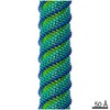
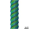
| Movie |
Movie viewer |
|---|---|
| Structure viewer | EM map: SurfViewMolmilJmol/JSmol |
| Supplemental images |
- Downloads & links
-EMDB archive
| Map data | emd_2237.map.gz | 165.7 MB | EMDB map data format | |
|---|---|---|---|---|
| Header (meta data) | emd-2237-v30.xmlemd-2237.xml | 12.8 KB 12.8 KB | Display Display | EMDB header |
| Images | emd_2237.tif | 246.1 KB | ||
| Archive directory |  http://ftp.pdbj.org/pub/emdb/structures/EMD-2237ftp://ftp.pdbj.org/pub/emdb/structures/EMD-2237 http://ftp.pdbj.org/pub/emdb/structures/EMD-2237ftp://ftp.pdbj.org/pub/emdb/structures/EMD-2237 | HTTPS FTP |
-Related structure data
| Related structure data |  3zeeMC  4i6pC M: atomic model generated by this map C: citing same article ( |
|---|---|
| Similar structure data |


-Links
| EMDB pages | EMDB (EBI/PDBe) / EMDataResource |
|---|
-Map
| File | Download / File: emd_2237.map.gz / Format: CCP4 / Size: 173.8 MB / Type: IMAGE STORED AS FLOATING POINT NUMBER (4 BYTES) | ||||||||||||||||||||||||||||||||||||||||||||||||||||||||||||||||||||
|---|---|---|---|---|---|---|---|---|---|---|---|---|---|---|---|---|---|---|---|---|---|---|---|---|---|---|---|---|---|---|---|---|---|---|---|---|---|---|---|---|---|---|---|---|---|---|---|---|---|---|---|---|---|---|---|---|---|---|---|---|---|---|---|---|---|---|---|---|---|
| Annotation | Reconstruction of Par3NTD | ||||||||||||||||||||||||||||||||||||||||||||||||||||||||||||||||||||
| Projections & slices | Image control
Images are generated by Spider. | ||||||||||||||||||||||||||||||||||||||||||||||||||||||||||||||||||||
| Voxel size | X=Y=Z: 0.933 Å | ||||||||||||||||||||||||||||||||||||||||||||||||||||||||||||||||||||
| Density |
| ||||||||||||||||||||||||||||||||||||||||||||||||||||||||||||||||||||
| Symmetry | Space group: 1 | ||||||||||||||||||||||||||||||||||||||||||||||||||||||||||||||||||||
| Details | EMDB XML:
CCP4 map header:
| ||||||||||||||||||||||||||||||||||||||||||||||||||||||||||||||||||||
 Z (Sec.)
 Y (Row.)
 X (Col.)


-Supplemental data
- Sample components
-Entire : Par-3 N-terminal DUF3534 domain
| Entire | Name: Par-3 N-terminal DUF3534 domain |
|---|---|
| Components |
|
-Supramolecule #1000: Par-3 N-terminal DUF3534 domain
| Supramolecule | Name: Par-3 N-terminal DUF3534 domain / type: sample / ID: 1000 / Oligomeric state: helical filament assembly / Number unique components: 95 |
|---|---|
| Molecular weight | Theoretical: 938 KDa |
-Macromolecule #1: Par-3 N-terminal DUF3534 domain
| Macromolecule | Name: Par-3 N-terminal DUF3534 domain / type: protein_or_peptide / ID: 1 / Name.synonym: Par-3 NTD / Number of copies: 95 / Oligomeric state: helical filament assembly / Recombinant expression: Yes |
|---|---|
| Source (natural) | Organism: |
| Molecular weight | Theoretical: 938 KDa |
| Recombinant expression | Organism:  |
-Experimental details
-Structure determination
| Method | cryo EM |
|---|---|
 Processing Processing | helical reconstruction |
| Aggregation state | filament |
-Sample preparation
| Concentration | 2.0 mg/mL |
|---|---|
| Buffer | pH: 8 / Details: 50 mM Tris, 100 mM NaCl, 1 mM DTT and 1 mM EDTA |
| Grid | Details: 300-mesh GiGTM holy carbon grid |
| Vitrification | Cryogen name: ETHANE / Chamber humidity: 100 % / Instrument: FEI VITROBOT MARK IV / Method: blotted 3.0 s with a blot force 3 |
- Electron microscopy
| Microscope | FEI TITAN KRIOS |
|---|---|
| Temperature | Average: 85 K |
| Alignment procedure | Legacy - Astigmatism: objective lens astigmatism was corrected at 96,000 times magnification Legacy - Electron beam tilt params: 0 |
| Date | Dec 1, 2010 |
| Image recording | Category: CCD / Film or detector model: GATAN ULTRASCAN 4000 (4k x 4k) / Number real images: 6460 / Average electron dose: 20 e/Å2 Details: The images were automatically collected by using leginon system. Bits/pixel: 32 |
| Tilt angle min | 0 |
| Tilt angle max | 0 |
| Electron beam | Acceleration voltage: 300 kV / Electron source:  FIELD EMISSION GUN FIELD EMISSION GUN |
| Electron optics | Illumination mode: FLOOD BEAM / Imaging mode: BRIGHT FIELD / Cs: 2.7 mm / Nominal defocus max: -2.5 µm / Nominal defocus min: -1.8 µm / Nominal magnification: 96000 |
| Sample stage | Specimen holder: Liquid nitrogen cooled / Specimen holder model: FEI TITAN KRIOS AUTOGRID HOLDER |
| Experimental equipment |  Model: Titan Krios / Image courtesy: FEI Company |
-Image processing
| Details | The initial model was obtained IHRSR. Then particles were aligned using EMAN1. |
|---|---|
| Final reconstruction | Applied symmetry - Helical parameters - Δz: 3.53 Å Applied symmetry - Helical parameters - Δ&Phi: 43.84 ° Applied symmetry - Helical parameters - Axial symmetry: C1 (asymmetric) Algorithm: OTHER / Resolution.type: BY AUTHOR / Resolution: 6.1 Å / Resolution method: OTHER / Software - Name: Spider, EMAN1 Details: The resolution was assessed by splitting original particles set into two halves and comparing two independent reconstructions. The final map was reconstructed by using the whole set. |
| CTF correction | Details: each image |
| Final angle assignment | Details: Euler angle system in EMAN1 |
-Atomic model buiding 1
| Initial model | PDB ID: Chain - #0 - Chain ID: A / Chain - #1 - Chain ID: B |
|---|---|
| Software | Name: Chimera and NAMD2 |
| Details | Protocol: Rigid body fitting first and then molecular dynamics flexible fitting. Crystal structure of the monomer was docked as a rigid body into the cryoEM map using UCSF Chimera and applied with the helical symmetry to build the initial model. Then the structural model was solvated in a box of water molecules with 150 mM NaCl in VMD, using 15 angstrom of padding in all directions. Extra ions were added to neutralize the systems. The simulations were performed with program NAMD 2.8, using the CHARMM27 force field with CMAP corrections. All the fitting procedure is the same as the application of symmetry-restrained MDFF to chaperonin reported previously. |
| Refinement | Space: REAL / Protocol: FLEXIBLE FIT / Target criteria: cross correlation coefficient |
| Output model | PDB-3zee: |
