 Movie
Movie Controller


+ Open data

- Basic information
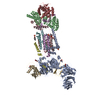
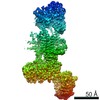
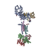
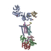
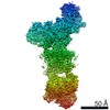
| Entry | Database: PDB / ID: 6z3w | |||||||||
|---|---|---|---|---|---|---|---|---|---|---|
| Title | Human ER membrane protein complex | |||||||||
 Components Components |
| |||||||||
 Keywords Keywords | MEMBRANE PROTEIN / Complex / insertase / PROTEIN TRANSPORT | |||||||||
| Function / homology |  Function and homology information Function and homology informationextrinsic component of endoplasmic reticulum membrane / EMC complex / : / omegasome membrane / protein insertion into ER membrane by stop-transfer membrane-anchor sequence / magnesium ion transport / tail-anchored membrane protein insertion into ER membrane / Miscellaneous transport and binding events / cobalt ion transmembrane transporter activity / ferrous iron transmembrane transporter activity ...extrinsic component of endoplasmic reticulum membrane / EMC complex / : / omegasome membrane / protein insertion into ER membrane by stop-transfer membrane-anchor sequence / magnesium ion transport / tail-anchored membrane protein insertion into ER membrane / Miscellaneous transport and binding events / cobalt ion transmembrane transporter activity / ferrous iron transmembrane transporter activity / copper ion transport / magnesium ion transmembrane transporter activity / RHOA GTPase cycle / autophagosome assembly / positive regulation of endothelial cell proliferation / positive regulation of endothelial cell migration / positive regulation of angiogenesis / carbohydrate binding / angiogenesis / early endosome membrane / early endosome / Golgi membrane / apoptotic process / endoplasmic reticulum membrane / Golgi apparatus / endoplasmic reticulum / protein-containing complex / extracellular region / membrane / plasma membrane / cytoplasm Similarity search - Function | |||||||||
| Biological species |  Homo sapiens (human) Homo sapiens (human) | |||||||||
| Method | ELECTRON MICROSCOPY / single particle reconstruction / cryo EM / Resolution: 6.4 Å | |||||||||
 Authors Authors | Hegde, R.S. / O'Donnell, J.P. | |||||||||
| Funding support |  United Kingdom, United Kingdom,  Germany, 2items Germany, 2items
| |||||||||
 Citation Citation | Journal: Elife / Year: 2020 Title: The architecture of EMC reveals a path for membrane protein insertion. Authors: John P O'Donnell / Ben P Phillips / Yuichi Yagita / Szymon Juszkiewicz / Armin Wagner / Duccio Malinverni / Robert J Keenan / Elizabeth A Miller / Ramanujan S Hegde /  Abstract: Approximately 25% of eukaryotic genes code for integral membrane proteins that are assembled at the endoplasmic reticulum. An abundant and widely conserved multi-protein complex termed EMC has been ...Approximately 25% of eukaryotic genes code for integral membrane proteins that are assembled at the endoplasmic reticulum. An abundant and widely conserved multi-protein complex termed EMC has been implicated in membrane protein biogenesis, but its mechanism of action is poorly understood. Here, we define the composition and architecture of human EMC using biochemical assays, crystallography of individual subunits, site-specific photocrosslinking, and cryo-EM reconstruction. Our results suggest that EMC's cytosolic domain contains a large, moderately hydrophobic vestibule that can bind a substrate's transmembrane domain (TMD). The cytosolic vestibule leads into a lumenally-sealed, lipid-exposed intramembrane groove large enough to accommodate a single substrate TMD. A gap between the cytosolic vestibule and intramembrane groove provides a potential path for substrate egress from EMC. These findings suggest how EMC facilitates energy-independent membrane insertion of TMDs, explain why only short lumenal domains are translocated by EMC, and constrain models of EMC's proposed chaperone function. | |||||||||
| History |
|
- Structure visualization
| Movie |
Movie viewer |
|---|---|
| Structure viewer | Molecule: MolmilJmol/JSmol |
- Downloads & links
-Download
| PDBx/mmCIF format | 6z3w.cif.gz | 278.3 KB | Display | PDBx/mmCIF format |
|---|---|---|---|---|
| PDB format | pdb6z3w.ent.gz | 188.2 KB | Display | PDB format |
| PDBx/mmJSON format | 6z3w.json.gz | Tree view | PDBx/mmJSON format | |
| Others |  Other downloads Other downloads |
-Validation report
| Arichive directory | https://data.pdbj.org/pub/pdb/validation_reports/z3/6z3wftp://data.pdbj.org/pub/pdb/validation_reports/z3/6z3w | HTTPS FTP |
|---|
-Related structure data
| Related structure data | 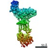 11058MC 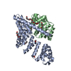 6y4lC C: citing same article ( M: map data used to model this data |
|---|---|
| Similar structure data |

-Links
 PDBj
PDBj


- Assembly
| Deposited unit | 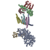
|
|---|---|
| 1 |
|
-Components
-ER membrane protein complex subunit ... , 8 types, 8 molecules ABCDFGHI
| #1: Protein | Mass: 86666.734 Da / Num. of mol.: 1 Source method: isolated from a genetically manipulated source Source: (gene. exp.) Homo sapiens (human) / Gene: EMC1, KIAA0090, PSEC0263 / Cell line (production host): HEK293 / Production host: Homo sapiens (human) / References: UniProt: Q8N766 |
|---|---|
| #2: Protein | Mass: 34882.531 Da / Num. of mol.: 1 Source method: isolated from a genetically manipulated source Source: (gene. exp.) Homo sapiens (human) / Cell line: HEK293 / Gene: EMC2, KIAA0103, TTC35 / Cell line (production host): HEK293 / Production host: Homo sapiens (human) / References: UniProt: Q15006 |
| #3: Protein | Mass: 29981.924 Da / Num. of mol.: 1 Source method: isolated from a genetically manipulated source Source: (gene. exp.) Homo sapiens (human) / Gene: EMC3, TMEM111 / Cell line (production host): HEK293 / Production host: Homo sapiens (human) / References: UniProt: Q9P0I2 |
| #4: Protein | Mass: 20104.572 Da / Num. of mol.: 1 Source method: isolated from a genetically manipulated source Source: (gene. exp.) Homo sapiens (human) / Gene: EMC4, TMEM85, HSPC184, PIG17 / Cell line (production host): HEK293 / Production host: Homo sapiens (human) / References: UniProt: Q5J8M3 |
| #6: Protein | Mass: 12029.248 Da / Num. of mol.: 1 Source method: isolated from a genetically manipulated source Source: (gene. exp.) Homo sapiens (human) / Gene: EMC6, TMEM93 / Cell line (production host): HEK293 / Production host: Homo sapiens (human) / References: UniProt: Q9BV81 |
| #7: Protein | Mass: 26501.586 Da / Num. of mol.: 1 Source method: isolated from a genetically manipulated source Source: (gene. exp.) Homo sapiens (human) / Gene: EMC7, C11orf3, C15orf24, HT022, UNQ905/PRO1926 / Cell line (production host): HEK293 / Production host: Homo sapiens (human) / References: UniProt: Q9NPA0 |
| #8: Protein | Mass: 23084.434 Da / Num. of mol.: 1 Source method: isolated from a genetically manipulated source Source: (gene. exp.) Homo sapiens (human) / Gene: EMC9, C14orf122, FAM158A, CGI-112 / Cell line (production host): HEK293 / Production host: Homo sapiens (human) / References: UniProt: Q9Y3B6 |
| #9: Protein | Mass: 27375.797 Da / Num. of mol.: 1 Source method: isolated from a genetically manipulated source Source: (gene. exp.) Homo sapiens (human) / Gene: EMC10, C19orf63, HSM1, INM02, UNQ764/PRO1556 / Cell line (production host): HEK293 / Production host: Homo sapiens (human) / References: UniProt: Q5UCC4 |
-Protein , 1 types, 1 molecules E
| #5: Protein | Mass: 14706.786 Da / Num. of mol.: 1 Source method: isolated from a genetically manipulated source Source: (gene. exp.) Homo sapiens (human) / Gene: MMGT1, EMC5, TMEM32 / Cell line (production host): HEK293 / Production host: Homo sapiens (human) / References: UniProt: Q8N4V1 |
|---|
-Experimental details
-Experiment
| Experiment | Method: ELECTRON MICROSCOPY |
|---|---|
| EM experiment | Aggregation state: PARTICLE / 3D reconstruction method: single particle reconstruction |
- Sample preparation
| Component | Name: Human ER membrane protein complex / Type: COMPLEX / Entity ID: all / Source: RECOMBINANT | |||||||||
|---|---|---|---|---|---|---|---|---|---|---|
| Molecular weight |
| |||||||||
| Source (natural) | Organism: Homo sapiens (human) | |||||||||
| Source (recombinant) | Organism: Homo sapiens (human) | |||||||||
| Buffer solution | pH: 7.5 | |||||||||
| Specimen | Embedding applied: NO / Shadowing applied: NO / Staining applied: NO / Vitrification applied: YES | |||||||||
| Vitrification | Cryogen name: ETHANE |
- Electron microscopy imaging
| Experimental equipment |  Model: Titan Krios / Image courtesy: FEI Company |
|---|---|
| Microscopy | Model: FEI TITAN KRIOS |
| Electron gun | Electron source:  FIELD EMISSION GUN / Accelerating voltage: 300 kV / Illumination mode: FLOOD BEAM FIELD EMISSION GUN / Accelerating voltage: 300 kV / Illumination mode: FLOOD BEAM |
| Electron lens | Mode: BRIGHT FIELD |
| Image recording | Electron dose: 40 e/Å2 / Film or detector model: GATAN K2 SUMMIT (4k x 4k) |
- Processing
| Software |
| ||||||||||||||||||||||||
|---|---|---|---|---|---|---|---|---|---|---|---|---|---|---|---|---|---|---|---|---|---|---|---|---|---|
| CTF correction | Type: PHASE FLIPPING AND AMPLITUDE CORRECTION | ||||||||||||||||||||||||
| 3D reconstruction | Resolution: 6.4 Å / Resolution method: FSC 0.143 CUT-OFF / Num. of particles: 167294 / Symmetry type: POINT | ||||||||||||||||||||||||
| Refinement | Cross valid method: NONE Stereochemistry target values: GeoStd + Monomer Library + CDL v1.2 | ||||||||||||||||||||||||
| Displacement parameters | Biso mean: 200 Å2 | ||||||||||||||||||||||||
| Refine LS restraints |
|
