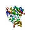
- PDB-6y4l: Crystal structure of human ER membrane protein complex subunits E... -
+
Open data
ID or keywords:
Loading...
-
Basic information
Entry
Database: PDB / ID: 6y4l
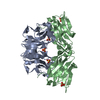
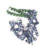
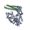
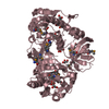
Title
Crystal structure of human ER membrane protein complex subunits EMC2 and EMC9
Components
(ER membrane protein complex subunit ...) x 2
Keywords
PROTEIN TRANSPORT / Complex / insertase
Function / homology
Function and homology information
extrinsic component of endoplasmic reticulum membrane / EMC complex / protein insertion into ER membrane by stop-transfer membrane-anchor sequence / tail-anchored membrane protein insertion into ER membrane / endoplasmic reticulum membrane / endoplasmic reticulum / cytoplasm Similarity search - Function
ER membrane protein complex subunit 8/9 / : / Uncharacterised protein family (UPF0172) / EMC2 TPR-like repeat domain / ER membrane protein complex subunit 2-like / TPR repeat region circular profile. / TPR repeat profile. / MPN domain / MPN domain profile. / Tetratricopeptide repeats ...ER membrane protein complex subunit 8/9 / : / Uncharacterised protein family (UPF0172) / EMC2 TPR-like repeat domain / ER membrane protein complex subunit 2-like / TPR repeat region circular profile. / TPR repeat profile. / MPN domain / MPN domain profile. / Tetratricopeptide repeats / Tetratricopeptide repeat / Tetratricopeptide-like helical domain superfamily Similarity search - Domain/homology
DI(HYDROXYETHYL)ETHER / ER membrane protein complex subunit 2 / ER membrane protein complex subunit 9 Similarity search - Component
Biological species
Homo sapiens (human)
Method
X-RAY DIFFRACTION / SYNCHROTRON / SAD / Resolution: 2.2 Å
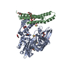
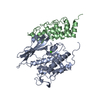
Journal: Elife / Year: 2020 Title: The architecture of EMC reveals a path for membrane protein insertion. Authors: John P O'Donnell / Ben P Phillips / Yuichi Yagita / Szymon Juszkiewicz / Armin Wagner / Duccio Malinverni / Robert J Keenan / Elizabeth A Miller / Ramanujan S Hegde / Abstract: Approximately 25% of eukaryotic genes code for integral membrane proteins that are assembled at the endoplasmic reticulum. An abundant and widely conserved multi-protein complex termed EMC has been ...Approximately 25% of eukaryotic genes code for integral membrane proteins that are assembled at the endoplasmic reticulum. An abundant and widely conserved multi-protein complex termed EMC has been implicated in membrane protein biogenesis, but its mechanism of action is poorly understood. Here, we define the composition and architecture of human EMC using biochemical assays, crystallography of individual subunits, site-specific photocrosslinking, and cryo-EM reconstruction. Our results suggest that EMC's cytosolic domain contains a large, moderately hydrophobic vestibule that can bind a substrate's transmembrane domain (TMD). The cytosolic vestibule leads into a lumenally-sealed, lipid-exposed intramembrane groove large enough to accommodate a single substrate TMD. A gap between the cytosolic vestibule and intramembrane groove provides a potential path for substrate egress from EMC. These findings suggest how EMC facilitates energy-independent membrane insertion of TMDs, explain why only short lumenal domains are translocated by EMC, and constrain models of EMC's proposed chaperone function.
In the structure databanks used in Yorodumi, some data are registered as the other names, "COVID-19 virus" and "2019-nCoV". Here are the details of the virus and the list of structure data.
Jan 31, 2019. EMDB accession codes are about to change! (news from PDBe EMDB page)
EMDB accession codes are about to change! (news from PDBe EMDB page)
The allocation of 4 digits for EMDB accession codes will soon come to an end. Whilst these codes will remain in use, new EMDB accession codes will include an additional digit and will expand incrementally as the available range of codes is exhausted. The current 4-digit format prefixed with “EMD-” (i.e. EMD-XXXX) will advance to a 5-digit format (i.e. EMD-XXXXX), and so on. It is currently estimated that the 4-digit codes will be depleted around Spring 2019, at which point the 5-digit format will come into force.
The EM Navigator/Yorodumi systems omit the EMD- prefix.
Related info.:Q: What is EMD? / ID/Accession-code notation in Yorodumi/EM Navigator
Yorodumi is a browser for structure data from EMDB, PDB, SASBDB, etc.
This page is also the successor to EM Navigator detail page, and also detail information page/front-end page for Omokage search.
The word "yorodu" (or yorozu) is an old Japanese word meaning "ten thousand". "mi" (miru) is to see.
Related info.:EMDB / PDB / SASBDB / Comparison of 3 databanks / Yorodumi Search / Aug 31, 2016. New EM Navigator & Yorodumi / Yorodumi Papers / Jmol/JSmol / Function and homology information / Changes in new EM Navigator and Yorodumi
 Movie
Movie Controller

 Yorodumi
 Open data
 Basic information
 Components
 Keywords
 Function and homology information
 Homo sapiens (human)
 X-RAY DIFFRACTION /
  Authors
 United Kingdom,
  Germany, 2items
  Citation
 Structure visualization
 Downloads & links
 Other downloads
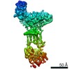


 PDBj
PDBj


 Assembly
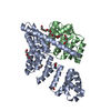


 Mass: 96.063 Da / Num. of mol.: 3 / Source method: obtained synthetically / Formula: SO4
 Mass: 62.068 Da / Num. of mol.: 3 / Source method: obtained synthetically / Formula: C2H6O2
 Mass: 354.436 Da / Num. of mol.: 1 / Source method: isolated from a natural source / Formula: C16H34O8 / Comment: precipitant*YM
 Mass: 106.120 Da / Num. of mol.: 3 / Source method: obtained synthetically / Formula: C4H10O3
 Sample preparation
 Processing