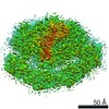
 Movie
Movie Controller


+ Open data

- Basic information
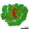
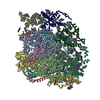
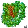
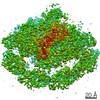
| Entry | Database: PDB / ID: 6yxr | |||||||||
|---|---|---|---|---|---|---|---|---|---|---|
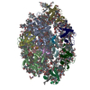
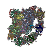
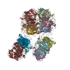
| Title | Dunaliella Minimal Photosystem I | |||||||||
 Components Components |
| |||||||||
 Keywords Keywords | PHOTOSYNTHESIS / membrane complex / photosystem I / dunaliella / light harvesting / excitation transfer | |||||||||
| Function / homology |  Function and homology information Function and homology informationphotosynthesis, light harvesting / photosystem I / photosynthetic electron transport in photosystem I / photosystem I / photosystem II / chlorophyll binding / chloroplast thylakoid membrane / photosynthesis / 4 iron, 4 sulfur cluster binding / electron transfer activity ...photosynthesis, light harvesting / photosystem I / photosynthetic electron transport in photosystem I / photosystem I / photosystem II / chlorophyll binding / chloroplast thylakoid membrane / photosynthesis / 4 iron, 4 sulfur cluster binding / electron transfer activity / oxidoreductase activity / magnesium ion binding / metal ion binding Similarity search - Function | |||||||||
| Biological species |  Dunaliella salina (plant) Dunaliella salina (plant) | |||||||||
| Method | ELECTRON MICROSCOPY / single particle reconstruction / cryo EM / Resolution: 3.4 Å | |||||||||
 Authors Authors | Nelson, N. / Caspy, I. / Malavath, T. / Klaiman, D. / Shkolinsky, Y. | |||||||||
| Funding support |  Israel, Israel,  Belgium, 2items Belgium, 2items
| |||||||||
 Citation Citation | Journal: Biochim Biophys Acta Bioenerg / Year: 2020 Title: Structure and energy transfer pathways of the Dunaliella Salina photosystem I supercomplex. Authors: Ido Caspy / Tirupathi Malavath / Daniel Klaiman / Maria Fadeeva / Yoel Shkolnisky / Nathan Nelson / Abstract: Oxygenic photosynthesis evolved more than 3 billion years ago in cyanobacteria. The increased complexity of photosystem I (PSI) became apparent from the high-resolution structures that were obtained ...Oxygenic photosynthesis evolved more than 3 billion years ago in cyanobacteria. The increased complexity of photosystem I (PSI) became apparent from the high-resolution structures that were obtained for the complexes that were isolated from various organisms, ranging from cyanobacteria to plants. These complexes are all evolutionarily linked. In this paper, the researchers have uncovered the increased complexity of PSI in a single organism demonstrated by the coexistance of two distinct PSI compositions. The Large Dunaliella PSI contains eight additional subunits, six in PSI core and two light harvesting complexes. Two additional chlorophyll a molecules pertinent for efficient excitation energy transfer in state II transition were identified in PsaL and PsaO. Short distances between these newly identified chlorophylls correspond with fast excitation transfer rates previously reported during state II transition. The apparent PSI conformations could be a coping mechanism for the high salinity. | |||||||||
| History |
|
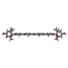
- Structure visualization
| Movie |
Movie viewer |
|---|---|
| Structure viewer | Molecule: MolmilJmol/JSmol |
- Downloads & links
-Download
| PDBx/mmCIF format | 6yxr.cif.gz | 969.1 KB | Display | PDBx/mmCIF format |
|---|---|---|---|---|
| PDB format | pdb6yxr.ent.gz | 769.8 KB | Display | PDB format |
| PDBx/mmJSON format | 6yxr.json.gz | Tree view | PDBx/mmJSON format | |
| Others |  Other downloads Other downloads |
-Validation report
| Arichive directory | https://data.pdbj.org/pub/pdb/validation_reports/yx/6yxrftp://data.pdbj.org/pub/pdb/validation_reports/yx/6yxr | HTTPS FTP |
|---|
-Related structure data
| Related structure data | 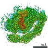 10995MC  6sl5C M: map data used to model this data C: citing same article ( |
|---|---|
| Similar structure data |


-Links
 PDBj
PDBj


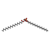
- Assembly
| Deposited unit | 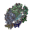
|
|---|---|
| 1 |
|
-Components
-Chlorophyll a-b binding protein, ... , 2 types, 2 molecules 13
| #1: Protein | Mass: 21305.230 Da / Num. of mol.: 1 / Source method: isolated from a natural source / Source: (natural) Dunaliella salina (plant) / References: UniProt: C1K003 |
|---|---|
| #3: Protein | Mass: 22732.824 Da / Num. of mol.: 1 / Source method: isolated from a natural source / Source: (natural) Dunaliella salina (plant) / References: UniProt: C1K004 |
-Protein , 5 types, 5 molecules 24DEF
| #2: Protein | Mass: 22813.822 Da / Num. of mol.: 1 / Source method: isolated from a natural source / Source: (natural) Dunaliella salina (plant) |
|---|---|
| #4: Protein | Mass: 23131.031 Da / Num. of mol.: 1 / Source method: isolated from a natural source / Source: (natural) Dunaliella salina (plant) |
| #8: Protein | Mass: 15883.294 Da / Num. of mol.: 1 / Source method: isolated from a natural source / Source: (natural) Dunaliella salina (plant) |
| #9: Protein | Mass: 7297.158 Da / Num. of mol.: 1 / Source method: isolated from a natural source / Source: (natural) Dunaliella salina (plant) |
| #10: Protein | Mass: 18212.902 Da / Num. of mol.: 1 / Source method: isolated from a natural source / Source: (natural) Dunaliella salina (plant) |
-Photosystem I ... , 4 types, 4 molecules ABCJ
| #5: Protein | Mass: 81748.172 Da / Num. of mol.: 1 / Source method: isolated from a natural source / Source: (natural) Dunaliella salina (plant) / References: UniProt: D0FXV2, photosystem I |
|---|---|
| #6: Protein | Mass: 81327.992 Da / Num. of mol.: 1 / Source method: isolated from a natural source / Source: (natural) Dunaliella salina (plant) / References: UniProt: D0FXZ0, photosystem I |
| #7: Protein | Mass: 8717.090 Da / Num. of mol.: 1 / Source method: isolated from a natural source / Source: (natural) Dunaliella salina (plant) / References: UniProt: D0FXW7, photosystem I |
| #11: Protein/peptide | Mass: 4467.188 Da / Num. of mol.: 1 / Source method: isolated from a natural source / Source: (natural) Dunaliella salina (plant) / References: UniProt: D0FXW0 |
-Sugars , 1 types, 1 molecules 
| #23: Sugar | ChemComp-DGD /  Type: saccharide / Mass: 949.299 Da / Num. of mol.: 1 / Source method: obtained synthetically / Formula: C51H96O15 Type: saccharide / Mass: 949.299 Da / Num. of mol.: 1 / Source method: obtained synthetically / Formula: C51H96O15 |
|---|
-Non-polymers , 11 types, 193 molecules 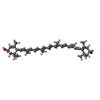


| #12: Chemical | ChemComp-LUT / (  Mass: 568.871 Da / Num. of mol.: 4 / Source method: obtained synthetically / Formula: C40H56O2 Mass: 568.871 Da / Num. of mol.: 4 / Source method: obtained synthetically / Formula: C40H56O2#13: Chemical | ChemComp-XAT / (  Mass: 600.870 Da / Num. of mol.: 4 / Source method: obtained synthetically / Formula: C40H56O4 Mass: 600.870 Da / Num. of mol.: 4 / Source method: obtained synthetically / Formula: C40H56O4#14: Chemical | ChemComp-BCR /  Mass: 536.873 Da / Num. of mol.: 23 / Source method: obtained synthetically / Formula: C40H56 Mass: 536.873 Da / Num. of mol.: 23 / Source method: obtained synthetically / Formula: C40H56#15: Chemical | ChemComp-CLA /  Mass: 893.489 Da / Num. of mol.: 134 / Source method: obtained synthetically / Formula: C55H72MgN4O5 Mass: 893.489 Da / Num. of mol.: 134 / Source method: obtained synthetically / Formula: C55H72MgN4O5#16: Chemical | ChemComp-CHL /  Mass: 907.472 Da / Num. of mol.: 10 / Source method: obtained synthetically / Formula: C55H70MgN4O6 Mass: 907.472 Da / Num. of mol.: 10 / Source method: obtained synthetically / Formula: C55H70MgN4O6#17: Chemical | ChemComp-LHG /  Mass: 722.970 Da / Num. of mol.: 6 / Source method: obtained synthetically / Formula: C38H75O10P / Comment: phospholipid*YM Mass: 722.970 Da / Num. of mol.: 6 / Source method: obtained synthetically / Formula: C38H75O10P / Comment: phospholipid*YM#18: Chemical | ChemComp-LMG /  Mass: 787.158 Da / Num. of mol.: 5 / Source method: obtained synthetically / Formula: C45H86O10 Mass: 787.158 Da / Num. of mol.: 5 / Source method: obtained synthetically / Formula: C45H86O10#19: Chemical | ChemComp-3PH / |  Mass: 704.998 Da / Num. of mol.: 1 / Source method: obtained synthetically / Formula: C39H77O8P Mass: 704.998 Da / Num. of mol.: 1 / Source method: obtained synthetically / Formula: C39H77O8P#20: Chemical | ChemComp-CL0 / |  Mass: 893.489 Da / Num. of mol.: 1 / Source method: obtained synthetically / Formula: C55H72MgN4O5 Mass: 893.489 Da / Num. of mol.: 1 / Source method: obtained synthetically / Formula: C55H72MgN4O5#21: Chemical |  Mass: 450.696 Da / Num. of mol.: 2 / Source method: obtained synthetically / Formula: C31H46O2 Mass: 450.696 Da / Num. of mol.: 2 / Source method: obtained synthetically / Formula: C31H46O2#22: Chemical |  Mass: 351.640 Da / Num. of mol.: 3 / Source method: obtained synthetically / Formula: Fe4S4 Mass: 351.640 Da / Num. of mol.: 3 / Source method: obtained synthetically / Formula: Fe4S4 |
|---|
-Details
| Has ligand of interest | N |
|---|---|
| Has protein modification | Y |
-Experimental details
-Experiment
| Experiment | Method: ELECTRON MICROSCOPY |
|---|---|
| EM experiment | Aggregation state: PARTICLE / 3D reconstruction method: single particle reconstruction |
- Sample preparation
| Component | Name: Large dunaliella salina photosystem I-LHC supercomplex Type: COMPLEX / Entity ID: #1-#11 / Source: NATURAL |
|---|---|
| Molecular weight | Experimental value: NO |
| Source (natural) | Organism: Dunaliella salina (plant) |
| Buffer solution | pH: 8 |
| Specimen | Embedding applied: NO / Shadowing applied: NO / Staining applied: NO / Vitrification applied: YES |
| Vitrification | Instrument: LEICA EM GP / Cryogen name: ETHANE / Humidity: 100 % / Chamber temperature: 277 K / Details: 2.5 sec blotting before plunging |
- Electron microscopy imaging
| Experimental equipment |  Model: Titan Krios / Image courtesy: FEI Company |
|---|---|
| Microscopy | Model: FEI TITAN KRIOS |
| Electron gun | Electron source:  FIELD EMISSION GUN / Accelerating voltage: 300 kV / Illumination mode: FLOOD BEAM FIELD EMISSION GUN / Accelerating voltage: 300 kV / Illumination mode: FLOOD BEAM |
| Electron lens | Mode: BRIGHT FIELD / Nominal defocus max: 3000 nm / Nominal defocus min: 900 nm |
| Image recording | Electron dose: 42.68 e/Å2 / Detector mode: COUNTING / Film or detector model: GATAN K2 SUMMIT (4k x 4k) / Num. of grids imaged: 1 / Num. of real images: 4306 |
- Processing
| Software |
| ||||||||||||||||||||||||||||||||||||||||
|---|---|---|---|---|---|---|---|---|---|---|---|---|---|---|---|---|---|---|---|---|---|---|---|---|---|---|---|---|---|---|---|---|---|---|---|---|---|---|---|---|---|
| EM software |
| ||||||||||||||||||||||||||||||||||||||||
| CTF correction | Type: NONE | ||||||||||||||||||||||||||||||||||||||||
| Particle selection | Num. of particles selected: 720711 | ||||||||||||||||||||||||||||||||||||||||
| Symmetry | Point symmetry: C1 (asymmetric) | ||||||||||||||||||||||||||||||||||||||||
| 3D reconstruction | Resolution: 3.4 Å / Resolution method: FSC 0.143 CUT-OFF / Num. of particles: 45969 / Symmetry type: POINT | ||||||||||||||||||||||||||||||||||||||||
| Atomic model building | Protocol: AB INITIO MODEL / Space: REAL | ||||||||||||||||||||||||||||||||||||||||
| Refinement | Cross valid method: NONE Stereochemistry target values: GeoStd + Monomer Library + CDL v1.2 | ||||||||||||||||||||||||||||||||||||||||
| Displacement parameters | Biso mean: 43.33 Å2 | ||||||||||||||||||||||||||||||||||||||||
| Refine LS restraints |
|
