Movie
Movie Controller


+ Open data

- Basic information
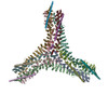
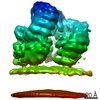
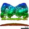
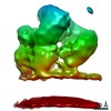
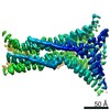
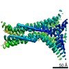
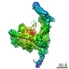
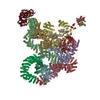
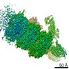
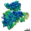
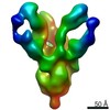
| Entry | Database: PDB / ID: 6yai | |||||||||
|---|---|---|---|---|---|---|---|---|---|---|
| Title | Clathrin with bound beta2 appendage of AP2 | |||||||||
 Components Components |
| |||||||||
 Keywords Keywords | ENDOCYTOSIS / clathrin / clathrin adaptor / ap2 / clathrin assembly | |||||||||
| Function / homology |  Function and homology information Function and homology informationLysosome Vesicle Biogenesis / WNT5A-dependent internalization of FZD4 / WNT5A-dependent internalization of FZD2, FZD5 and ROR2 / Cargo recognition for clathrin-mediated endocytosis / Gap junction degradation / Formation of annular gap junctions / Clathrin-mediated endocytosis / clathrin coat of trans-Golgi network vesicle / Nef Mediated CD8 Down-regulation / clathrin light chain binding ...Lysosome Vesicle Biogenesis / WNT5A-dependent internalization of FZD4 / WNT5A-dependent internalization of FZD2, FZD5 and ROR2 / Cargo recognition for clathrin-mediated endocytosis / Gap junction degradation / Formation of annular gap junctions / Clathrin-mediated endocytosis / clathrin coat of trans-Golgi network vesicle / Nef Mediated CD8 Down-regulation / clathrin light chain binding / WNT5A-dependent internalization of FZD2, FZD5 and ROR2 / clathrin complex / Trafficking of GluR2-containing AMPA receptors / WNT5A-dependent internalization of FZD4 / clathrin adaptor complex / extrinsic component of presynaptic endocytic zone membrane / clathrin coat of coated pit / postsynaptic endocytic zone / postsynaptic neurotransmitter receptor internalization / AP-2 adaptor complex / Retrograde neurotrophin signalling / clathrin-coated endocytic vesicle / clathrin coat assembly / LDL clearance / clathrin-dependent endocytosis / signal sequence receptor activity / Nef Mediated CD4 Down-regulation / coronary vasculature development / positive regulation of protein localization to membrane / endolysosome membrane / neurotransmitter secretion / clathrin-coated vesicle / ventricular septum development / aorta development / ciliary membrane / clathrin binding / Recycling pathway of L1 / EPH-ephrin mediated repulsion of cells / positive regulation of endocytosis / synaptic vesicle endocytosis / vesicle-mediated transport / MHC class II antigen presentation / receptor-mediated endocytosis / VLDLR internalisation and degradation / ribosomal large subunit biogenesis / kidney development / trans-Golgi network / clathrin-coated endocytic vesicle membrane / intracellular protein transport / spindle / cytoplasmic side of plasma membrane / disordered domain specific binding / synaptic vesicle / endocytic vesicle membrane / Cargo recognition for clathrin-mediated endocytosis / mitotic cell cycle / Clathrin-mediated endocytosis / Potential therapeutics for SARS / protein-containing complex binding / nucleolus / structural molecule activity / glutamatergic synapse / nucleoplasm / membrane / plasma membrane / cytosol Similarity search - Function | |||||||||
| Biological species |  Homo sapiens (human) Homo sapiens (human) | |||||||||
| Method | ELECTRON MICROSCOPY / subtomogram averaging / cryo EM / Resolution: 9.2 Å | |||||||||
 Authors Authors | Kovtun, O. / Kane Dickson, V. / Kelly, B.T. / Owen, D. / Briggs, J.A.G. | |||||||||
| Funding support |  United Kingdom, 2items United Kingdom, 2items
| |||||||||
 Citation Citation | Journal: Sci Adv / Year: 2020 Title: Architecture of the AP2/clathrin coat on the membranes of clathrin-coated vesicles. Authors: Oleksiy Kovtun / Veronica Kane Dickson / Bernard T Kelly / David J Owen / John A G Briggs /  Abstract: Clathrin-mediated endocytosis (CME) is crucial for modulating the protein composition of a cell's plasma membrane. Clathrin forms a cage-like, polyhedral outer scaffold around a vesicle, to which ...Clathrin-mediated endocytosis (CME) is crucial for modulating the protein composition of a cell's plasma membrane. Clathrin forms a cage-like, polyhedral outer scaffold around a vesicle, to which cargo-selecting clathrin adaptors are attached. Adaptor protein complex (AP2) is the key adaptor in CME. Crystallography has shown AP2 to adopt a range of conformations. Here, we used cryo-electron microscopy, tomography, and subtomogram averaging to determine structures, interactions, and arrangements of clathrin and AP2 at the key steps of coat assembly, from AP2 in solution to membrane-assembled clathrin-coated vesicles (CCVs). AP2 binds cargo and PtdIns(4,5) (phosphatidylinositol 4,5-bisphosphate)-containing membranes via multiple interfaces, undergoing conformational rearrangement from its cytosolic state. The binding mode of AP2 β2 appendage into the clathrin lattice in CCVs and buds implies how the adaptor structurally modulates coat curvature and coat disassembly. | |||||||||
| History |
|
- Structure visualization
| Movie |
Movie viewer |
|---|---|
| Structure viewer | Molecule: MolmilJmol/JSmol |
- Downloads & links
-Download
| PDBx/mmCIF format | 6yai.cif.gz | 951.1 KB | Display | PDBx/mmCIF format |
|---|---|---|---|---|
| PDB format | pdb6yai.ent.gz | 661.3 KB | Display | PDB format |
| PDBx/mmJSON format | 6yai.json.gz | Tree view | PDBx/mmJSON format | |
| Others |  Other downloads Other downloads |
-Validation report
| Arichive directory | https://data.pdbj.org/pub/pdb/validation_reports/ya/6yaiftp://data.pdbj.org/pub/pdb/validation_reports/ya/6yai | HTTPS FTP |
|---|
-Related structure data
| Related structure data | 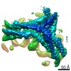 10754MC 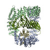 6yaeC 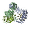 6yafC 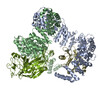 6yahC C: citing same article ( M: map data used to model this data |
|---|---|
| Similar structure data |


-Links
 PDBj
PDBj


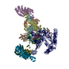
- Assembly
| Deposited unit | 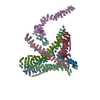
|
|---|---|
| 1 |
|
-Components
| #1: Antibody | Mass: 187145.125 Da / Num. of mol.: 8 / Source method: isolated from a natural source / Source: (natural) #2: Protein | | Mass: 26429.457 Da / Num. of mol.: 1 Source method: isolated from a genetically manipulated source Source: (gene. exp.) Homo sapiens (human) / Gene: AP2B1, ADTB2, CLAPB1Production host:  References: UniProt: P63010 #3: Antibody | | Mass: 187117.094 Da / Num. of mol.: 1 / Source method: isolated from a natural source / Source: (natural) #4: Protein | Mass: 25218.500 Da / Num. of mol.: 4 / Source method: isolated from a natural source / Source: (natural) |
|---|
-Experimental details
-Experiment
| Experiment | Method: ELECTRON MICROSCOPY |
|---|---|
| EM experiment | Aggregation state: 3D ARRAY / 3D reconstruction method: subtomogram averaging |
- Sample preparation
| Component |
| ||||||||||||||||||||||||||||
|---|---|---|---|---|---|---|---|---|---|---|---|---|---|---|---|---|---|---|---|---|---|---|---|---|---|---|---|---|---|
| Molecular weight |
| ||||||||||||||||||||||||||||
| Source (natural) |
| ||||||||||||||||||||||||||||
| Source (recombinant) | Organism: | ||||||||||||||||||||||||||||
| Buffer solution | pH: 7.2 | ||||||||||||||||||||||||||||
| Buffer component |
| ||||||||||||||||||||||||||||
| Specimen | Conc.: 0.7 mg/ml / Embedding applied: NO / Shadowing applied: NO / Staining applied: NO / Vitrification applied: YES Details: The sample (in vitro budding reaction) contained AP2, clathrin and 400 nm extruded liposomes | ||||||||||||||||||||||||||||
| Specimen support | Grid type: C-flat-2/2 | ||||||||||||||||||||||||||||
| Vitrification | Instrument: LEICA EM GP / Cryogen name: ETHANE / Humidity: 98 % / Chamber temperature: 291 K Details: The sample was supplemented with 10 nm nanogold fiducials, and 3 ul of the mixture was backside blotted for 3 seconds. |
- Electron microscopy imaging
| Experimental equipment |  Model: Titan Krios / Image courtesy: FEI Company |
|---|---|
| Microscopy | Model: FEI TITAN KRIOS |
| Electron gun | Electron source:  FIELD EMISSION GUN / Accelerating voltage: 300 kV / Illumination mode: FLOOD BEAM FIELD EMISSION GUN / Accelerating voltage: 300 kV / Illumination mode: FLOOD BEAM |
| Electron lens | Mode: BRIGHT FIELD / Nominal magnification: 81000 X / Calibrated magnification: 81000 X / Nominal defocus max: 6500 nm / Nominal defocus min: 1500 nm / Calibrated defocus min: 1500 nm / Calibrated defocus max: 6500 nm / Cs: 2.7 mm / C2 aperture diameter: 100 µm / Alignment procedure: ZEMLIN TABLEAU |
| Specimen holder | Cryogen: NITROGEN / Specimen holder model: FEI TITAN KRIOS AUTOGRID HOLDER |
| Image recording | Average exposure time: 0.2 sec. / Electron dose: 3.2 e/Å2 / Detector mode: COUNTING / Film or detector model: GATAN K2 QUANTUM (4k x 4k) / Num. of grids imaged: 2 Details: The images were collected in movie mode at 10 frames per second |
| EM imaging optics | Energyfilter name: GIF Quantum LS / Energyfilter slit width: 20 eV |
| Image scans | Width: 3838 / Height: 3710 / Movie frames/image: 10 / Used frames/image: 1-10 |
- Processing
| EM software |
| ||||||||||||||||||||||||||||||||||||||||||||||||
|---|---|---|---|---|---|---|---|---|---|---|---|---|---|---|---|---|---|---|---|---|---|---|---|---|---|---|---|---|---|---|---|---|---|---|---|---|---|---|---|---|---|---|---|---|---|---|---|---|---|
| Image processing | Details: The images were low pass filtered according to the cumulative radiation dose. | ||||||||||||||||||||||||||||||||||||||||||||||||
| CTF correction | Details: CTF correction in novaCTF with by multiplication / Type: PHASE FLIPPING AND AMPLITUDE CORRECTION | ||||||||||||||||||||||||||||||||||||||||||||||||
| Symmetry | Point symmetry: C1 (asymmetric) | ||||||||||||||||||||||||||||||||||||||||||||||||
| 3D reconstruction | Resolution: 9.2 Å / Resolution method: FSC 0.143 CUT-OFF / Num. of particles: 12076 / Symmetry type: POINT | ||||||||||||||||||||||||||||||||||||||||||||||||
| EM volume selection | Method: geometrically defined initial positions Details: To define initial positions of subtomograms, centres and radii of coated vesicles, buds were manually marked in bin4 tomograms. These measurements were used to define spheres. Subtomogram ...Details: To define initial positions of subtomograms, centres and radii of coated vesicles, buds were manually marked in bin4 tomograms. These measurements were used to define spheres. Subtomogram positions were then defined on the surface of these spheres with uniform sampling. The orientations were calculated to be normal to the surfaces of the spheres with random in-plane rotation. Num. of tomograms: 58121 / Num. of volumes extracted: 198871 / Reference model: reference-free | ||||||||||||||||||||||||||||||||||||||||||||||||
| Atomic model building | Protocol: FLEXIBLE FIT / Space: REAL / Target criteria: correlation coefficient | ||||||||||||||||||||||||||||||||||||||||||||||||
| Atomic model building | 3D fitting-ID: 1 / Source name: PDB / Type: experimental model
|