Movie
Movie Controller


+ Open data

- Basic information
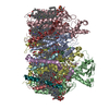
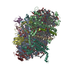
| Entry | Database: PDB / ID: 6xnz | ||||||
|---|---|---|---|---|---|---|---|
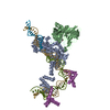
| Title | Structure of RAG1 (R848M/E649V)-RAG2-DNA Target Capture Complex | ||||||
 Components Components |
| ||||||
 Keywords Keywords | RECOMBINATION / DNA Transposase | ||||||
| Function / homology |  Function and homology information Function and homology informationmature B cell differentiation involved in immune response / DNA recombinase complex / B cell homeostatic proliferation / endodeoxyribonuclease complex / negative regulation of T cell differentiation in thymus / pre-B cell allelic exclusion / positive regulation of organ growth / V(D)J recombination / negative regulation of T cell apoptotic process / negative regulation of thymocyte apoptotic process ...mature B cell differentiation involved in immune response / DNA recombinase complex / B cell homeostatic proliferation / endodeoxyribonuclease complex / negative regulation of T cell differentiation in thymus / pre-B cell allelic exclusion / positive regulation of organ growth / V(D)J recombination / negative regulation of T cell apoptotic process / negative regulation of thymocyte apoptotic process / phosphatidylinositol-3,4-bisphosphate binding / regulation of behavioral fear response / histone H3K4me3 reader activity / phosphatidylinositol-3,5-bisphosphate binding / regulation of T cell differentiation / positive regulation of T cell differentiation / T cell lineage commitment / B cell lineage commitment / T cell homeostasis / phosphatidylinositol-3,4,5-trisphosphate binding / T cell differentiation / protein autoubiquitination / phosphatidylinositol-4,5-bisphosphate binding / phosphatidylinositol binding / B cell differentiation / thymus development / RING-type E3 ubiquitin transferase / visual learning / ubiquitin-protein transferase activity / T cell differentiation in thymus / ubiquitin protein ligase activity / chromatin organization / endonuclease activity / DNA recombination / histone binding / sequence-specific DNA binding / Hydrolases; Acting on ester bonds / adaptive immune response / defense response to bacterium / hydrolase activity / chromatin binding / protein homodimerization activity / zinc ion binding / nucleoplasm / metal ion binding / identical protein binding / nucleus Similarity search - Function | ||||||
| Biological species |  | ||||||
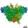
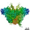
| Method | ELECTRON MICROSCOPY / single particle reconstruction / cryo EM / Resolution: 3.8 Å | ||||||
 Authors Authors | Zhang, Y. / Corbett, E. / Wu, S. / Schatz, D.G. | ||||||
| Funding support |  United States, 1items United States, 1items
| ||||||
 Citation Citation | Journal: EMBO J / Year: 2020 Title: Structural basis for the activation and suppression of transposition during evolution of the RAG recombinase. Authors: Yuhang Zhang / Elizabeth Corbett / Shenping Wu / David G Schatz / Abstract: Jawed vertebrate adaptive immunity relies on the RAG1/RAG2 (RAG) recombinase, a domesticated transposase, for assembly of antigen receptor genes. Using an integration-activated form of RAG1 with ...Jawed vertebrate adaptive immunity relies on the RAG1/RAG2 (RAG) recombinase, a domesticated transposase, for assembly of antigen receptor genes. Using an integration-activated form of RAG1 with methionine at residue 848 and cryo-electron microscopy, we determined structures that capture RAG engaged with transposon ends and U-shaped target DNA prior to integration (the target capture complex) and two forms of the RAG strand transfer complex that differ based on whether target site DNA is annealed or dynamic. Target site DNA base unstacking, flipping, and melting by RAG1 methionine 848 explain how this residue activates transposition, how RAG can stabilize sharp bends in target DNA, and why replacement of residue 848 by arginine during RAG domestication led to suppression of transposition activity. RAG2 extends a jawed vertebrate-specific loop to interact with target site DNA, and functional assays demonstrate that this loop represents another evolutionary adaptation acquired during RAG domestication to inhibit transposition. Our findings identify mechanistic principles of the final step in cut-and-paste transposition and the molecular and structural logic underlying the transformation of RAG from transposase to recombinase. | ||||||
| History |
|
- Structure visualization
| Movie |
Movie viewer |
|---|---|
| Structure viewer | Molecule: MolmilJmol/JSmol |
- Downloads & links
-Download
| PDBx/mmCIF format | 6xnz.cif.gz | 389.2 KB | Display | PDBx/mmCIF format |
|---|---|---|---|---|
| PDB format | pdb6xnz.ent.gz | 297.2 KB | Display | PDB format |
| PDBx/mmJSON format | 6xnz.json.gz | Tree view | PDBx/mmJSON format | |
| Others |  Other downloads Other downloads |
-Validation report
| Arichive directory | https://data.pdbj.org/pub/pdb/validation_reports/xn/6xnzftp://data.pdbj.org/pub/pdb/validation_reports/xn/6xnz | HTTPS FTP |
|---|
-Related structure data
| Related structure data | 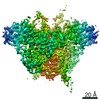 22274MC  6xnxC  6xnyC M: map data used to model this data C: citing same article ( |
|---|---|
| Similar structure data |


-Links
 PDBj
PDBj


- Assembly
| Deposited unit | 
|
|---|---|
| 1 |
|
-Components
-V(D)J recombination-activating protein ... , 2 types, 4 molecules ACBD
| #1: Protein | Mass: 85750.781 Da / Num. of mol.: 2 Source method: isolated from a genetically manipulated source Source: (gene. exp.)  Homo sapiens (human) Homo sapiens (human)References: UniProt: P15919, Hydrolases; Acting on ester bonds, RING-type E3 ubiquitin transferase #2: Protein | Mass: 40416.984 Da / Num. of mol.: 2 Source method: isolated from a genetically manipulated source Source: (gene. exp.) Homo sapiens (human) / References: UniProt: P21784 |
|---|
-DNA chain , 6 types, 6 molecules IJxMyL
| #3: DNA chain | Mass: 11368.285 Da / Num. of mol.: 1 / Source method: obtained synthetically / Source: (synth.) |
|---|---|
| #4: DNA chain | Mass: 11408.310 Da / Num. of mol.: 1 / Source method: obtained synthetically / Source: (synth.) |
| #5: DNA chain | Mass: 10456.710 Da / Num. of mol.: 1 / Source method: obtained synthetically / Source: (synth.) |
| #6: DNA chain | Mass: 10461.758 Da / Num. of mol.: 1 / Source method: obtained synthetically / Source: (synth.) |
| #7: DNA chain | Mass: 13837.883 Da / Num. of mol.: 1 / Source method: obtained synthetically / Source: (synth.) |
| #8: DNA chain | Mass: 13877.906 Da / Num. of mol.: 1 / Source method: obtained synthetically / Source: (synth.) |
-Non-polymers , 1 types, 2 molecules 
| #9: Chemical |  Mass: 65.409 Da / Num. of mol.: 2 / Source method: obtained synthetically / Formula: Zn / Feature type: SUBJECT OF INVESTIGATION Mass: 65.409 Da / Num. of mol.: 2 / Source method: obtained synthetically / Formula: Zn / Feature type: SUBJECT OF INVESTIGATION |
|---|
-Details
| Has ligand of interest | Y |
|---|
-Experimental details
-Experiment
| Experiment | Method: ELECTRON MICROSCOPY |
|---|---|
| EM experiment | Aggregation state: PARTICLE / 3D reconstruction method: single particle reconstruction |
- Sample preparation
| Component |
| ||||||||||||||||||||||||
|---|---|---|---|---|---|---|---|---|---|---|---|---|---|---|---|---|---|---|---|---|---|---|---|---|---|
| Molecular weight | Units: KILODALTONS/NANOMETER / Experimental value: NO | ||||||||||||||||||||||||
| Source (natural) |
| ||||||||||||||||||||||||
| Source (recombinant) |
| ||||||||||||||||||||||||
| Buffer solution | pH: 7.6 Details: 20 mM HEPES pH7.6, 0.5 mM TCEP, 5 mM MgCl2 and 150 mM KCl | ||||||||||||||||||||||||
| Specimen | Conc.: 0.4 mg/ml / Embedding applied: NO / Shadowing applied: NO / Staining applied: NO / Vitrification applied: YES | ||||||||||||||||||||||||
| Vitrification | Instrument: FEI VITROBOT MARK IV / Cryogen name: ETHANE / Humidity: 100 % / Chamber temperature: 283 K |
- Electron microscopy imaging
| Experimental equipment |  Model: Titan Krios / Image courtesy: FEI Company |
|---|---|
| Microscopy | Model: FEI TITAN KRIOS |
| Electron gun | Electron source:  FIELD EMISSION GUN / Accelerating voltage: 300 kV / Illumination mode: FLOOD BEAM FIELD EMISSION GUN / Accelerating voltage: 300 kV / Illumination mode: FLOOD BEAM |
| Electron lens | Mode: BRIGHT FIELD / Nominal magnification: 130000 X / Nominal defocus max: 2300 nm / Nominal defocus min: 800 nm / Cs: 2.7 mm / C2 aperture diameter: 50 µm |
| Specimen holder | Cryogen: NITROGEN / Specimen holder model: FEI TITAN KRIOS AUTOGRID HOLDER |
| Image recording | Average exposure time: 11 sec. / Electron dose: 72.8 e/Å2 / Detector mode: SUPER-RESOLUTION / Film or detector model: GATAN K2 SUMMIT (4k x 4k) / Num. of real images: 3527 |
- Processing
| EM software |
| ||||||||||||||||||||||||||||||||||||
|---|---|---|---|---|---|---|---|---|---|---|---|---|---|---|---|---|---|---|---|---|---|---|---|---|---|---|---|---|---|---|---|---|---|---|---|---|---|
| CTF correction | Type: PHASE FLIPPING AND AMPLITUDE CORRECTION | ||||||||||||||||||||||||||||||||||||
| Particle selection | Num. of particles selected: 636773 | ||||||||||||||||||||||||||||||||||||
| 3D reconstruction | Resolution: 3.8 Å / Resolution method: FSC 0.143 CUT-OFF / Num. of particles: 22270 / Num. of class averages: 1 / Symmetry type: POINT | ||||||||||||||||||||||||||||||||||||
| Atomic model building | Protocol: RIGID BODY FIT / Space: REAL | ||||||||||||||||||||||||||||||||||||
| Atomic model building | PDB-ID: 5ZDZ Accession code: 5ZDZ / Source name: PDB / Type: experimental model |