 ムービー
ムービー コントローラー


+ データを開く

- 基本情報
基本情報
| 登録情報 | データベース: PDB / ID: 6x5z | ||||||||||||||||||||||||
|---|---|---|---|---|---|---|---|---|---|---|---|---|---|---|---|---|---|---|---|---|---|---|---|---|---|
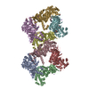
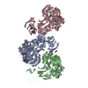
| タイトル | Bovine Cardiac Myosin in Complex with Chicken Skeletal Actin and Human Cardiac Tropomyosin in the Rigor State | ||||||||||||||||||||||||
 要素 要素 |
| ||||||||||||||||||||||||
 キーワード キーワード | CONTRACTILE PROTEIN / myosin / tropomyosin / actin / cardiac | ||||||||||||||||||||||||
| 機能・相同性 |  機能・相同性情報 機能・相同性情報positive regulation of heart rate by epinephrine / muscle thin filament tropomyosin / bleb / Striated Muscle Contraction / negative regulation of vascular associated smooth muscle cell migration / regulation of muscle contraction / muscle filament sliding / myosin filament / adult heart development / ruffle organization ...positive regulation of heart rate by epinephrine / muscle thin filament tropomyosin / bleb / Striated Muscle Contraction / negative regulation of vascular associated smooth muscle cell migration / regulation of muscle contraction / muscle filament sliding / myosin filament / adult heart development / ruffle organization / negative regulation of vascular associated smooth muscle cell proliferation / positive regulation of ATP-dependent activity / Striated Muscle Contraction / myosin II complex / structural constituent of muscle / ventricular cardiac muscle tissue morphogenesis / regulation of heart contraction / sarcomere organization / microfilament motor activity / myofibril / skeletal muscle thin filament assembly / striated muscle thin filament / positive regulation of cell adhesion / Smooth Muscle Contraction / skeletal muscle fiber development / cardiac muscle contraction / stress fiber / positive regulation of stress fiber assembly / cytoskeleton organization / cytoskeletal protein binding / sarcomere / negative regulation of cell migration / actin filament organization / actin filament / ruffle membrane / 加水分解酵素; 酸無水物に作用; 酸無水物に作用・細胞または細胞小器官の運動に関与 / wound healing / cellular response to reactive oxygen species / structural constituent of cytoskeleton / actin filament binding / actin cytoskeleton / actin binding / regulation of cell shape / calmodulin binding / cytoskeleton / hydrolase activity / protein heterodimerization activity / protein homodimerization activity / ATP binding / identical protein binding / cytoplasm / cytosol 類似検索 - 分子機能 | ||||||||||||||||||||||||
| 生物種 |  Homo sapiens (ヒト) Homo sapiens (ヒト)  | ||||||||||||||||||||||||
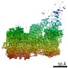
| 手法 | 電子顕微鏡法 / らせん対称体再構成法 / クライオ電子顕微鏡法 / 解像度: 4.24 Å | ||||||||||||||||||||||||
 データ登録者 データ登録者 | Doran, M.H. / Lehman, W. / Rynkiewicz, M.J. / Bullitt, E. | ||||||||||||||||||||||||
| 資金援助 |  米国, European Union, 7件 米国, European Union, 7件
| ||||||||||||||||||||||||
 引用 引用 | ジャーナル: Biophys J / 年: 2020 タイトル: Cryo-EM and Molecular Docking Shows Myosin Loop 4 Contacts Actin and Tropomyosin on Thin Filaments. 著者: Matthew H Doran / Elumalai Pavadai / Michael J Rynkiewicz / Jonathan Walklate / Esther Bullitt / Jeffrey R Moore / Michael Regnier / Michael A Geeves / William Lehman /  要旨: The motor protein myosin drives muscle and nonmuscle motility by binding to and moving along actin of thin filaments. Myosin binding to actin also modulates interactions of the regulatory protein, ...The motor protein myosin drives muscle and nonmuscle motility by binding to and moving along actin of thin filaments. Myosin binding to actin also modulates interactions of the regulatory protein, tropomyosin, on thin filaments, and conversely tropomyosin affects myosin binding to actin. Insight into this reciprocity will facilitate a molecular level elucidation of tropomyosin regulation of myosin interaction with actin in muscle contraction, and in turn, promote better understanding of nonmuscle cell motility. Indeed, experimental approaches such as fiber diffraction, cryoelectron microscopy, and three-dimensional reconstruction have long been used to define regulatory interaction of tropomyosin and myosin on actin at a structural level. However, their limited resolution has not proven sufficient to determine tropomyosin and myosin contacts at an atomic-level and thus to fully substantiate possible functional contributions. To overcome this deficiency, we have followed a hybrid approach by performing new cryogenic electron microscopy reconstruction of myosin-S1-decorated F-actin-tropomyosin together with atomic scale protein-protein docking of tropomyosin to the EM models. Here, cryo-EM data were derived from filaments reconstituted with α1-actin, cardiac αα-tropomyosin, and masseter muscle β-myosin complexes; masseter myosin, which shares sequence identity with β-cardiac myosin-heavy chain, was used because of its stability in vitro. The data were used to build an atomic model of the tropomyosin cable that fits onto the actin filament between the tip of the myosin head and a cleft on the innermost edge of actin subunits. The docking and atomic scale fitting showed multiple discrete interactions of myosin loop 4 and acidic residues on successive 39-42 residue-long tropomyosin pseudorepeats. The contacts between S1 and tropomyosin on actin appear to compete with and displace ones normally found between actin and tropomyosin on myosin-free thin filaments in relaxed muscle, thus restructuring the filament during myosin-induced activation. | ||||||||||||||||||||||||
| 履歴 |
|
- 構造の表示
構造の表示
| ムービー |
ムービービューア |
|---|---|
| 構造ビューア | 分子: MolmilJmol/JSmol |
- ダウンロードとリンク
-ダウンロード
| PDBx/mmCIF形式 | 6x5z.cif.gz | 634.8 KB | 表示 | PDBx/mmCIF形式 |
|---|---|---|---|---|
| PDB形式 | pdb6x5z.ent.gz | 510.9 KB | 表示 | PDB形式 |
| PDBx/mmJSON形式 | 6x5z.json.gz | ツリー表示 | PDBx/mmJSON形式 | |
| その他 |  その他のダウンロード その他のダウンロード |
-検証レポート
| 文書・要旨 | 6x5z_validation.pdf.gz | 1.1 MB | 表示 | wwPDB検証レポート |
|---|---|---|---|---|
| 文書・詳細版 | 6x5z_full_validation.pdf.gz | 1.2 MB | 表示 | |
| XML形式データ | 6x5z_validation.xml.gz | 113 KB | 表示 | |
| CIF形式データ | 6x5z_validation.cif.gz | 167 KB | 表示 | |
| アーカイブディレクトリ | https://data.pdbj.org/pub/pdb/validation_reports/x5/6x5zftp://data.pdbj.org/pub/pdb/validation_reports/x5/6x5z | HTTPS FTP |
-関連構造データ


-リンク
 PDBj
PDBj


- 集合体
集合体
| 登録構造単位 | 
|
|---|---|
| 1 |
|
| 対称性 | らせん対称: (回転対称性: 1 / Dyad axis: no / N subunits divisor: 1 / Num. of operations: 2 / Rise per n subunits: 27.5 Å / Rotation per n subunits: -166.4 °) |
-要素
| #1: タンパク質 | 分子量: 42096.953 Da / 分子数: 3 / 由来タイプ: 天然 詳細: Residues 1-9 and 377 were disordered and were not modeled. 由来: (天然) #2: タンパク質 | 分子量: 32763.621 Da / 分子数: 2 / 由来タイプ: 組換発現 詳細: Only the central section (residues 45-210) was modeled. 由来: (組換発現) Homo sapiens (ヒト) / 遺伝子: TPM1, C15orf13, TMSA / 発現宿主:  #3: タンパク質 | 分子量: 97446.898 Da / 分子数: 3 / Fragment: S1 fragment (UNP residues 1-850) / 由来タイプ: 天然 詳細: Residues 1-35, 200-216, 624-641, and 777-850 were disordered and not modeled. 由来: (天然) #4: 化合物 |   分子量: 427.201 Da / 分子数: 3 / 由来タイプ: 合成 / 式: C10H15N5O10P2 / コメント: ADP, エネルギー貯蔵分子*YM 分子量: 427.201 Da / 分子数: 3 / 由来タイプ: 合成 / 式: C10H15N5O10P2 / コメント: ADP, エネルギー貯蔵分子*YM#5: 化合物 |   分子量: 24.305 Da / 分子数: 3 / 由来タイプ: 合成 / 式: Mg 分子量: 24.305 Da / 分子数: 3 / 由来タイプ: 合成 / 式: Mg研究の焦点であるリガンドがあるか | N | |
|---|
-実験情報
-実験
| 実験 | 手法: 電子顕微鏡法 |
|---|---|
| EM実験 | 試料の集合状態: HELICAL ARRAY / 3次元再構成法: らせん対称体再構成法 |
- 試料調製
試料調製
| 構成要素 |
| |||||||||||||||||||||||||||||||||||
|---|---|---|---|---|---|---|---|---|---|---|---|---|---|---|---|---|---|---|---|---|---|---|---|---|---|---|---|---|---|---|---|---|---|---|---|---|
| 分子量 | 値: 0.40322 MDa / 実験値: NO | |||||||||||||||||||||||||||||||||||
| 由来(天然) |
| |||||||||||||||||||||||||||||||||||
| 由来(組換発現) | 生物種: | |||||||||||||||||||||||||||||||||||
| 緩衝液 | pH: 7 | |||||||||||||||||||||||||||||||||||
| 緩衝液成分 |
| |||||||||||||||||||||||||||||||||||
| 試料 | 濃度: 0.525 mg/ml / 包埋: NO / シャドウイング: NO / 染色: NO / 凍結: YES | |||||||||||||||||||||||||||||||||||
| 試料支持 | 詳細: Glow discharged using a PELCO easiGlow station / グリッドの材料: COPPER / グリッドのサイズ: 200 divisions/in. / グリッドのタイプ: Quantifoil R2/1 | |||||||||||||||||||||||||||||||||||
| 急速凍結 | 装置: FEI VITROBOT MARK III / 凍結剤: ETHANE / 湿度: 100 % / 凍結前の試料温度: 283 K 詳細: Thin filaments were reconstituted by first mixing F-actin and tropomyosin to final concentrations of 10 uM actin and 7 uM tropomyosin. Just before applying 1.5 uL actin-tropomyosin to a ...詳細: Thin filaments were reconstituted by first mixing F-actin and tropomyosin to final concentrations of 10 uM actin and 7 uM tropomyosin. Just before applying 1.5 uL actin-tropomyosin to a freshly glow discharged holey-carbon grid, the surfactant octyl B-D-glucopyranoside was added to the protein solution to a concentration of 12 nM. The grid sample was manually blotted for 1 second and a 1.5 uL drop of 7.5 uM myosin-S1 sub-fragment was then applied to the blotted grid sample. The sample was immediately blotted for 4 seconds and plunge-frozen in liquid ethane. |
- 電子顕微鏡撮影
| 実験機器 |  モデル: Titan Krios / 画像提供: FEI Company |
|---|---|
| 顕微鏡 | モデル: FEI TITAN KRIOS |
| 電子銃 | 電子線源:  FIELD EMISSION GUN / 加速電圧: 300 kV / 照射モード: SPOT SCAN FIELD EMISSION GUN / 加速電圧: 300 kV / 照射モード: SPOT SCAN |
| 電子レンズ | モード: BRIGHT FIELD / 倍率(補正後): 130000 X / 最大 デフォーカス(公称値): 2500 nm / 最小 デフォーカス(公称値): 1500 nm |
| 試料ホルダ | 試料ホルダーモデル: FEI TITAN KRIOS AUTOGRID HOLDER |
| 撮影 | 平均露光時間: 7 sec. / 電子線照射量: 53 e/Å2 / 検出モード: COUNTING フィルム・検出器のモデル: GATAN K2 SUMMIT (4k x 4k) 撮影したグリッド数: 1 / 実像数: 2496 |
| 電子光学装置 | エネルギーフィルター名称: In-column Omega Filter エネルギーフィルタースリット幅: 20 eV |
| 画像スキャン | 横: 3838 / 縦: 3710 / 動画フレーム数/画像: 35 / 利用したフレーム数/画像: 1-35 |
- 解析
解析
| EMソフトウェア |
| ||||||||||||||||||||||||||||
|---|---|---|---|---|---|---|---|---|---|---|---|---|---|---|---|---|---|---|---|---|---|---|---|---|---|---|---|---|---|
| CTF補正 | タイプ: PHASE FLIPPING AND AMPLITUDE CORRECTION | ||||||||||||||||||||||||||||
| らせん対称 | 回転角度/サブユニット: -166.4 ° / 軸方向距離/サブユニット: 27.5 Å / らせん対称軸の対称性: C1 | ||||||||||||||||||||||||||||
| 粒子像の選択 | 選択した粒子像数: 45040 | ||||||||||||||||||||||||||||
| 3次元再構成 | 解像度: 4.24 Å / 解像度の算出法: FSC 0.143 CUT-OFF / 粒子像の数: 32158 / 詳細: Performed in the Relion post-processing step / 対称性のタイプ: HELICAL | ||||||||||||||||||||||||||||
| 原子モデル構築 |
| ||||||||||||||||||||||||||||
| 原子モデル構築 | 3D fitting-ID: 1 / Source name: PDB / タイプ: experimental model
|


