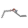
Movie
Movie Controller


+ Open data

- Basic information
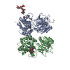
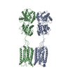
| Entry | Database: PDB / ID: 6wiv | |||||||||||||||||||||
|---|---|---|---|---|---|---|---|---|---|---|---|---|---|---|---|---|---|---|---|---|---|---|
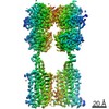
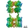
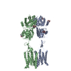
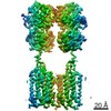
| Title | Structure of human GABA(B) receptor in an inactive state | |||||||||||||||||||||
 Components Components | (Gamma-aminobutyric acid type B receptor subunit ...) x 2 | |||||||||||||||||||||
 Keywords Keywords | MEMBRANE PROTEIN / G protein coupled receptor (GPCR) / GABA / GABA(B) receptor / Class C GPCR / Phospholipids / Inhibitory neurotransmission. | |||||||||||||||||||||
| Function / homology |  Function and homology information Function and homology informationnegative regulation of gamma-aminobutyric acid secretion / GABA B receptor activation / G protein-coupled GABA receptor complex / : / neuron-glial cell signaling / G protein-coupled receptor heterodimeric complex / G protein-coupled GABA receptor activity / : / negative regulation of epinephrine secretion / negative regulation of dopamine secretion ...negative regulation of gamma-aminobutyric acid secretion / GABA B receptor activation / G protein-coupled GABA receptor complex / : / neuron-glial cell signaling / G protein-coupled receptor heterodimeric complex / G protein-coupled GABA receptor activity / : / negative regulation of epinephrine secretion / negative regulation of dopamine secretion / positive regulation of growth hormone secretion / extracellular matrix protein binding / negative regulation of adenylate cyclase activity / GABA receptor complex / negative regulation of synaptic transmission / Class C/3 (Metabotropic glutamate/pheromone receptors) / positive regulation of glutamate secretion / gamma-aminobutyric acid signaling pathway / synaptic transmission, GABAergic / axolemma / dendritic shaft / response to nicotine / Schaffer collateral - CA1 synapse / GABA-ergic synapse / mitochondrial membrane / adenylate cyclase-inhibiting G protein-coupled receptor signaling pathway / osteoblast differentiation / Activation of G protein gated Potassium channels / Inhibition of voltage gated Ca2+ channels via Gbeta/gamma subunits / synaptic vesicle / transmembrane signaling receptor activity / presynaptic membrane / G alpha (i) signalling events / dendritic spine / chemical synaptic transmission / response to ethanol / postsynaptic membrane / neuron projection / G protein-coupled receptor signaling pathway / protein heterodimerization activity / negative regulation of cell population proliferation / neuronal cell body / endoplasmic reticulum membrane / glutamatergic synapse / : / plasma membrane / cytoplasm Similarity search - Function | |||||||||||||||||||||
| Biological species |  Homo sapiens (human) Homo sapiens (human) | |||||||||||||||||||||
| Method | ELECTRON MICROSCOPY / single particle reconstruction / cryo EM / Resolution: 3.3 Å | |||||||||||||||||||||
 Authors Authors | Park, J. / Fu, Z. / Frangaj, A. / Liu, J. / Mosyak, L. / Shen, T. / Slavkovich, V.N. / Ray, K.M. / Taura, J. / Cao, B. ...Park, J. / Fu, Z. / Frangaj, A. / Liu, J. / Mosyak, L. / Shen, T. / Slavkovich, V.N. / Ray, K.M. / Taura, J. / Cao, B. / Geng, Y. / Zuo, H. / Kou, Y. / Grassucci, R. / Chen, S. / Liu, Z. / Lin, X. / Williams, J.P. / Rice, W.J. / Eng, E.T. / Huang, R.K. / Soni, R.K. / Kloss, B. / Yu, Z. / Javitch, J.A. / Hendrickson, W.A. / Slesinger, P.A. / Quick, M. / Graziano, J. / Yu, H. / Fiehn, O. / Clarke, O.B. / Frank, J. / Fan, Q.R. | |||||||||||||||||||||
| Funding support |  United States, 6items United States, 6items
| |||||||||||||||||||||
 Citation Citation | Journal: Nature / Year: 2020 Title: Structure of human GABA receptor in an inactive state. Authors: Jinseo Park / Ziao Fu / Aurel Frangaj / Jonathan Liu / Lidia Mosyak / Tong Shen / Vesna N Slavkovich / Kimberly M Ray / Jaume Taura / Baohua Cao / Yong Geng / Hao Zuo / Yongjun Kou / Robert ...Authors: Jinseo Park / Ziao Fu / Aurel Frangaj / Jonathan Liu / Lidia Mosyak / Tong Shen / Vesna N Slavkovich / Kimberly M Ray / Jaume Taura / Baohua Cao / Yong Geng / Hao Zuo / Yongjun Kou / Robert Grassucci / Shaoxia Chen / Zheng Liu / Xin Lin / Justin P Williams / William J Rice / Edward T Eng / Rick K Huang / Rajesh K Soni / Brian Kloss / Zhiheng Yu / Jonathan A Javitch / Wayne A Hendrickson / Paul A Slesinger / Matthias Quick / Joseph Graziano / Hongtao Yu / Oliver Fiehn / Oliver B Clarke / Joachim Frank / Qing R Fan /   Abstract: The human GABA receptor-a member of the class C family of G-protein-coupled receptors (GPCRs)-mediates inhibitory neurotransmission and has been implicated in epilepsy, pain and addiction. A unique ...The human GABA receptor-a member of the class C family of G-protein-coupled receptors (GPCRs)-mediates inhibitory neurotransmission and has been implicated in epilepsy, pain and addiction. A unique GPCR that is known to require heterodimerization for function, the GABA receptor has two subunits, GABA and GABA, that are structurally homologous but perform distinct and complementary functions. GABA recognizes orthosteric ligands, while GABA couples with G proteins. Each subunit is characterized by an extracellular Venus flytrap (VFT) module, a descending peptide linker, a seven-helix transmembrane domain and a cytoplasmic tail. Although the VFT heterodimer structure has been resolved, the structure of the full-length receptor and its transmembrane signalling mechanism remain unknown. Here we present a near full-length structure of the GABA receptor, captured in an inactive state by cryo-electron microscopy. Our structure reveals several ligands that preassociate with the receptor, including two large endogenous phospholipids that are embedded within the transmembrane domains to maintain receptor integrity and modulate receptor function. We also identify a previously unknown heterodimer interface between transmembrane helices 3 and 5 of both subunits, which serves as a signature of the inactive conformation. A unique 'intersubunit latch' within this transmembrane interface maintains the inactive state, and its disruption leads to constitutive receptor activity. | |||||||||||||||||||||
| History |
|
- Structure visualization
| Movie |
Movie viewer |
|---|---|
| Structure viewer | Molecule: MolmilJmol/JSmol |
- Downloads & links
-Download
| PDBx/mmCIF format | 6wiv.cif.gz | 267.6 KB | Display | PDBx/mmCIF format |
|---|---|---|---|---|
| PDB format | pdb6wiv.ent.gz | 208 KB | Display | PDB format |
| PDBx/mmJSON format | 6wiv.json.gz | Tree view | PDBx/mmJSON format | |
| Others |  Other downloads Other downloads |
-Validation report
| Arichive directory | https://data.pdbj.org/pub/pdb/validation_reports/wi/6wivftp://data.pdbj.org/pub/pdb/validation_reports/wi/6wiv | HTTPS FTP |
|---|
-Related structure data
| Related structure data | 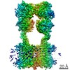 21685MC M: map data used to model this data C: citing same article ( |
|---|---|
| Similar structure data | |
| EM raw data | EMPIAR-10410 (Title: Structure of human GABA(B) receptor in an inactive state Data size: 854.1 Data #1: Unaligned multi-frame micrographs of GABA(B) receptor in the inactive state [micrographs - multiframe]) |


-Links
 PDBj
PDBj


- Assembly
| Deposited unit | 
|
|---|---|
| 1 |
|
-Components
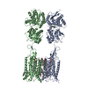
-Gamma-aminobutyric acid type B receptor subunit ... , 2 types, 2 molecules AB
| #1: Protein | Mass: 91585.867 Da / Num. of mol.: 1 Source method: isolated from a genetically manipulated source Source: (gene. exp.) Homo sapiens (human) / Gene: GABBR1, GPRC3A / Cell line (production host): HEK293 GnTl- / Production host: Homo sapiens (human) / References: UniProt: Q9UBS5 |
|---|---|
| #2: Protein | Mass: 93278.781 Da / Num. of mol.: 1 Source method: isolated from a genetically manipulated source Source: (gene. exp.) Homo sapiens (human) / Gene: GABBR2, GPR51, GPRC3B / Cell line (production host): HEK293 GnTl- / Production host: Homo sapiens (human) / References: UniProt: O75899 |
-Sugars , 1 types, 4 molecules 
| #3: Sugar | ChemComp-NAG /  Type: D-saccharide, beta linking / Mass: 221.208 Da / Num. of mol.: 4 / Source method: obtained synthetically / Formula: C8H15NO6 / Feature type: SUBJECT OF INVESTIGATION Type: D-saccharide, beta linking / Mass: 221.208 Da / Num. of mol.: 4 / Source method: obtained synthetically / Formula: C8H15NO6 / Feature type: SUBJECT OF INVESTIGATION |
|---|
-Non-polymers , 4 types, 13 molecules 


| #4: Chemical | ChemComp-CA /  Mass: 40.078 Da / Num. of mol.: 1 / Source method: obtained synthetically / Formula: Ca / Feature type: SUBJECT OF INVESTIGATION Mass: 40.078 Da / Num. of mol.: 1 / Source method: obtained synthetically / Formula: Ca / Feature type: SUBJECT OF INVESTIGATION | ||
|---|---|---|---|
| #5: Chemical | ChemComp-U3G / ( Mass: 766.039 Da / Num. of mol.: 1 / Source method: obtained synthetically / Formula: C43H76NO8P / Feature type: SUBJECT OF INVESTIGATION Mass: 766.039 Da / Num. of mol.: 1 / Source method: obtained synthetically / Formula: C43H76NO8P / Feature type: SUBJECT OF INVESTIGATION | ||
| #6: Chemical | ChemComp-CLR /  Mass: 386.654 Da / Num. of mol.: 10 / Source method: obtained synthetically / Formula: C27H46O / Feature type: SUBJECT OF INVESTIGATION Mass: 386.654 Da / Num. of mol.: 10 / Source method: obtained synthetically / Formula: C27H46O / Feature type: SUBJECT OF INVESTIGATION#7: Chemical | ChemComp-U3D / [( |  Mass: 814.167 Da / Num. of mol.: 1 / Source method: obtained synthetically / Formula: C46H88NO8P / Feature type: SUBJECT OF INVESTIGATION Mass: 814.167 Da / Num. of mol.: 1 / Source method: obtained synthetically / Formula: C46H88NO8P / Feature type: SUBJECT OF INVESTIGATION |
-Details
| Has ligand of interest | Y |
|---|---|
| Has protein modification | Y |
-Experimental details
-Experiment
| Experiment | Method: ELECTRON MICROSCOPY |
|---|---|
| EM experiment | Aggregation state: PARTICLE / 3D reconstruction method: single particle reconstruction |
- Sample preparation
| Component | Name: Heterodimer human GABA(B) receptor composed of GABA(B1b) and GABA(B2) subunits. Type: COMPLEX / Entity ID: #1-#2 / Source: RECOMBINANT | |||||||||||||||||||||||||
|---|---|---|---|---|---|---|---|---|---|---|---|---|---|---|---|---|---|---|---|---|---|---|---|---|---|---|
| Molecular weight | Value: 0.193 MDa / Experimental value: YES | |||||||||||||||||||||||||
| Source (natural) | Organism: Homo sapiens (human) | |||||||||||||||||||||||||
| Source (recombinant) | Organism: Homo sapiens (human) / Cell: HEK 293 GnTI- / Plasmid: modified bacMam | |||||||||||||||||||||||||
| Buffer solution | pH: 7.5 Details: Solutions were made fresh from concentrated and filtered to avoid microbial contamination. | |||||||||||||||||||||||||
| Buffer component |
| |||||||||||||||||||||||||
| Specimen | Conc.: 0.3 mg/ml / Embedding applied: NO / Shadowing applied: NO / Staining applied: NO / Vitrification applied: YES / Details: This sample was monodisperse | |||||||||||||||||||||||||
| Specimen support | Grid material: GOLD / Grid mesh size: 300 divisions/in. / Grid type: Quantifoil R0.6/1 | |||||||||||||||||||||||||
| Vitrification | Instrument: FEI VITROBOT MARK IV / Cryogen name: ETHANE-PROPANE / Humidity: 100 % / Chamber temperature: 277 K / Details: blot for 4 seconds before plunging |
- Electron microscopy imaging
| Experimental equipment |  Model: Titan Krios / Image courtesy: FEI Company |
|---|---|
| Microscopy | Model: FEI TITAN KRIOS / Details: Preliminary grid screening was performed manually. |
| Electron gun | Electron source:  FIELD EMISSION GUN / Accelerating voltage: 300 kV / Illumination mode: FLOOD BEAM FIELD EMISSION GUN / Accelerating voltage: 300 kV / Illumination mode: FLOOD BEAM |
| Electron lens | Mode: BRIGHT FIELD / Nominal magnification: 130000 X / Nominal defocus max: -2000 nm / Nominal defocus min: -500 nm / Cs: 2.7 mm / C2 aperture diameter: 70 µm / Alignment procedure: COMA FREE |
| Specimen holder | Cryogen: NITROGEN / Specimen holder model: FEI TITAN KRIOS AUTOGRID HOLDER / Temperature (max): 100 K |
| Image recording | Average exposure time: 12 sec. / Electron dose: 85 e/Å2 / Detector mode: COUNTING / Film or detector model: GATAN K2 SUMMIT (4k x 4k) / Num. of grids imaged: 1 / Num. of real images: 3435 |
| EM imaging optics | Energyfilter slit width: 20 eV |
| Image scans | Movie frames/image: 60 / Used frames/image: 1-60 |
- Processing
| EM software |
| |||||||||||||||||||||||||||||||||||||||||||||
|---|---|---|---|---|---|---|---|---|---|---|---|---|---|---|---|---|---|---|---|---|---|---|---|---|---|---|---|---|---|---|---|---|---|---|---|---|---|---|---|---|---|---|---|---|---|---|
| CTF correction | Type: PHASE FLIPPING AND AMPLITUDE CORRECTION | |||||||||||||||||||||||||||||||||||||||||||||
| Particle selection | Num. of particles selected: 1048241 | |||||||||||||||||||||||||||||||||||||||||||||
| Symmetry | Point symmetry: C1 (asymmetric) | |||||||||||||||||||||||||||||||||||||||||||||
| 3D reconstruction | Resolution: 3.3 Å / Resolution method: FSC 0.143 CUT-OFF / Num. of particles: 233737 / Num. of class averages: 1 / Symmetry type: POINT | |||||||||||||||||||||||||||||||||||||||||||||
| Atomic model building | Protocol: FLEXIBLE FIT / Space: REAL | |||||||||||||||||||||||||||||||||||||||||||||
| Atomic model building | 3D fitting-ID: 1 / Accession code: 4MQE / Initial refinement model-ID: 1 / PDB-ID: 4MQE / Source name: PDB / Type: experimental model
| |||||||||||||||||||||||||||||||||||||||||||||
| Refinement | Stereochemistry target values: GeoStd + Monomer Library + CDL v1.2 | |||||||||||||||||||||||||||||||||||||||||||||
| Displacement parameters | Biso mean: 68.95 Å2 | |||||||||||||||||||||||||||||||||||||||||||||
| Refine LS restraints |
|