- EMDB-21685: Structure of human GABA(B) receptor in an inactive state -
+
Open data
ID or keywords:
Loading...
-
Basic information
Entry
Database: EMDB / ID: EMD-21685
Title
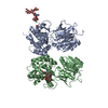
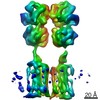
Structure of human GABA(B) receptor in an inactive state
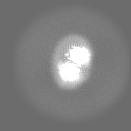
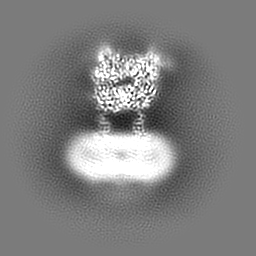
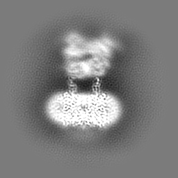
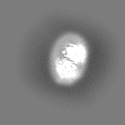
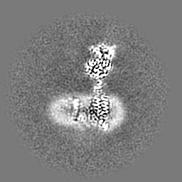
Map data
Final composite map combining local reconstructions for the extracellular and transmembrane domains. Map was re-sampled in Chimera with pixel spacing 1.1 angstrom.
Sample
Complex: Heterodimer human GABA(B) receptor composed of GABA(B1b) and GABA(B2) subunits.
Protein or peptide: Gamma-aminobutyric acid type B receptor subunit 1
Protein or peptide: Gamma-aminobutyric acid type B receptor subunit 2
National Institutes of Health/National Institute of General Medical Sciences (NIH/NIGMS)
R01GM088454
United States
National Institutes of Health/National Institute of General Medical Sciences (NIH/NIGMS)
R01GM125801
United States
National Institutes of Health/National Institute of General Medical Sciences (NIH/NIGMS)
U2CES030158
United States
National Institutes of Health/National Institute of General Medical Sciences (NIH/NIGMS)
P41GM116799
United States
National Institutes of Health/Office of the Director
R01GM107462
United States
National Institutes of Health/National Institute of General Medical Sciences (NIH/NIGMS)
P41GM103310
United States
Citation
Journal: Nature / Year: 2020 Title: Structure of human GABA receptor in an inactive state. Authors: Jinseo Park / Ziao Fu / Aurel Frangaj / Jonathan Liu / Lidia Mosyak / Tong Shen / Vesna N Slavkovich / Kimberly M Ray / Jaume Taura / Baohua Cao / Yong Geng / Hao Zuo / Yongjun Kou / Robert ...Authors: Jinseo Park / Ziao Fu / Aurel Frangaj / Jonathan Liu / Lidia Mosyak / Tong Shen / Vesna N Slavkovich / Kimberly M Ray / Jaume Taura / Baohua Cao / Yong Geng / Hao Zuo / Yongjun Kou / Robert Grassucci / Shaoxia Chen / Zheng Liu / Xin Lin / Justin P Williams / William J Rice / Edward T Eng / Rick K Huang / Rajesh K Soni / Brian Kloss / Zhiheng Yu / Jonathan A Javitch / Wayne A Hendrickson / Paul A Slesinger / Matthias Quick / Joseph Graziano / Hongtao Yu / Oliver Fiehn / Oliver B Clarke / Joachim Frank / Qing R Fan / Abstract: The human GABA receptor-a member of the class C family of G-protein-coupled receptors (GPCRs)-mediates inhibitory neurotransmission and has been implicated in epilepsy, pain and addiction. A unique ...The human GABA receptor-a member of the class C family of G-protein-coupled receptors (GPCRs)-mediates inhibitory neurotransmission and has been implicated in epilepsy, pain and addiction. A unique GPCR that is known to require heterodimerization for function, the GABA receptor has two subunits, GABA and GABA, that are structurally homologous but perform distinct and complementary functions. GABA recognizes orthosteric ligands, while GABA couples with G proteins. Each subunit is characterized by an extracellular Venus flytrap (VFT) module, a descending peptide linker, a seven-helix transmembrane domain and a cytoplasmic tail. Although the VFT heterodimer structure has been resolved, the structure of the full-length receptor and its transmembrane signalling mechanism remain unknown. Here we present a near full-length structure of the GABA receptor, captured in an inactive state by cryo-electron microscopy. Our structure reveals several ligands that preassociate with the receptor, including two large endogenous phospholipids that are embedded within the transmembrane domains to maintain receptor integrity and modulate receptor function. We also identify a previously unknown heterodimer interface between transmembrane helices 3 and 5 of both subunits, which serves as a signature of the inactive conformation. A unique 'intersubunit latch' within this transmembrane interface maintains the inactive state, and its disruption leads to constitutive receptor activity.
History
Deposition
Apr 10, 2020
-
Header (metadata) release
Jul 1, 2020
-
Map release
Jul 1, 2020
-
Update
Oct 9, 2024
-
Current status
Oct 9, 2024
Processing site: RCSB / Status: Released
-
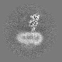
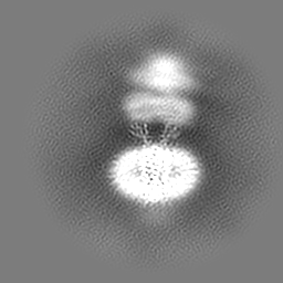
Structure visualization
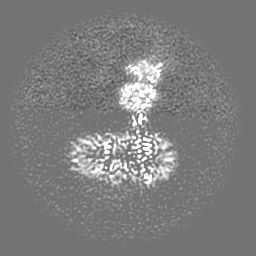
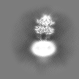
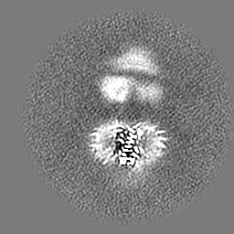
Movie
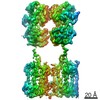
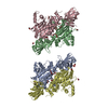
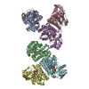
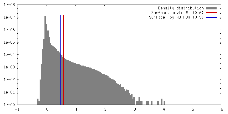
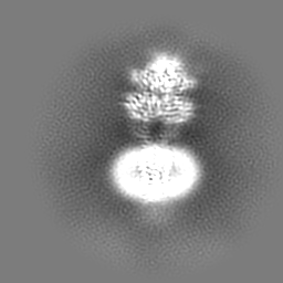
Surface view with section colored by density value
EMPIAR-10410 (Title: Structure of human GABA(B) receptor in an inactive state Data size: 854.1 Data #1: Unaligned multi-frame micrographs of GABA(B) receptor in the inactive state [micrographs - multiframe])
Download / File: emd_21685.map.gz / Format: CCP4 / Size: 64 MB / Type: IMAGE STORED AS FLOATING POINT NUMBER (4 BYTES)
Annotation
Final composite map combining local reconstructions for the extracellular and transmembrane domains. Map was re-sampled in Chimera with pixel spacing 1.1 angstrom.
Details: Solutions were made fresh from concentrated and filtered to avoid microbial contamination.
Grid
Model: Quantifoil R0.6/1 / Material: GOLD / Mesh: 300 / Support film - Material: CARBON / Support film - topology: HOLEY / Support film - Film thickness: 50 / Pretreatment - Type: PLASMA CLEANING / Pretreatment - Time: 25 sec. / Pretreatment - Atmosphere: OTHER
Vitrification
Cryogen name: ETHANE-PROPANE / Chamber humidity: 100 % / Chamber temperature: 277 K / Instrument: FEI VITROBOT MARK IV / Details: blot for 4 seconds before plunging.
Details
This sample was monodisperse
-
Electron microscopy
Microscope
FEI TITAN KRIOS
Temperature
Max: 100.0 K
Specialist optics
Energy filter - Slit width: 20 eV
Details
Preliminary grid screening was performed manually.
Image recording
Film or detector model: GATAN K2 SUMMIT (4k x 4k) / Detector mode: COUNTING / Digitization - Frames/image: 1-60 / Number grids imaged: 1 / Number real images: 3435 / Average exposure time: 12.0 sec. / Average electron dose: 85.0 e/Å2
Electron beam
Acceleration voltage: 300 kV / Electron source: FIELD EMISSION GUN
In the structure databanks used in Yorodumi, some data are registered as the other names, "COVID-19 virus" and "2019-nCoV". Here are the details of the virus and the list of structure data.
Jan 31, 2019. EMDB accession codes are about to change! (news from PDBe EMDB page)
EMDB accession codes are about to change! (news from PDBe EMDB page)
The allocation of 4 digits for EMDB accession codes will soon come to an end. Whilst these codes will remain in use, new EMDB accession codes will include an additional digit and will expand incrementally as the available range of codes is exhausted. The current 4-digit format prefixed with “EMD-” (i.e. EMD-XXXX) will advance to a 5-digit format (i.e. EMD-XXXXX), and so on. It is currently estimated that the 4-digit codes will be depleted around Spring 2019, at which point the 5-digit format will come into force.
The EM Navigator/Yorodumi systems omit the EMD- prefix.
Related info.:Q: What is EMD? / ID/Accession-code notation in Yorodumi/EM Navigator
Yorodumi is a browser for structure data from EMDB, PDB, SASBDB, etc.
This page is also the successor to EM Navigator detail page, and also detail information page/front-end page for Omokage search.
The word "yorodu" (or yorozu) is an old Japanese word meaning "ten thousand". "mi" (miru) is to see.
Related info.:EMDB / PDB / SASBDB / Comparison of 3 databanks / Yorodumi Search / Aug 31, 2016. New EM Navigator & Yorodumi / Yorodumi Papers / Jmol/JSmol / Function and homology information / Changes in new EM Navigator and Yorodumi
 Movie
Movie Controller


 Open data
 Basic information
 Map data
 Sample
 Keywords
 Function and homology information
 Homo sapiens (human)
 Authors
 United States, 6 items
  Citation

 Structure visualization
 Downloads & links
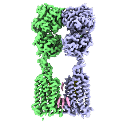 emd_21685.png
 http://ftp.pdbj.org/pub/emdb/structures/EMD-21685
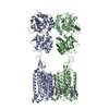


 Z (Sec.)
 Y (Row.)
 X (Col.)


 Sample components


 Processing
 Electron microscopy
 FIELD EMISSION GUN