 Movie
Movie Controller

[English] 日本語
 Yorodumi
- PDB-6vz1: Cryo-EM structure of human diacylglycerol O-acyltransferase 1 com... -
+ Open data

- Basic information
| Entry | Database: PDB / ID: 6vz1 | |||||||||||||||||||||||||||||||||
|---|---|---|---|---|---|---|---|---|---|---|---|---|---|---|---|---|---|---|---|---|---|---|---|---|---|---|---|---|---|---|---|---|---|---|
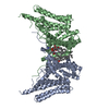
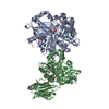
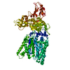
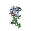
| Title | Cryo-EM structure of human diacylglycerol O-acyltransferase 1 complexed with acyl-CoA substrate | |||||||||||||||||||||||||||||||||
 Components Components | Diacylglycerol O-acyltransferase 1 | |||||||||||||||||||||||||||||||||
 Keywords Keywords | TRANSFERASE / Triglyceride Biosynthesis / Lipid Storage / Lipid Metabolism | |||||||||||||||||||||||||||||||||
| Function / homology |  Function and homology information Function and homology informationretinol O-fatty-acyltransferase / 2-acylglycerol O-acyltransferase activity / retinol O-fatty-acyltransferase activity / monoacylglycerol biosynthetic process / Triglyceride biosynthesis / diacylglycerol metabolic process / long-chain fatty-acyl-CoA metabolic process / Acyl chain remodeling of DAG and TAG / diacylglycerol O-acyltransferase / diacylglycerol O-acyltransferase activity ...retinol O-fatty-acyltransferase / 2-acylglycerol O-acyltransferase activity / retinol O-fatty-acyltransferase activity / monoacylglycerol biosynthetic process / Triglyceride biosynthesis / diacylglycerol metabolic process / long-chain fatty-acyl-CoA metabolic process / Acyl chain remodeling of DAG and TAG / diacylglycerol O-acyltransferase / diacylglycerol O-acyltransferase activity / very-low-density lipoprotein particle assembly / triglyceride biosynthetic process / lipid storage / triglyceride metabolic process / acyltransferase activity / fatty acid homeostasis / specific granule membrane / Neutrophil degranulation / endoplasmic reticulum membrane / membrane / identical protein binding / plasma membrane Similarity search - Function | |||||||||||||||||||||||||||||||||
| Biological species |  Homo sapiens (human) Homo sapiens (human) | |||||||||||||||||||||||||||||||||
| Method | ELECTRON MICROSCOPY / single particle reconstruction / cryo EM / Resolution: 3.2 Å | |||||||||||||||||||||||||||||||||
 Authors Authors | Sui, X. / Wang, K. / Gluchowski, N. / Liao, M. / Walther, C.T. / Farese Jr., V.R. | |||||||||||||||||||||||||||||||||
| Funding support |  United States, 4items United States, 4items
| |||||||||||||||||||||||||||||||||
 Citation Citation | Journal: Nature / Year: 2020 Title: Structure and catalytic mechanism of a human triacylglycerol-synthesis enzyme. Authors: Xuewu Sui / Kun Wang / Nina L Gluchowski / Shane D Elliott / Maofu Liao / Tobias C Walther / Robert V Farese / Abstract: Triacylglycerols store metabolic energy in organisms and have industrial uses as foods and fuels. Excessive accumulation of triacylglycerols in humans causes obesity and is associated with metabolic ...Triacylglycerols store metabolic energy in organisms and have industrial uses as foods and fuels. Excessive accumulation of triacylglycerols in humans causes obesity and is associated with metabolic diseases. Triacylglycerol synthesis is catalysed by acyl-CoA diacylglycerol acyltransferase (DGAT) enzymes, the structures and catalytic mechanisms of which remain unknown. Here we determined the structure of dimeric human DGAT1, a member of the membrane-bound O-acyltransferase (MBOAT) family, by cryo-electron microscopy at approximately 3.0 Å resolution. DGAT1 forms a homodimer through N-terminal segments and a hydrophobic interface, with putative active sites within the membrane region. A structure obtained with oleoyl-CoA substrate resolved at approximately 3.2 Å shows that the CoA moiety binds DGAT1 on the cytosolic side and the acyl group lies deep within a hydrophobic channel, positioning the acyl-CoA thioester bond near an invariant catalytic histidine residue. The reaction centre is located inside a large cavity, which opens laterally to the membrane bilayer, providing lipid access to the active site. A lipid-like density-possibly representing an acyl-acceptor molecule-is located within the reaction centre, orthogonal to acyl-CoA. Insights provided by the DGAT1 structures, together with mutagenesis and functional studies, provide the basis for a model of the catalysis of triacylglycerol synthesis by DGAT. | |||||||||||||||||||||||||||||||||
| History |
|
- Structure visualization
| Movie |
Movie viewer |
|---|---|
| Structure viewer | Molecule: MolmilJmol/JSmol |
- Downloads & links
-Download
| PDBx/mmCIF format | 6vz1.cif.gz | 169.1 KB | Display | PDBx/mmCIF format |
|---|---|---|---|---|
| PDB format | pdb6vz1.ent.gz | 132.2 KB | Display | PDB format |
| PDBx/mmJSON format | 6vz1.json.gz | Tree view | PDBx/mmJSON format | |
| Others |  Other downloads Other downloads |
-Validation report
| Arichive directory | https://data.pdbj.org/pub/pdb/validation_reports/vz/6vz1ftp://data.pdbj.org/pub/pdb/validation_reports/vz/6vz1 | HTTPS FTP |
|---|
-Related structure data
| Related structure data |  21481MC  6vyiC M: map data used to model this data C: citing same article ( |
|---|---|
| Similar structure data |


-Links
 PDBj
PDBj- Assembly
| Deposited unit | 
|
|---|---|
| 1 |
|
-Components
| #1: Protein | Mass: 55339.133 Da / Num. of mol.: 2 Source method: isolated from a genetically manipulated source Source: (gene. exp.) Homo sapiens (human) / Gene: DGAT1, AGRP1, DGAT / Plasmid: pFasBacMam / Production host: Homo sapiens (human)References: UniProt: O75907, diacylglycerol O-acyltransferase, retinol O-fatty-acyltransferase #2: Chemical | ChemComp-6OU / [( 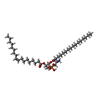  Mass: 717.996 Da / Num. of mol.: 4 / Source method: obtained synthetically / Formula: C39H76NO8P / Comment: phospholipid*YM Mass: 717.996 Da / Num. of mol.: 4 / Source method: obtained synthetically / Formula: C39H76NO8P / Comment: phospholipid*YM#3: Chemical |   Mass: 1031.980 Da / Num. of mol.: 2 / Source method: obtained synthetically / Formula: C39H68N7O17P3S / Feature type: SUBJECT OF INVESTIGATION Mass: 1031.980 Da / Num. of mol.: 2 / Source method: obtained synthetically / Formula: C39H68N7O17P3S / Feature type: SUBJECT OF INVESTIGATIONHas ligand of interest | Y | Has protein modification | N | |
|---|
-Experimental details
-Experiment
| Experiment | Method: ELECTRON MICROSCOPY |
|---|---|
| EM experiment | Aggregation state: PARTICLE / 3D reconstruction method: single particle reconstruction |
- Sample preparation
| Component | Name: human diacylglycerol O-acyltransferase 1 / Type: COMPLEX Details: human diacylglycerol O-acyltransferase 1 complexed with oleoyl-CoA substrate Entity ID: #1 / Source: RECOMBINANT |
|---|---|
| Molecular weight | Experimental value: NO |
| Source (natural) | Organism: Homo sapiens (human) |
| Source (recombinant) | Organism: Homo sapiens (human) / Plasmid: pFasBacMam |
| Buffer solution | pH: 7.5 |
| Specimen | Conc.: 5 mg/ml / Embedding applied: NO / Shadowing applied: NO / Staining applied: NO / Vitrification applied: YES / Details: Protein sample was monodisperse. |
| Vitrification | Instrument: GATAN CRYOPLUNGE 3 / Cryogen name: ETHANE / Humidity: 90 % |
- Electron microscopy imaging
| Experimental equipment |  Model: Titan Krios / Image courtesy: FEI Company |
|---|---|
| Microscopy | Model: FEI TITAN KRIOS |
| Electron gun | Electron source:  FIELD EMISSION GUN / Accelerating voltage: 300 kV / Illumination mode: FLOOD BEAM FIELD EMISSION GUN / Accelerating voltage: 300 kV / Illumination mode: FLOOD BEAM |
| Electron lens | Mode: BRIGHT FIELD |
| Image recording | Electron dose: 43 e/Å2 / Detector mode: SUPER-RESOLUTION / Film or detector model: GATAN K3 (6k x 4k) |
- Processing
| Software | Name: PHENIX / Version: 1.16_3549: / Classification: refinement | ||||||||||||||||||||||||
|---|---|---|---|---|---|---|---|---|---|---|---|---|---|---|---|---|---|---|---|---|---|---|---|---|---|
| EM software | Name: PHENIX / Category: model refinement | ||||||||||||||||||||||||
| CTF correction | Type: PHASE FLIPPING AND AMPLITUDE CORRECTION | ||||||||||||||||||||||||
| 3D reconstruction | Resolution: 3.2 Å / Resolution method: FSC 0.143 CUT-OFF / Num. of particles: 28165 / Symmetry type: POINT | ||||||||||||||||||||||||
| Refine LS restraints |
|
