ムービー
ムービー コントローラー


+ データを開く

- 基本情報
基本情報
| 登録情報 | データベース: PDB / ID: 6vcc | ||||||
|---|---|---|---|---|---|---|---|
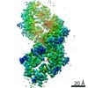
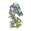
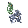
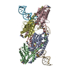
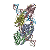
| タイトル | Cryo-EM structure of the Dvl2 DIX filament | ||||||
 要素 要素 | Segment polarity protein dishevelled homolog DVL-2 | ||||||
 キーワード キーワード | SIGNALING PROTEIN / Dishevelled / DIX domain / Wnt signaling | ||||||
| 機能・相同性 |  機能・相同性情報 機能・相同性情報WNT mediated activation of DVL / convergent extension involved in organogenesis / PCP/CE pathway / Signaling by Hippo / convergent extension involved in neural plate elongation / Disassembly of the destruction complex and recruitment of AXIN to the membrane / segmentation / planar cell polarity pathway involved in neural tube closure / Asymmetric localization of PCP proteins / cochlea morphogenesis ...WNT mediated activation of DVL / convergent extension involved in organogenesis / PCP/CE pathway / Signaling by Hippo / convergent extension involved in neural plate elongation / Disassembly of the destruction complex and recruitment of AXIN to the membrane / segmentation / planar cell polarity pathway involved in neural tube closure / Asymmetric localization of PCP proteins / cochlea morphogenesis / WNT5A-dependent internalization of FZD4 / Degradation of DVL / segment specification / positive regulation of neuron projection arborization / RHO GTPases Activate Formins / Cargo recognition for clathrin-mediated endocytosis / Clathrin-mediated endocytosis / frizzled binding / aggresome / Wnt signaling pathway, planar cell polarity pathway / clathrin-coated vesicle / heart looping / outflow tract morphogenesis / lateral plasma membrane / canonical Wnt signaling pathway / heart morphogenesis / positive regulation of JUN kinase activity / : / positive regulation of GTPase activity / neural tube closure / positive regulation of JNK cascade / Wnt signaling pathway / small GTPase binding / protein localization / apical part of cell / protein-macromolecule adaptor activity / heart development / regulation of cell population proliferation / cell cortex / cytoplasmic vesicle / cytoskeleton / nuclear body / intracellular signal transduction / protein domain specific binding / protein kinase binding / positive regulation of transcription by RNA polymerase II / identical protein binding / nucleus / plasma membrane / cytoplasm / cytosol 類似検索 - 分子機能 | ||||||
| 生物種 |  | ||||||
| 手法 | 電子顕微鏡法 / らせん対称体再構成法 / クライオ電子顕微鏡法 / 解像度: 3.6 Å | ||||||
 データ登録者 データ登録者 | Enos, M. / Kan, W. / Muennich, S. / Chen, D.H. / Skiniotis, G. / Weis, W.I. | ||||||
| 資金援助 |  米国, 1件 米国, 1件
| ||||||
 引用 引用 | ジャーナル: Elife / 年: 2020 タイトル: Limited dishevelled/Axin oligomerization determines efficiency of Wnt/β-catenin signal transduction. 著者: Wei Kan / Michael D Enos / Elgin Korkmazhan / Stefan Muennich / Dong-Hua Chen / Melissa V Gammons / Mansi Vasishtha / Mariann Bienz / Alexander R Dunn / Georgios Skiniotis / William I Weis /  要旨: In Wnt/β-catenin signaling, the transcriptional coactivator β-catenin is regulated by its phosphorylation in a complex that includes the scaffold protein Axin and associated kinases. Wnt binding to ...In Wnt/β-catenin signaling, the transcriptional coactivator β-catenin is regulated by its phosphorylation in a complex that includes the scaffold protein Axin and associated kinases. Wnt binding to its coreceptors activates the cytosolic effector Dishevelled (Dvl), leading to the recruitment of Axin and the inhibition of β-catenin phosphorylation. This process requires interaction of homologous DIX domains present in Dvl and Axin, but is mechanistically undefined. We show that Dvl DIX forms antiparallel, double-stranded oligomers in vitro, and that Dvl in cells forms oligomers typically <10 molecules at endogenous expression levels. Axin DIX (DAX) forms small single-stranded oligomers, but its self-association is stronger than that of DIX. DAX caps the ends of DIX oligomers, such that a DIX oligomer has at most four DAX binding sites. The relative affinities and stoichiometry of the DIX-DAX interaction provide a mechanism for efficient inhibition of β-catenin phosphorylation upon Axin recruitment to the Wnt receptor complex. | ||||||
| 履歴 |
|
- 構造の表示
構造の表示
| ムービー |
ムービービューア |
|---|---|
| 構造ビューア | 分子: MolmilJmol/JSmol |
- ダウンロードとリンク
-ダウンロード
| PDBx/mmCIF形式 | 6vcc.cif.gz | 171.1 KB | 表示 | PDBx/mmCIF形式 |
|---|---|---|---|---|
| PDB形式 | pdb6vcc.ent.gz | 138.7 KB | 表示 | PDB形式 |
| PDBx/mmJSON形式 | 6vcc.json.gz | ツリー表示 | PDBx/mmJSON形式 | |
| その他 |  その他のダウンロード その他のダウンロード |
-検証レポート
| 文書・要旨 | 6vcc_validation.pdf.gz | 1.3 MB | 表示 | wwPDB検証レポート |
|---|---|---|---|---|
| 文書・詳細版 | 6vcc_full_validation.pdf.gz | 1.3 MB | 表示 | |
| XML形式データ | 6vcc_validation.xml.gz | 32.3 KB | 表示 | |
| CIF形式データ | 6vcc_validation.cif.gz | 50.2 KB | 表示 | |
| アーカイブディレクトリ | https://data.pdbj.org/pub/pdb/validation_reports/vc/6vccftp://data.pdbj.org/pub/pdb/validation_reports/vc/6vcc | HTTPS FTP |
-関連構造データ

-リンク
 PDBj
PDBj


- 集合体
集合体
| 登録構造単位 | 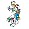
|
|---|---|
| 1 |
|
| 対称性 | らせん対称: (回転対称性: 1 / Dyad axis: no / N subunits divisor: 1 / Num. of operations: 6 / Rise per n subunits: 13.5 Å / Rotation per n subunits: 40 °) |
-要素
| #1: タンパク質 | 分子量: 9333.547 Da / 分子数: 12 / 断片: DIX domain (UNP residues 12-92) / 由来タイプ: 組換発現 / 由来: (組換発現)  |
|---|
-実験情報
-実験
| 実験 | 手法: 電子顕微鏡法 |
|---|---|
| EM実験 | 試料の集合状態: HELICAL ARRAY / 3次元再構成法: らせん対称体再構成法 |
- 試料調製
試料調製
| 構成要素 | 名称: Segment polarity protein dishevelled homolog DVL-2 / タイプ: COMPLEX 詳細: Protein spontaneously formed a double-stranded, antiparallel helical filament upon removal of the MBP tag with TEV protease. Entity ID: all / 由来: RECOMBINANT | |||||||||||||||||||||||||
|---|---|---|---|---|---|---|---|---|---|---|---|---|---|---|---|---|---|---|---|---|---|---|---|---|---|---|
| 分子量 | 実験値: NO | |||||||||||||||||||||||||
| 由来(天然) | 生物種: | |||||||||||||||||||||||||
| 由来(組換発現) | 生物種: | |||||||||||||||||||||||||
| 緩衝液 | pH: 8 / 詳細: Dithiothreitol was added fresh the day of use. | |||||||||||||||||||||||||
| 緩衝液成分 |
| |||||||||||||||||||||||||
| 試料 | 濃度: 0.12 mg/ml / 包埋: NO / シャドウイング: NO / 染色: NO / 凍結: YES 詳細: Sample was taken from a single fraction of a gel filtration run performed immediately before grid preparation. | |||||||||||||||||||||||||
| 試料支持 | グリッドの材料: COPPER / グリッドのサイズ: 200 divisions/in. | |||||||||||||||||||||||||
| 急速凍結 | 装置: LEICA EM GP / 凍結剤: ETHANE / 湿度: 95 % / 凍結前の試料温度: 295 K 詳細: The grid was blotted for 2.0 s using Whatman #1 filter paper (GE Healthcare). |
- 電子顕微鏡撮影
| 実験機器 |  モデル: Titan Krios / 画像提供: FEI Company |
|---|---|
| 顕微鏡 | モデル: FEI TITAN KRIOS |
| 電子銃 | 電子線源:  FIELD EMISSION GUN / 加速電圧: 300 kV / 照射モード: FLOOD BEAM FIELD EMISSION GUN / 加速電圧: 300 kV / 照射モード: FLOOD BEAM |
| 電子レンズ | モード: BRIGHT FIELD / 倍率(補正後): 29000 X / Calibrated defocus min: 500 nm / 最大 デフォーカス(補正後): 2000 nm / Cs: 2.7 mm / アライメント法: COMA FREE |
| 試料ホルダ | 凍結剤: NITROGEN 試料ホルダーモデル: FEI TITAN KRIOS AUTOGRID HOLDER |
| 撮影 | 平均露光時間: 10 sec. / 電子線照射量: 98 e/Å2 / 検出モード: COUNTING フィルム・検出器のモデル: GATAN K2 SUMMIT (4k x 4k) 実像数: 540 |
| 画像スキャン | 横: 3838 / 縦: 3710 / 動画フレーム数/画像: 50 / 利用したフレーム数/画像: 4-50 |
- 解析
解析
| ソフトウェア | 名称: PHENIX / バージョン: 1.17.1_3660: / 分類: 精密化 | ||||||||||||||||||||||||||||||||||||||||||||
|---|---|---|---|---|---|---|---|---|---|---|---|---|---|---|---|---|---|---|---|---|---|---|---|---|---|---|---|---|---|---|---|---|---|---|---|---|---|---|---|---|---|---|---|---|---|
| EMソフトウェア |
| ||||||||||||||||||||||||||||||||||||||||||||
| CTF補正 | タイプ: PHASE FLIPPING AND AMPLITUDE CORRECTION | ||||||||||||||||||||||||||||||||||||||||||||
| らせん対称 | 回転角度/サブユニット: 48 ° / 軸方向距離/サブユニット: 13.5 Å / らせん対称軸の対称性: C1 | ||||||||||||||||||||||||||||||||||||||||||||
| 粒子像の選択 | 選択した粒子像数: 437872 | ||||||||||||||||||||||||||||||||||||||||||||
| 3次元再構成 | 解像度: 3.6 Å / 解像度の算出法: FSC 0.143 CUT-OFF / 粒子像の数: 110105 / アルゴリズム: BACK PROJECTION / クラス平均像の数: 2 / 対称性のタイプ: HELICAL | ||||||||||||||||||||||||||||||||||||||||||||
| 原子モデル構築 | プロトコル: RIGID BODY FIT / 空間: REAL 詳細: The two mutations in the source (Y27W and C80S) were reverted to Trp and Ser, respectively, before fitting. Model refinement was carried out through alternating rounds of real-space ...詳細: The two mutations in the source (Y27W and C80S) were reverted to Trp and Ser, respectively, before fitting. Model refinement was carried out through alternating rounds of real-space refinement in PHENIX and manual building in Coot. | ||||||||||||||||||||||||||||||||||||||||||||
| 原子モデル構築 | PDB-ID: 6IW3 PDB chain-ID: A / Accession code: 6IW3 / Pdb chain residue range: 12-92 / Source name: PDB / タイプ: experimental model | ||||||||||||||||||||||||||||||||||||||||||||
| 拘束条件 |
|