Movie
Movie Controller


+ Open data

- Basic information
| Entry | Database: PDB / ID: 6vcc | ||||||
|---|---|---|---|---|---|---|---|
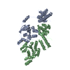
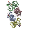
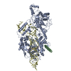
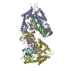
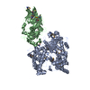
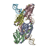
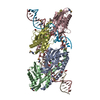
| Title | Cryo-EM structure of the Dvl2 DIX filament | ||||||
 Components Components | Segment polarity protein dishevelled homolog DVL-2 | ||||||
 Keywords Keywords | SIGNALING PROTEIN / Dishevelled / DIX domain / Wnt signaling | ||||||
| Function / homology |  Function and homology information Function and homology informationWNT mediated activation of DVL / convergent extension involved in organogenesis / PCP/CE pathway / Signaling by Hippo / convergent extension involved in neural plate elongation / Disassembly of the destruction complex and recruitment of AXIN to the membrane / segmentation / planar cell polarity pathway involved in neural tube closure / Asymmetric localization of PCP proteins / cochlea morphogenesis ...WNT mediated activation of DVL / convergent extension involved in organogenesis / PCP/CE pathway / Signaling by Hippo / convergent extension involved in neural plate elongation / Disassembly of the destruction complex and recruitment of AXIN to the membrane / segmentation / planar cell polarity pathway involved in neural tube closure / Asymmetric localization of PCP proteins / cochlea morphogenesis / WNT5A-dependent internalization of FZD4 / Degradation of DVL / segment specification / positive regulation of neuron projection arborization / RHO GTPases Activate Formins / Cargo recognition for clathrin-mediated endocytosis / Clathrin-mediated endocytosis / frizzled binding / aggresome / Wnt signaling pathway, planar cell polarity pathway / clathrin-coated vesicle / heart looping / outflow tract morphogenesis / lateral plasma membrane / canonical Wnt signaling pathway / heart morphogenesis / positive regulation of JUN kinase activity / : / positive regulation of GTPase activity / neural tube closure / positive regulation of JNK cascade / Wnt signaling pathway / small GTPase binding / protein localization / apical part of cell / protein-macromolecule adaptor activity / heart development / regulation of cell population proliferation / cell cortex / cytoplasmic vesicle / cytoskeleton / nuclear body / intracellular signal transduction / protein domain specific binding / protein kinase binding / positive regulation of transcription by RNA polymerase II / identical protein binding / nucleus / plasma membrane / cytoplasm / cytosol Similarity search - Function | ||||||
| Biological species |  | ||||||
| Method | ELECTRON MICROSCOPY / helical reconstruction / cryo EM / Resolution: 3.6 Å | ||||||
 Authors Authors | Enos, M. / Kan, W. / Muennich, S. / Chen, D.H. / Skiniotis, G. / Weis, W.I. | ||||||
| Funding support |  United States, 1items United States, 1items
| ||||||
 Citation Citation | Journal: Elife / Year: 2020 Title: Limited dishevelled/Axin oligomerization determines efficiency of Wnt/β-catenin signal transduction. Authors: Wei Kan / Michael D Enos / Elgin Korkmazhan / Stefan Muennich / Dong-Hua Chen / Melissa V Gammons / Mansi Vasishtha / Mariann Bienz / Alexander R Dunn / Georgios Skiniotis / William I Weis /  Abstract: In Wnt/β-catenin signaling, the transcriptional coactivator β-catenin is regulated by its phosphorylation in a complex that includes the scaffold protein Axin and associated kinases. Wnt binding to ...In Wnt/β-catenin signaling, the transcriptional coactivator β-catenin is regulated by its phosphorylation in a complex that includes the scaffold protein Axin and associated kinases. Wnt binding to its coreceptors activates the cytosolic effector Dishevelled (Dvl), leading to the recruitment of Axin and the inhibition of β-catenin phosphorylation. This process requires interaction of homologous DIX domains present in Dvl and Axin, but is mechanistically undefined. We show that Dvl DIX forms antiparallel, double-stranded oligomers in vitro, and that Dvl in cells forms oligomers typically <10 molecules at endogenous expression levels. Axin DIX (DAX) forms small single-stranded oligomers, but its self-association is stronger than that of DIX. DAX caps the ends of DIX oligomers, such that a DIX oligomer has at most four DAX binding sites. The relative affinities and stoichiometry of the DIX-DAX interaction provide a mechanism for efficient inhibition of β-catenin phosphorylation upon Axin recruitment to the Wnt receptor complex. | ||||||
| History |
|
- Structure visualization
| Movie |
Movie viewer |
|---|---|
| Structure viewer | Molecule: MolmilJmol/JSmol |
- Downloads & links
-Download
| PDBx/mmCIF format | 6vcc.cif.gz | 171.1 KB | Display | PDBx/mmCIF format |
|---|---|---|---|---|
| PDB format | pdb6vcc.ent.gz | 138.7 KB | Display | PDB format |
| PDBx/mmJSON format | 6vcc.json.gz | Tree view | PDBx/mmJSON format | |
| Others |  Other downloads Other downloads |
-Validation report
| Summary document | 6vcc_validation.pdf.gz | 1.3 MB | Display | wwPDB validaton report |
|---|---|---|---|---|
| Full document | 6vcc_full_validation.pdf.gz | 1.3 MB | Display | |
| Data in XML | 6vcc_validation.xml.gz | 32.3 KB | Display | |
| Data in CIF | 6vcc_validation.cif.gz | 50.2 KB | Display | |
| Arichive directory | https://data.pdbj.org/pub/pdb/validation_reports/vc/6vccftp://data.pdbj.org/pub/pdb/validation_reports/vc/6vcc | HTTPS FTP |
-Related structure data
| Related structure data | 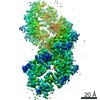 21148MC M: map data used to model this data C: citing same article ( |
|---|---|
| Similar structure data |

-Links
 PDBj
PDBj


- Assembly
| Deposited unit | 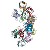
|
|---|---|
| 1 |
|
| Symmetry | Helical symmetry: (Circular symmetry: 1 / Dyad axis: no / N subunits divisor: 1 / Num. of operations: 6 / Rise per n subunits: 13.5 Å / Rotation per n subunits: 40 °) |
-Components
| #1: Protein | Mass: 9333.547 Da / Num. of mol.: 12 / Fragment: DIX domain (UNP residues 12-92) Source method: isolated from a genetically manipulated source Source: (gene. exp.)  |
|---|
-Experimental details
-Experiment
| Experiment | Method: ELECTRON MICROSCOPY |
|---|---|
| EM experiment | Aggregation state: HELICAL ARRAY / 3D reconstruction method: helical reconstruction |
- Sample preparation
| Component | Name: Segment polarity protein dishevelled homolog DVL-2 / Type: COMPLEX Details: Protein spontaneously formed a double-stranded, antiparallel helical filament upon removal of the MBP tag with TEV protease. Entity ID: all / Source: RECOMBINANT | |||||||||||||||||||||||||
|---|---|---|---|---|---|---|---|---|---|---|---|---|---|---|---|---|---|---|---|---|---|---|---|---|---|---|
| Molecular weight | Experimental value: NO | |||||||||||||||||||||||||
| Source (natural) | Organism: | |||||||||||||||||||||||||
| Source (recombinant) | Organism: | |||||||||||||||||||||||||
| Buffer solution | pH: 8 / Details: Dithiothreitol was added fresh the day of use. | |||||||||||||||||||||||||
| Buffer component |
| |||||||||||||||||||||||||
| Specimen | Conc.: 0.12 mg/ml / Embedding applied: NO / Shadowing applied: NO / Staining applied: NO / Vitrification applied: YES Details: Sample was taken from a single fraction of a gel filtration run performed immediately before grid preparation. | |||||||||||||||||||||||||
| Specimen support | Grid material: COPPER / Grid mesh size: 200 divisions/in. | |||||||||||||||||||||||||
| Vitrification | Instrument: LEICA EM GP / Cryogen name: ETHANE / Humidity: 95 % / Chamber temperature: 295 K Details: The grid was blotted for 2.0 s using Whatman #1 filter paper (GE Healthcare). |
- Electron microscopy imaging
| Experimental equipment |  Model: Titan Krios / Image courtesy: FEI Company |
|---|---|
| Microscopy | Model: FEI TITAN KRIOS |
| Electron gun | Electron source:  FIELD EMISSION GUN / Accelerating voltage: 300 kV / Illumination mode: FLOOD BEAM FIELD EMISSION GUN / Accelerating voltage: 300 kV / Illumination mode: FLOOD BEAM |
| Electron lens | Mode: BRIGHT FIELD / Calibrated magnification: 29000 X / Calibrated defocus min: 500 nm / Calibrated defocus max: 2000 nm / Cs: 2.7 mm / Alignment procedure: COMA FREE |
| Specimen holder | Cryogen: NITROGEN / Specimen holder model: FEI TITAN KRIOS AUTOGRID HOLDER |
| Image recording | Average exposure time: 10 sec. / Electron dose: 98 e/Å2 / Detector mode: COUNTING / Film or detector model: GATAN K2 SUMMIT (4k x 4k) / Num. of real images: 540 |
| Image scans | Width: 3838 / Height: 3710 / Movie frames/image: 50 / Used frames/image: 4-50 |
- Processing
| Software | Name: PHENIX / Version: 1.17.1_3660: / Classification: refinement | ||||||||||||||||||||||||||||||||||||||||||||
|---|---|---|---|---|---|---|---|---|---|---|---|---|---|---|---|---|---|---|---|---|---|---|---|---|---|---|---|---|---|---|---|---|---|---|---|---|---|---|---|---|---|---|---|---|---|
| EM software |
| ||||||||||||||||||||||||||||||||||||||||||||
| CTF correction | Type: PHASE FLIPPING AND AMPLITUDE CORRECTION | ||||||||||||||||||||||||||||||||||||||||||||
| Helical symmerty | Angular rotation/subunit: 48 ° / Axial rise/subunit: 13.5 Å / Axial symmetry: C1 | ||||||||||||||||||||||||||||||||||||||||||||
| Particle selection | Num. of particles selected: 437872 | ||||||||||||||||||||||||||||||||||||||||||||
| 3D reconstruction | Resolution: 3.6 Å / Resolution method: FSC 0.143 CUT-OFF / Num. of particles: 110105 / Algorithm: BACK PROJECTION / Num. of class averages: 2 / Symmetry type: HELICAL | ||||||||||||||||||||||||||||||||||||||||||||
| Atomic model building | Protocol: RIGID BODY FIT / Space: REAL Details: The two mutations in the source (Y27W and C80S) were reverted to Trp and Ser, respectively, before fitting. Model refinement was carried out through alternating rounds of real-space ...Details: The two mutations in the source (Y27W and C80S) were reverted to Trp and Ser, respectively, before fitting. Model refinement was carried out through alternating rounds of real-space refinement in PHENIX and manual building in Coot. | ||||||||||||||||||||||||||||||||||||||||||||
| Atomic model building | PDB-ID: 6IW3 Pdb chain-ID: A / Accession code: 6IW3 / Pdb chain residue range: 12-92 / Source name: PDB / Type: experimental model | ||||||||||||||||||||||||||||||||||||||||||||
| Refine LS restraints |
|