+ Open data
- Basic information
| Entry | Database: PDB / ID: 6sp2 | |||||||||
|---|---|---|---|---|---|---|---|---|---|---|
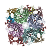
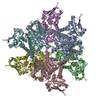
| Title | CryoEM structure of SERINC from Drosophila melanogaster | |||||||||
 Components Components | Membrane protein TMS1d | |||||||||
 Keywords Keywords | MEMBRANE PROTEIN / Anti-retroviral / TM10 / SERINC fold / novel fold | |||||||||
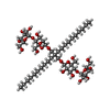
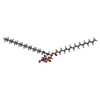
| Function / homology | Serine incorporator/TMS membrane protein / Serine incorporator (Serinc) / membrane / CARDIOLIPIN / Chem-P5S / Membrane protein TMS1d Function and homology information Function and homology information | |||||||||
| Biological species |  | |||||||||
| Method | ELECTRON MICROSCOPY / single particle reconstruction / cryo EM / Resolution: 3.33 Å | |||||||||
 Authors Authors | Pye, V.E. / Nans, A. / Cherepanov, P. | |||||||||
| Funding support |  United Kingdom, United Kingdom,  United States, 2items United States, 2items
| |||||||||
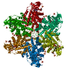
 Citation Citation |  Journal: Nat Struct Mol Biol / Year: 2020 Journal: Nat Struct Mol Biol / Year: 2020Title: A bipartite structural organization defines the SERINC family of HIV-1 restriction factors. Authors: Valerie E Pye / Annachiara Rosa / Cinzia Bertelli / Weston B Struwe / Sarah L Maslen / Robin Corey / Idlir Liko / Mark Hassall / Giada Mattiuzzo / Allison Ballandras-Colas / Andrea Nans / ...Authors: Valerie E Pye / Annachiara Rosa / Cinzia Bertelli / Weston B Struwe / Sarah L Maslen / Robin Corey / Idlir Liko / Mark Hassall / Giada Mattiuzzo / Allison Ballandras-Colas / Andrea Nans / Yasuhiro Takeuchi / Phillip J Stansfeld / J Mark Skehel / Carol V Robinson / Massimo Pizzato / Peter Cherepanov /   Abstract: The human integral membrane protein SERINC5 potently restricts HIV-1 infectivity and sensitizes the virus to antibody-mediated neutralization. Here, using cryo-EM, we determine the structures of ...The human integral membrane protein SERINC5 potently restricts HIV-1 infectivity and sensitizes the virus to antibody-mediated neutralization. Here, using cryo-EM, we determine the structures of human SERINC5 and its orthologue from Drosophila melanogaster at subnanometer and near-atomic resolution, respectively. The structures reveal a novel fold comprised of ten transmembrane helices organized into two subdomains and bisected by a long diagonal helix. A lipid binding groove and clusters of conserved residues highlight potential functional sites. A structure-based mutagenesis scan identified surface-exposed regions and the interface between the subdomains of SERINC5 as critical for HIV-1-restriction activity. The same regions are also important for viral sensitization to neutralizing antibodies, directly linking the antiviral activity of SERINC5 with remodeling of the HIV-1 envelope glycoprotein. | |||||||||
| History |
|
- Structure visualization
| Movie |
 Movie viewer Movie viewer |
|---|---|
| Structure viewer | Molecule:  Molmil Molmil Jmol/JSmol Jmol/JSmol |
- Downloads & links
- Download
| PDBx/mmCIF format |  6sp2.cif.gz 6sp2.cif.gz | 418.2 KB | Display |  PDBx/mmCIF format PDBx/mmCIF format |
|---|---|---|---|---|
| PDB format |  pdb6sp2.ent.gz pdb6sp2.ent.gz | 344.9 KB | Display |  PDB format PDB format |
| PDBx/mmJSON format |  6sp2.json.gz 6sp2.json.gz | Tree view |  PDBx/mmJSON format PDBx/mmJSON format | |
| Others |  Other downloads Other downloads |
-Validation report
| Summary document |  6sp2_validation.pdf.gz 6sp2_validation.pdf.gz | 2.2 MB | Display |  wwPDB validaton report wwPDB validaton report |
|---|---|---|---|---|
| Full document |  6sp2_full_validation.pdf.gz 6sp2_full_validation.pdf.gz | 2.2 MB | Display | |
| Data in XML |  6sp2_validation.xml.gz 6sp2_validation.xml.gz | 83.1 KB | Display | |
| Data in CIF |  6sp2_validation.cif.gz 6sp2_validation.cif.gz | 101.3 KB | Display | |
| Arichive directory |  https://data.pdbj.org/pub/pdb/validation_reports/sp/6sp2 https://data.pdbj.org/pub/pdb/validation_reports/sp/6sp2 ftp://data.pdbj.org/pub/pdb/validation_reports/sp/6sp2 ftp://data.pdbj.org/pub/pdb/validation_reports/sp/6sp2 | HTTPS FTP |
-Related structure data
| Related structure data |  10279MC C: citing same article ( M: map data used to model this data |
|---|---|
| Similar structure data |
- Links
Links
- Assembly
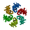
| Deposited unit | 
|
|---|---|
| 1 |
|
- Components
| #1: Protein | Mass: 54721.738 Da / Num. of mol.: 6 Source method: isolated from a genetically manipulated source Source: (gene. exp.)   #2: Chemical | ChemComp-LMN / #3: Chemical | ChemComp-P5S / #4: Chemical | ChemComp-CDL / Has ligand of interest | N | Has protein modification | Y | |
|---|
-Experimental details
-Experiment
| Experiment | Method: ELECTRON MICROSCOPY |
|---|---|
| EM experiment | Aggregation state: PARTICLE / 3D reconstruction method: single particle reconstruction |
- Sample preparation
| Component | Name: SERINC homo-hexamer / Type: COMPLEX Details: homo-hexamer of SERINC from Drosophila melanogaster recombinantly expressed and purified in detergent micelle; imaged as single particle. Entity ID: #1 / Source: RECOMBINANT | ||||||||||||||||||||||||||||||
|---|---|---|---|---|---|---|---|---|---|---|---|---|---|---|---|---|---|---|---|---|---|---|---|---|---|---|---|---|---|---|---|
| Molecular weight | Value: 0.32805646 MDa / Experimental value: NO | ||||||||||||||||||||||||||||||
| Source (natural) | Organism:  | ||||||||||||||||||||||||||||||
| Source (recombinant) | Organism:  | ||||||||||||||||||||||||||||||
| Buffer solution | pH: 7.5 | ||||||||||||||||||||||||||||||
| Buffer component |
| ||||||||||||||||||||||||||||||
| Specimen | Conc.: 0.5 mg/ml / Embedding applied: NO / Shadowing applied: NO / Staining applied: NO / Vitrification applied: YES / Details: Freshly purified mono-dispersed sample | ||||||||||||||||||||||||||||||
| Specimen support | Grid material: COPPER / Grid mesh size: 400 divisions/in. / Grid type: Quantifoil R1.2/1.3 | ||||||||||||||||||||||||||||||
| Vitrification | Instrument: FEI VITROBOT MARK IV / Cryogen name: ETHANE / Humidity: 100 % / Chamber temperature: 293 K / Details: wait 60 seconds, blot for 3-4 seconds |
- Electron microscopy imaging
| Experimental equipment |  Model: Titan Krios / Image courtesy: FEI Company |
|---|---|
| Microscopy | Model: FEI TITAN KRIOS |
| Electron gun | Electron source:  FIELD EMISSION GUN / Accelerating voltage: 300 kV / Illumination mode: FLOOD BEAM FIELD EMISSION GUN / Accelerating voltage: 300 kV / Illumination mode: FLOOD BEAM |
| Electron lens | Mode: BRIGHT FIELD |
| Image recording | Electron dose: 50 e/Å2 / Detector mode: COUNTING / Film or detector model: GATAN K2 QUANTUM (4k x 4k) / Num. of grids imaged: 1 / Details: 5,807 movies were collected |
- Processing
| EM software |
| ||||||||||||||||||||||||||||||||||||||||
|---|---|---|---|---|---|---|---|---|---|---|---|---|---|---|---|---|---|---|---|---|---|---|---|---|---|---|---|---|---|---|---|---|---|---|---|---|---|---|---|---|---|
| CTF correction | Type: NONE | ||||||||||||||||||||||||||||||||||||||||
| Particle selection | Num. of particles selected: 1857080 Details: A sub-set of particles semi-automatically picked in EMAN2 Boxer were used to generate the starting 2D class averages, which, upon low-pass filtering to 20 A, served as templates for auto- ...Details: A sub-set of particles semi-automatically picked in EMAN2 Boxer were used to generate the starting 2D class averages, which, upon low-pass filtering to 20 A, served as templates for auto-picking of the entire dataset. | ||||||||||||||||||||||||||||||||||||||||
| Symmetry | Point symmetry: C6 (6 fold cyclic) | ||||||||||||||||||||||||||||||||||||||||
| 3D reconstruction | Resolution: 3.33 Å / Resolution method: FSC 0.143 CUT-OFF / Num. of particles: 159252 / Algorithm: FOURIER SPACE / Num. of class averages: 1 / Symmetry type: POINT | ||||||||||||||||||||||||||||||||||||||||
| Atomic model building | B value: 150.6 / Protocol: AB INITIO MODEL / Space: REAL / Target criteria: Cross-correlation coefficient |
 Movie
Movie Controller


 PDBj
PDBj
