 ムービー
ムービー コントローラー


+ データを開く

- 基本情報
基本情報
| 登録情報 | データベース: PDB / ID: 6l3h | |||||||||||||||||||||
|---|---|---|---|---|---|---|---|---|---|---|---|---|---|---|---|---|---|---|---|---|---|---|
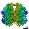
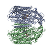
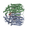
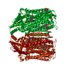
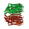
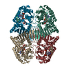
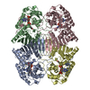
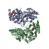
| タイトル | Cryo-EM structure of dimeric quinol dependent Nitric Oxide Reductase (qNOR) from the pathogen Neisseria meninigitidis | |||||||||||||||||||||
 要素 要素 | Nitric-oxide reductase | |||||||||||||||||||||
 キーワード キーワード | OXIDOREDUCTASE / Neisseria meningitidis / quinol-dependent electrogenic Nitric Oxide Reductase (qNOR) | |||||||||||||||||||||
| 機能・相同性 |  機能・相同性情報 機能・相同性情報nitric-oxide reductase / cytochrome bo3 ubiquinol oxidase activity / cytochrome-c oxidase activity / electron transport coupled proton transport / aerobic respiration / respiratory electron transport chain / heme binding / membrane / metal ion binding 類似検索 - 分子機能 | |||||||||||||||||||||
| 生物種 |  Neisseria meningitidis alpha14 (髄膜炎菌) Neisseria meningitidis alpha14 (髄膜炎菌) | |||||||||||||||||||||
| 手法 | 電子顕微鏡法 / 単粒子再構成法 / クライオ電子顕微鏡法 / 解像度: 3.06 Å | |||||||||||||||||||||
 データ登録者 データ登録者 | Jamali, M.M.A. / Gopalasingam, C.C. / Johnson, R.M. / Tosha, T. / Muench, S.P. / Muramoto, K. / Antonyuk, S.V. / Shiro, Y. / Hasnain, S.S. | |||||||||||||||||||||
| 資金援助 |  日本, 日本,  英国, 6件 英国, 6件
| |||||||||||||||||||||
 引用 引用 | ジャーナル: IUCrJ / 年: 2020 タイトル: The active form of quinol-dependent nitric oxide reductase from is a dimer. 著者: M Arif M Jamali / Chai C Gopalasingam / Rachel M Johnson / Takehiko Tosha / Kazumasa Muramoto / Stephen P Muench / Svetlana V Antonyuk / Yoshitsugu Shiro / Samar S Hasnain / 要旨: is carried by nearly a billion humans, causing developmental impairment and over 100 000 deaths a year. A quinol-dependent nitric oxide reductase (qNOR) plays a critical role in the survival of ... is carried by nearly a billion humans, causing developmental impairment and over 100 000 deaths a year. A quinol-dependent nitric oxide reductase (qNOR) plays a critical role in the survival of the bacterium in the human host. X-ray crystallographic analyses of qNOR, including that from (qNOR) reported here at 3.15 Å resolution, show monomeric assemblies, despite the more active dimeric sample being used for crystallization. Cryo-electron microscopic analysis of the same chromatographic fraction of qNOR, however, revealed a dimeric assembly at 3.06 Å resolution. It is shown that zinc (which is used in crystallization) binding near the dimer-stabilizing TMII region contributes to the disruption of the dimer. A similar destabilization is observed in the monomeric (∼85 kDa) cryo-EM structure of a mutant (Glu494Ala) qNOR from the opportunistic pathogen () , which primarily migrates as a monomer. The monomer-dimer transition of qNORs seen in the cryo-EM and crystallographic structures has wider implications for structural studies of multimeric membrane proteins. X-ray crystallographic and cryo-EM structural analyses have been performed on the same chromatographic fraction of qNOR to high resolution. This represents one of the first examples in which the two approaches have been used to reveal a monomeric assembly and a dimeric assembly in vitrified cryo-EM grids. A number of factors have been identified that may trigger the destabilization of helices that are necessary to preserve the integrity of the dimer. These include zinc binding near the entry of the putative proton-transfer channel and the preservation of the conformational integrity of the active site. The mutation near the active site results in disruption of the active site, causing an additional destabilization of helices (TMIX and TMX) that flank the proton-transfer channel helices, creating an inert monomeric enzyme. | |||||||||||||||||||||
| 履歴 |
|
- 構造の表示
構造の表示
| ムービー |
ムービービューア |
|---|---|
| 構造ビューア | 分子: MolmilJmol/JSmol |
- ダウンロードとリンク
-ダウンロード
| PDBx/mmCIF形式 | 6l3h.cif.gz | 270.2 KB | 表示 | PDBx/mmCIF形式 |
|---|---|---|---|---|
| PDB形式 | pdb6l3h.ent.gz | 218.4 KB | 表示 | PDB形式 |
| PDBx/mmJSON形式 | 6l3h.json.gz | ツリー表示 | PDBx/mmJSON形式 | |
| その他 |  その他のダウンロード その他のダウンロード |
-検証レポート
| 文書・要旨 | 6l3h_validation.pdf.gz | 1.2 MB | 表示 | wwPDB検証レポート |
|---|---|---|---|---|
| 文書・詳細版 | 6l3h_full_validation.pdf.gz | 1.2 MB | 表示 | |
| XML形式データ | 6l3h_validation.xml.gz | 45.2 KB | 表示 | |
| CIF形式データ | 6l3h_validation.cif.gz | 69.3 KB | 表示 | |
| アーカイブディレクトリ | https://data.pdbj.org/pub/pdb/validation_reports/l3/6l3hftp://data.pdbj.org/pub/pdb/validation_reports/l3/6l3h | HTTPS FTP |
-関連構造データ


-リンク
 PDBj
PDBj

- 集合体
集合体
| 登録構造単位 | 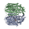
|
|---|---|
| 1 |
|
-要素
| #1: タンパク質 | 分子量: 84389.211 Da / 分子数: 2 / 由来タイプ: 組換発現 由来: (組換発現) Neisseria meningitidis alpha14 (髄膜炎菌)株: alpha14 / 遺伝子: norB, NMO_1451 / プラスミド: pRSET-C / 発現宿主: #2: 化合物 | ChemComp-HEM /   分子量: 616.487 Da / 分子数: 4 / 由来タイプ: 合成 / 式: C34H32FeN4O4 分子量: 616.487 Da / 分子数: 4 / 由来タイプ: 合成 / 式: C34H32FeN4O4#3: 化合物 |   分子量: 55.845 Da / 分子数: 2 / 由来タイプ: 合成 / 式: Fe 分子量: 55.845 Da / 分子数: 2 / 由来タイプ: 合成 / 式: Fe#4: 化合物 |   分子量: 40.078 Da / 分子数: 2 / 由来タイプ: 合成 / 式: Ca 分子量: 40.078 Da / 分子数: 2 / 由来タイプ: 合成 / 式: Ca研究の焦点であるリガンドがあるか | N | |
|---|
-実験情報
-実験
| 実験 | 手法: 電子顕微鏡法 |
|---|---|
| EM実験 | 試料の集合状態: PARTICLE / 3次元再構成法: 単粒子再構成法 |
- 試料調製
試料調製
| 構成要素 | 名称: Dimeric quinol dependent Nitric Oxide Reductase / タイプ: COMPLEX / Entity ID: #1 / 由来: RECOMBINANT |
|---|---|
| 分子量 | 値: 0.15 MDa / 実験値: NO |
| 由来(天然) | 生物種: Neisseria meningitidis alpha14 (髄膜炎菌) |
| 由来(組換発現) | 生物種: |
| 緩衝液 | pH: 8 |
| 試料 | 濃度: 4 mg/ml / 包埋: NO / シャドウイング: NO / 染色: NO / 凍結: YES |
| 試料支持 | グリッドの材料: COPPER / グリッドのタイプ: Quantifoil R1.2/1.3 |
| 急速凍結 | 装置: FEI VITROBOT MARK IV / 凍結剤: ETHANE / 湿度: 100 % / 凍結前の試料温度: 277 K 詳細: Blot time of 6 seconds, with accompanying blot force of 6. |
- 電子顕微鏡撮影
| 実験機器 |  モデル: Titan Krios / 画像提供: FEI Company |
|---|---|
| 顕微鏡 | モデル: FEI TITAN KRIOS |
| 電子銃 | 電子線源:  FIELD EMISSION GUN / 加速電圧: 300 kV / 照射モード: FLOOD BEAM FIELD EMISSION GUN / 加速電圧: 300 kV / 照射モード: FLOOD BEAM |
| 電子レンズ | モード: BRIGHT FIELD / 倍率(公称値): 130000 X / 最大 デフォーカス(公称値): -3500 nm / 最小 デフォーカス(公称値): -1000 nm / Cs: 2.7 mm / C2レンズ絞り径: 70 µm |
| 試料ホルダ | 凍結剤: NITROGEN 試料ホルダーモデル: FEI TITAN KRIOS AUTOGRID HOLDER |
| 撮影 | 平均露光時間: 8 sec. / 電子線照射量: 69.44 e/Å2 / 検出モード: COUNTING フィルム・検出器のモデル: GATAN K2 SUMMIT (4k x 4k) 撮影したグリッド数: 1 / 実像数: 3182 |
| 画像スキャン | 動画フレーム数/画像: 40 / 利用したフレーム数/画像: 1-40 |
- 解析
解析
| ソフトウェア | 名称: PHENIX / バージョン: 1.16_3549: / 分類: 精密化 | ||||||||||||||||||||||||||||||||
|---|---|---|---|---|---|---|---|---|---|---|---|---|---|---|---|---|---|---|---|---|---|---|---|---|---|---|---|---|---|---|---|---|---|
| EMソフトウェア |
| ||||||||||||||||||||||||||||||||
| CTF補正 | タイプ: PHASE FLIPPING AND AMPLITUDE CORRECTION | ||||||||||||||||||||||||||||||||
| 粒子像の選択 | 選択した粒子像数: 970000 詳細: 2D templates generated from 2,000 manually picked particles. Templates (low pass filtered to 20 angstrom) then used for automated picking of micrographs. | ||||||||||||||||||||||||||||||||
| 対称性 | 点対称性: C2 (2回回転対称) | ||||||||||||||||||||||||||||||||
| 3次元再構成 | 解像度: 3.06 Å / 解像度の算出法: FSC 0.143 CUT-OFF / 粒子像の数: 233556 / アルゴリズム: BACK PROJECTION / クラス平均像の数: 1 / 対称性のタイプ: POINT | ||||||||||||||||||||||||||||||||
| 原子モデル構築 | プロトコル: FLEXIBLE FIT / 空間: REAL | ||||||||||||||||||||||||||||||||
| 原子モデル構築 | PDB-ID: 6L1X Accession code: 6L1X / Source name: PDB / タイプ: experimental model | ||||||||||||||||||||||||||||||||
| 拘束条件 |
|
