 Movie
Movie Controller

[English] 日本語
 Yorodumi
- PDB-6l3h: Cryo-EM structure of dimeric quinol dependent Nitric Oxide Reduct... -
+ Open data

- Basic information
| Entry | Database: PDB / ID: 6l3h | |||||||||||||||||||||
|---|---|---|---|---|---|---|---|---|---|---|---|---|---|---|---|---|---|---|---|---|---|---|
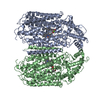
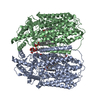
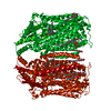
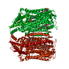
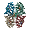
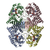
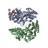
| Title | Cryo-EM structure of dimeric quinol dependent Nitric Oxide Reductase (qNOR) from the pathogen Neisseria meninigitidis | |||||||||||||||||||||
 Components Components | Nitric-oxide reductase | |||||||||||||||||||||
 Keywords Keywords | OXIDOREDUCTASE / Neisseria meningitidis / quinol-dependent electrogenic Nitric Oxide Reductase (qNOR) | |||||||||||||||||||||
| Function / homology |  Function and homology information Function and homology informationnitric-oxide reductase / cytochrome-c oxidase activity / aerobic respiration / oxidoreductase activity / heme binding / membrane / metal ion binding Similarity search - Function | |||||||||||||||||||||
| Biological species |  Neisseria meningitidis alpha14 (bacteria) Neisseria meningitidis alpha14 (bacteria) | |||||||||||||||||||||
| Method | ELECTRON MICROSCOPY / single particle reconstruction / cryo EM / Resolution: 3.06 Å | |||||||||||||||||||||
 Authors Authors | Jamali, M.M.A. / Gopalasingam, C.C. / Johnson, R.M. / Tosha, T. / Muench, S.P. / Muramoto, K. / Antonyuk, S.V. / Shiro, Y. / Hasnain, S.S. | |||||||||||||||||||||
| Funding support |  Japan, Japan,  United Kingdom, 6items United Kingdom, 6items
| |||||||||||||||||||||
 Citation Citation | Journal: IUCrJ / Year: 2020 Title: The active form of quinol-dependent nitric oxide reductase from is a dimer. Authors: M Arif M Jamali / Chai C Gopalasingam / Rachel M Johnson / Takehiko Tosha / Kazumasa Muramoto / Stephen P Muench / Svetlana V Antonyuk / Yoshitsugu Shiro / Samar S Hasnain / Abstract: is carried by nearly a billion humans, causing developmental impairment and over 100 000 deaths a year. A quinol-dependent nitric oxide reductase (qNOR) plays a critical role in the survival of ... is carried by nearly a billion humans, causing developmental impairment and over 100 000 deaths a year. A quinol-dependent nitric oxide reductase (qNOR) plays a critical role in the survival of the bacterium in the human host. X-ray crystallographic analyses of qNOR, including that from (qNOR) reported here at 3.15 Å resolution, show monomeric assemblies, despite the more active dimeric sample being used for crystallization. Cryo-electron microscopic analysis of the same chromatographic fraction of qNOR, however, revealed a dimeric assembly at 3.06 Å resolution. It is shown that zinc (which is used in crystallization) binding near the dimer-stabilizing TMII region contributes to the disruption of the dimer. A similar destabilization is observed in the monomeric (∼85 kDa) cryo-EM structure of a mutant (Glu494Ala) qNOR from the opportunistic pathogen () , which primarily migrates as a monomer. The monomer-dimer transition of qNORs seen in the cryo-EM and crystallographic structures has wider implications for structural studies of multimeric membrane proteins. X-ray crystallographic and cryo-EM structural analyses have been performed on the same chromatographic fraction of qNOR to high resolution. This represents one of the first examples in which the two approaches have been used to reveal a monomeric assembly and a dimeric assembly in vitrified cryo-EM grids. A number of factors have been identified that may trigger the destabilization of helices that are necessary to preserve the integrity of the dimer. These include zinc binding near the entry of the putative proton-transfer channel and the preservation of the conformational integrity of the active site. The mutation near the active site results in disruption of the active site, causing an additional destabilization of helices (TMIX and TMX) that flank the proton-transfer channel helices, creating an inert monomeric enzyme. | |||||||||||||||||||||
| History |
|
- Structure visualization
| Movie |
Movie viewer |
|---|---|
| Structure viewer | Molecule: MolmilJmol/JSmol |
- Downloads & links
-Download
| PDBx/mmCIF format | 6l3h.cif.gz | 270.2 KB | Display | PDBx/mmCIF format |
|---|---|---|---|---|
| PDB format | pdb6l3h.ent.gz | 218.4 KB | Display | PDB format |
| PDBx/mmJSON format | 6l3h.json.gz | Tree view | PDBx/mmJSON format | |
| Others |  Other downloads Other downloads |
-Validation report
| Arichive directory | https://data.pdbj.org/pub/pdb/validation_reports/l3/6l3hftp://data.pdbj.org/pub/pdb/validation_reports/l3/6l3h | HTTPS FTP |
|---|
-Related structure data
| Related structure data | 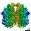 0822MC  6l1xC  6t6vC M: map data used to model this data C: citing same article ( |
|---|---|
| Similar structure data |


-Links
 PDBj
PDBj

- Assembly
| Deposited unit | 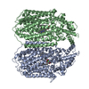
|
|---|---|
| 1 |
|
-Components
| #1: Protein | Mass: 84389.211 Da / Num. of mol.: 2 Source method: isolated from a genetically manipulated source Source: (gene. exp.) Neisseria meningitidis alpha14 (bacteria)Strain: alpha14 / Gene: norB, NMO_1451 / Plasmid: pRSET-C / Production host: #2: Chemical | ChemComp-HEM /   Mass: 616.487 Da / Num. of mol.: 4 / Source method: obtained synthetically / Formula: C34H32FeN4O4 Mass: 616.487 Da / Num. of mol.: 4 / Source method: obtained synthetically / Formula: C34H32FeN4O4#3: Chemical |   Mass: 55.845 Da / Num. of mol.: 2 / Source method: obtained synthetically / Formula: Fe Mass: 55.845 Da / Num. of mol.: 2 / Source method: obtained synthetically / Formula: Fe#4: Chemical |   Mass: 40.078 Da / Num. of mol.: 2 / Source method: obtained synthetically / Formula: Ca Mass: 40.078 Da / Num. of mol.: 2 / Source method: obtained synthetically / Formula: CaHas ligand of interest | N | |
|---|
-Experimental details
-Experiment
| Experiment | Method: ELECTRON MICROSCOPY |
|---|---|
| EM experiment | Aggregation state: PARTICLE / 3D reconstruction method: single particle reconstruction |
- Sample preparation
| Component | Name: Dimeric quinol dependent Nitric Oxide Reductase / Type: COMPLEX / Entity ID: #1 / Source: RECOMBINANT |
|---|---|
| Molecular weight | Value: 0.15 MDa / Experimental value: NO |
| Source (natural) | Organism: Neisseria meningitidis alpha14 (bacteria) |
| Source (recombinant) | Organism: |
| Buffer solution | pH: 8 |
| Specimen | Conc.: 4 mg/ml / Embedding applied: NO / Shadowing applied: NO / Staining applied: NO / Vitrification applied: YES |
| Specimen support | Grid material: COPPER / Grid type: Quantifoil R1.2/1.3 |
| Vitrification | Instrument: FEI VITROBOT MARK IV / Cryogen name: ETHANE / Humidity: 100 % / Chamber temperature: 277 K Details: Blot time of 6 seconds, with accompanying blot force of 6. |
- Electron microscopy imaging
| Experimental equipment |  Model: Titan Krios / Image courtesy: FEI Company |
|---|---|
| Microscopy | Model: FEI TITAN KRIOS |
| Electron gun | Electron source:  FIELD EMISSION GUN / Accelerating voltage: 300 kV / Illumination mode: FLOOD BEAM FIELD EMISSION GUN / Accelerating voltage: 300 kV / Illumination mode: FLOOD BEAM |
| Electron lens | Mode: BRIGHT FIELD / Nominal magnification: 130000 X / Nominal defocus max: -3500 nm / Nominal defocus min: -1000 nm / Cs: 2.7 mm / C2 aperture diameter: 70 µm |
| Specimen holder | Cryogen: NITROGEN / Specimen holder model: FEI TITAN KRIOS AUTOGRID HOLDER |
| Image recording | Average exposure time: 8 sec. / Electron dose: 69.44 e/Å2 / Detector mode: COUNTING / Film or detector model: GATAN K2 SUMMIT (4k x 4k) / Num. of grids imaged: 1 / Num. of real images: 3182 |
| Image scans | Movie frames/image: 40 / Used frames/image: 1-40 |
- Processing
| Software | Name: PHENIX / Version: 1.16_3549: / Classification: refinement | ||||||||||||||||||||||||||||||||
|---|---|---|---|---|---|---|---|---|---|---|---|---|---|---|---|---|---|---|---|---|---|---|---|---|---|---|---|---|---|---|---|---|---|
| EM software |
| ||||||||||||||||||||||||||||||||
| CTF correction | Type: PHASE FLIPPING AND AMPLITUDE CORRECTION | ||||||||||||||||||||||||||||||||
| Particle selection | Num. of particles selected: 970000 Details: 2D templates generated from 2,000 manually picked particles. Templates (low pass filtered to 20 angstrom) then used for automated picking of micrographs. | ||||||||||||||||||||||||||||||||
| Symmetry | Point symmetry: C2 (2 fold cyclic) | ||||||||||||||||||||||||||||||||
| 3D reconstruction | Resolution: 3.06 Å / Resolution method: FSC 0.143 CUT-OFF / Num. of particles: 233556 / Algorithm: BACK PROJECTION / Num. of class averages: 1 / Symmetry type: POINT | ||||||||||||||||||||||||||||||||
| Atomic model building | Protocol: FLEXIBLE FIT / Space: REAL | ||||||||||||||||||||||||||||||||
| Atomic model building | PDB-ID: 6L1X Accession code: 6L1X / Source name: PDB / Type: experimental model | ||||||||||||||||||||||||||||||||
| Refine LS restraints |
|
