[English] 日本語
 Yorodumi
- PDB-6k7l: Cryo-EM structure of the human P4-type flippase ATP8A1-CDC50 (E2P... -
+ Open data
- Basic information
| Entry | Database: PDB / ID: 6k7l | |||||||||
|---|---|---|---|---|---|---|---|---|---|---|
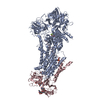
| Title | Cryo-EM structure of the human P4-type flippase ATP8A1-CDC50 (E2P state class2) | |||||||||
 Components Components |
| |||||||||
 Keywords Keywords | MEMBRANE PROTEIN / flippase | |||||||||
| Function / homology |  Function and homology information Function and homology informationaminophospholipid translocation / positive regulation of phospholipid translocation / aminophospholipid transport / aminophospholipid flippase activity / chromaffin granule membrane / phosphatidylserine flippase activity / protein localization to endosome / ATPase-coupled intramembrane lipid transporter activity / phospholipid-translocating ATPase complex / positive regulation of protein exit from endoplasmic reticulum ...aminophospholipid translocation / positive regulation of phospholipid translocation / aminophospholipid transport / aminophospholipid flippase activity / chromaffin granule membrane / phosphatidylserine flippase activity / protein localization to endosome / ATPase-coupled intramembrane lipid transporter activity / phospholipid-translocating ATPase complex / positive regulation of protein exit from endoplasmic reticulum / phosphatidylserine floppase activity / xenobiotic transmembrane transport / phospholipid transport / ATPase-coupled monoatomic cation transmembrane transporter activity / P-type phospholipid transporter / phospholipid translocation / azurophil granule membrane / transport vesicle membrane / organelle membrane / Ion transport by P-type ATPases / synaptic vesicle endocytosis / transport across blood-brain barrier / specific granule membrane / endomembrane system / learning / trans-Golgi network / positive regulation of neuron projection development / synaptic vesicle membrane / late endosome membrane / early endosome membrane / cytoplasmic vesicle / monoatomic ion transmembrane transport / positive regulation of cell migration / apical plasma membrane / intracellular membrane-bounded organelle / Neutrophil degranulation / glutamatergic synapse / structural molecule activity / Golgi apparatus / magnesium ion binding / endoplasmic reticulum / ATP hydrolysis activity / extracellular exosome / ATP binding / membrane / plasma membrane / cytosol Similarity search - Function | |||||||||
| Biological species |  Homo sapiens (human) Homo sapiens (human) | |||||||||
| Method | ELECTRON MICROSCOPY / single particle reconstruction / cryo EM / Resolution: 2.83 Å | |||||||||
 Authors Authors | Hiraizumi, M. / Yamashita, K. / Nishizawa, T. / Nureki, O. | |||||||||
 Citation Citation |  Journal: Science / Year: 2019 Journal: Science / Year: 2019Title: Cryo-EM structures capture the transport cycle of the P4-ATPase flippase. Authors: Masahiro Hiraizumi / Keitaro Yamashita / Tomohiro Nishizawa / Osamu Nureki /  Abstract: In eukaryotic membranes, type IV P-type adenosine triphosphatases (P4-ATPases) mediate the translocation of phospholipids from the outer to the inner leaflet and maintain lipid asymmetry, which is ...In eukaryotic membranes, type IV P-type adenosine triphosphatases (P4-ATPases) mediate the translocation of phospholipids from the outer to the inner leaflet and maintain lipid asymmetry, which is critical for membrane trafficking and signaling pathways. Here, we report the cryo-electron microscopy structures of six distinct intermediates of the human ATP8A1-CDC50a heterocomplex at resolutions of 2.6 to 3.3 angstroms, elucidating the lipid translocation cycle of this P4-ATPase. ATP-dependent phosphorylation induces a large rotational movement of the actuator domain around the phosphorylation site in the phosphorylation domain, accompanied by lateral shifts of the first and second transmembrane helices, thereby allowing phosphatidylserine binding. The phospholipid head group passes through the hydrophilic cleft, while the acyl chain is exposed toward the lipid environment. These findings advance our understanding of the flippase mechanism and the disease-associated mutants of P4-ATPases. | |||||||||
| History |
|
- Structure visualization
| Movie |
 Movie viewer Movie viewer |
|---|---|
| Structure viewer | Molecule:  Molmil Molmil Jmol/JSmol Jmol/JSmol |
- Downloads & links
- Download
| PDBx/mmCIF format |  6k7l.cif.gz 6k7l.cif.gz | 263.6 KB | Display |  PDBx/mmCIF format PDBx/mmCIF format |
|---|---|---|---|---|
| PDB format |  pdb6k7l.ent.gz pdb6k7l.ent.gz | 196 KB | Display |  PDB format PDB format |
| PDBx/mmJSON format |  6k7l.json.gz 6k7l.json.gz | Tree view |  PDBx/mmJSON format PDBx/mmJSON format | |
| Others |  Other downloads Other downloads |
-Validation report
| Summary document |  6k7l_validation.pdf.gz 6k7l_validation.pdf.gz | 1.3 MB | Display |  wwPDB validaton report wwPDB validaton report |
|---|---|---|---|---|
| Full document |  6k7l_full_validation.pdf.gz 6k7l_full_validation.pdf.gz | 1.4 MB | Display | |
| Data in XML |  6k7l_validation.xml.gz 6k7l_validation.xml.gz | 49.6 KB | Display | |
| Data in CIF |  6k7l_validation.cif.gz 6k7l_validation.cif.gz | 75.4 KB | Display | |
| Arichive directory |  https://data.pdbj.org/pub/pdb/validation_reports/k7/6k7l https://data.pdbj.org/pub/pdb/validation_reports/k7/6k7l ftp://data.pdbj.org/pub/pdb/validation_reports/k7/6k7l ftp://data.pdbj.org/pub/pdb/validation_reports/k7/6k7l | HTTPS FTP |
-Related structure data
| Related structure data |  9939MC  9931C  9932C  9933C  9934C  9935C  9936C  9937C  9938C  9940C  9941C  9942C  6k7gC  6k7hC  6k7iC  6k7jC  6k7kC  6k7mC  6k7nC M: map data used to model this data C: citing same article ( |
|---|---|
| Similar structure data | |
| EM raw data |  EMPIAR-10303 (Title: Cryo-EM structures of human P4-ATPase flippase / Data size: 11.0 TB EMPIAR-10303 (Title: Cryo-EM structures of human P4-ATPase flippase / Data size: 11.0 TBData #1: Unaligned movies for E2P class 1,2,3 [micrographs - multiframe] Data #2: Unaligned movies for E2Pi-PL and E1P [micrographs - multiframe] Data #3: Unaligned movies for E1P-ADP [micrographs - multiframe] Data #4: Unaligned movies for E1 class1,2,3 [micrographs - multiframe] Data #5: Unaligned movies for E1-ATP class 1,2,3 [micrographs - multiframe]) |
- Links
Links
- Assembly
| Deposited unit | 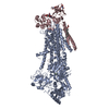
|
|---|---|
| 1 |
|
- Components
-Protein , 2 types, 2 molecules AC
| #1: Protein | Mass: 130537.852 Da / Num. of mol.: 1 Source method: isolated from a genetically manipulated source Source: (gene. exp.)  Homo sapiens (human) / Plasmid: pcDNA3.4 / Cell line (production host): Expi293F / Production host: Homo sapiens (human) / Plasmid: pcDNA3.4 / Cell line (production host): Expi293F / Production host:  Homo sapiens (human) Homo sapiens (human)References: UniProt: Q59EX4, UniProt: Q9Y2Q0*PLUS, P-type phospholipid transporter |
|---|---|
| #2: Protein | Mass: 40727.527 Da / Num. of mol.: 1 Source method: isolated from a genetically manipulated source Source: (gene. exp.)  Homo sapiens (human) / Gene: TMEM30A / Plasmid: pcDNA3.4 / Cell line (production host): Expi293F / Production host: Homo sapiens (human) / Gene: TMEM30A / Plasmid: pcDNA3.4 / Cell line (production host): Expi293F / Production host:  Homo sapiens (human) / References: UniProt: Q9NV96 Homo sapiens (human) / References: UniProt: Q9NV96 |
-Sugars , 2 types, 3 molecules 
| #3: Polysaccharide | alpha-D-mannopyranose-(1-4)-2-acetamido-2-deoxy-beta-D-glucopyranose-(1-4)-2-acetamido-2-deoxy-beta- ...alpha-D-mannopyranose-(1-4)-2-acetamido-2-deoxy-beta-D-glucopyranose-(1-4)-2-acetamido-2-deoxy-beta-D-glucopyranose Source method: isolated from a genetically manipulated source |
|---|---|
| #7: Sugar |
-Non-polymers , 3 types, 3 molecules 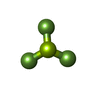


| #4: Chemical | ChemComp-BEF / |
|---|---|
| #5: Chemical | ChemComp-MG / |
| #6: Chemical | ChemComp-Y01 / |
-Details
| Has ligand of interest | Y |
|---|---|
| Has protein modification | Y |
-Experimental details
-Experiment
| Experiment | Method: ELECTRON MICROSCOPY |
|---|---|
| EM experiment | Aggregation state: PARTICLE / 3D reconstruction method: single particle reconstruction |
- Sample preparation
| Component | Name: ATP8A1-CDC50a / Type: COMPLEX / Entity ID: #1-#2 / Source: RECOMBINANT |
|---|---|
| Molecular weight | Experimental value: NO |
| Source (natural) | Organism:  Homo sapiens (human) Homo sapiens (human) |
| Source (recombinant) | Organism:  Homo sapiens (human) / Cell: Expi293F / Plasmid: pcDNA3.4 Homo sapiens (human) / Cell: Expi293F / Plasmid: pcDNA3.4 |
| Buffer solution | pH: 7.5 |
| Specimen | Conc.: 4 mg/ml / Embedding applied: NO / Shadowing applied: NO / Staining applied: NO / Vitrification applied: YES |
| Vitrification | Cryogen name: ETHANE |
- Electron microscopy imaging
| Experimental equipment |  Model: Titan Krios / Image courtesy: FEI Company |
|---|---|
| Microscopy | Model: FEI TITAN KRIOS |
| Electron gun | Electron source:  FIELD EMISSION GUN / Accelerating voltage: 300 kV / Illumination mode: OTHER FIELD EMISSION GUN / Accelerating voltage: 300 kV / Illumination mode: OTHER |
| Electron lens | Mode: BRIGHT FIELD |
| Image recording | Electron dose: 64 e/Å2 / Film or detector model: GATAN K3 BIOQUANTUM (6k x 4k) |
- Processing
| Software |
| ||||||||||||||||||||||||
|---|---|---|---|---|---|---|---|---|---|---|---|---|---|---|---|---|---|---|---|---|---|---|---|---|---|
| CTF correction | Type: PHASE FLIPPING AND AMPLITUDE CORRECTION | ||||||||||||||||||||||||
| 3D reconstruction | Resolution: 2.83 Å / Resolution method: FSC 0.143 CUT-OFF / Num. of particles: 193240 / Symmetry type: POINT | ||||||||||||||||||||||||
| Refinement | Stereochemistry target values: CDL v1.2 | ||||||||||||||||||||||||
| Refine LS restraints |
|
 Movie
Movie Controller







 PDBj
PDBj


