 Movie
Movie Controller


+ Open data

- Basic information
| Entry | Database: PDB / ID: 6roh | |||||||||||||||||||||
|---|---|---|---|---|---|---|---|---|---|---|---|---|---|---|---|---|---|---|---|---|---|---|
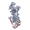
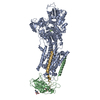
| Title | Cryo-EM structure of the autoinhibited Drs2p-Cdc50p | |||||||||||||||||||||
 Components Components |
| |||||||||||||||||||||
 Keywords Keywords | LIPID TRANSPORT / Lipid Flippase / P-type ATPase / PS Transport. | |||||||||||||||||||||
| Function / homology |  Function and homology information Function and homology informationCdc50p-Drs2p complex / actin cortical patch localization / aminophospholipid translocation / phosphatidylcholine flippase activity / Ion transport by P-type ATPases / post-Golgi vesicle-mediated transport / phosphatidylserine flippase activity / phospholipid-translocating ATPase complex / phosphatidylserine floppase activity / ATPase-coupled intramembrane lipid carrier activity ...Cdc50p-Drs2p complex / actin cortical patch localization / aminophospholipid translocation / phosphatidylcholine flippase activity / Ion transport by P-type ATPases / post-Golgi vesicle-mediated transport / phosphatidylserine flippase activity / phospholipid-translocating ATPase complex / phosphatidylserine floppase activity / ATPase-coupled intramembrane lipid carrier activity / phosphatidylethanolamine flippase activity / endocytic recycling / P-type phospholipid transporter / phosphatidylinositol-4-phosphate binding / retrograde transport, endosome to Golgi / phospholipid translocation / Neutrophil degranulation / trans-Golgi network / intracellular protein transport / endocytosis / late endosome membrane / endosome membrane / magnesium ion binding / Golgi apparatus / endoplasmic reticulum / ATP hydrolysis activity / ATP binding / plasma membrane / cytosol Similarity search - Function | |||||||||||||||||||||
| Biological species |  | |||||||||||||||||||||
| Method | ELECTRON MICROSCOPY / single particle reconstruction / cryo EM / Resolution: 2.8 Å | |||||||||||||||||||||
 Authors Authors | Timcenko, M. / Lyons, J.A. / Januliene, D. / Ulstrup, J.J. / Dieudonne, T. / Montigny, C. / Ash, M.R. / Karlsen, J.L. / Boesen, T. / Kuhlbrandt, W. ...Timcenko, M. / Lyons, J.A. / Januliene, D. / Ulstrup, J.J. / Dieudonne, T. / Montigny, C. / Ash, M.R. / Karlsen, J.L. / Boesen, T. / Kuhlbrandt, W. / Lenoir, G. / Moeller, A. / Nissen, P. | |||||||||||||||||||||
| Funding support |  Denmark, Denmark,  France, 6items France, 6items
| |||||||||||||||||||||
 Citation Citation | Journal: Nature / Year: 2019 Title: Structure and autoregulation of a P4-ATPase lipid flippase. Authors: Milena Timcenko / Joseph A Lyons / Dovile Januliene / Jakob J Ulstrup / Thibaud Dieudonné / Cédric Montigny / Miriam-Rose Ash / Jesper Lykkegaard Karlsen / Thomas Boesen / Werner ...Authors: Milena Timcenko / Joseph A Lyons / Dovile Januliene / Jakob J Ulstrup / Thibaud Dieudonné / Cédric Montigny / Miriam-Rose Ash / Jesper Lykkegaard Karlsen / Thomas Boesen / Werner Kühlbrandt / Guillaume Lenoir / Arne Moeller / Poul Nissen /  Abstract: Type 4 P-type ATPases (P4-ATPases) are lipid flippases that drive the active transport of phospholipids from exoplasmic or luminal leaflets to cytosolic leaflets of eukaryotic membranes. The ...Type 4 P-type ATPases (P4-ATPases) are lipid flippases that drive the active transport of phospholipids from exoplasmic or luminal leaflets to cytosolic leaflets of eukaryotic membranes. The molecular architecture of P4-ATPases and the mechanism through which they recognize and transport lipids have remained unknown. Here we describe the cryo-electron microscopy structure of the P4-ATPase Drs2p-Cdc50p, a Saccharomyces cerevisiae lipid flippase that is specific to phosphatidylserine and phosphatidylethanolamine. Drs2p-Cdc50p is autoinhibited by the C-terminal tail of Drs2p, and activated by the lipid phosphatidylinositol-4-phosphate (PtdIns4P or PI4P). We present three structures that represent the complex in an autoinhibited, an intermediate and a fully activated state. The analysis highlights specific features of P4-ATPases and reveals sites of autoinhibition and PI4P-dependent activation. We also observe a putative lipid translocation pathway in this flippase that involves a conserved PISL motif in transmembrane segment 4 and polar residues of transmembrane segments 2 and 5, in particular Lys1018, in the centre of the lipid bilayer. | |||||||||||||||||||||
| History |
|
- Structure visualization
| Movie |
Movie viewer |
|---|---|
| Structure viewer | Molecule: MolmilJmol/JSmol |
- Downloads & links
-Download
| PDBx/mmCIF format | 6roh.cif.gz | 284 KB | Display | PDBx/mmCIF format |
|---|---|---|---|---|
| PDB format | pdb6roh.ent.gz | 218.7 KB | Display | PDB format |
| PDBx/mmJSON format | 6roh.json.gz | Tree view | PDBx/mmJSON format | |
| Others |  Other downloads Other downloads |
-Validation report
| Arichive directory | https://data.pdbj.org/pub/pdb/validation_reports/ro/6rohftp://data.pdbj.org/pub/pdb/validation_reports/ro/6roh | HTTPS FTP |
|---|
-Related structure data
| Related structure data |  4972MC  4973C  4974C 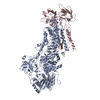 6roiC 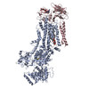 6rojC M: map data used to model this data C: citing same article ( |
|---|---|
| Similar structure data |


-Links
 PDBj
PDBj


- Assembly
| Deposited unit | 
|
|---|---|
| 1 |
|
-Components
-Protein , 2 types, 2 molecules AC
| #1: Protein | Mass: 163730.766 Da / Num. of mol.: 1 Source method: isolated from a genetically manipulated source Details: D560 described by Aspartate beryllium trifluoride (BFD) engineered C-terminal GGGG-LVPRGS-BAD-tag Residues 1-104 are removed by proteolysis with thrombin Residues 1364-1460 are removed after ...Details: D560 described by Aspartate beryllium trifluoride (BFD) engineered C-terminal GGGG-LVPRGS-BAD-tag Residues 1-104 are removed by proteolysis with thrombin Residues 1364-1460 are removed after cleaving of the purification tag by thrombin Source: (gene. exp.) Strain: ATCC 204508 / S288c / Gene: DRS2, YAL026C, FUN38 / Plasmid: pYedp60 / Production host: References: UniProt: P39524, P-type phospholipid transporter |
|---|---|
| #2: Protein | Mass: 47371.797 Da / Num. of mol.: 1 Source method: isolated from a genetically manipulated source Details: engineered C-terminal GGGG-LVPRGS-GG-10xHistag Source: (gene. exp.) Strain: ATCC 204508 / S288c / Gene: CDC50, YCR094W, YCR94W / Plasmid: pYedp60 / Production host: |
-Sugars , 3 types, 3 molecules 
| #3: Polysaccharide | 2-acetamido-2-deoxy-beta-D-glucopyranose-(1-4)-2-acetamido-2-deoxy-beta-D-glucopyranose Source method: isolated from a genetically manipulated source |
|---|---|
| #4: Polysaccharide | beta-D-mannopyranose-(1-3)-beta-D-mannopyranose-(1-4)-2-acetamido-2-deoxy-beta-D-glucopyranose-(1-4) ...beta-D-mannopyranose-(1-3)-beta-D-mannopyranose-(1-4)-2-acetamido-2-deoxy-beta-D-glucopyranose-(1-4)-2-acetamido-2-deoxy-beta-D-glucopyranose Source method: isolated from a genetically manipulated source |
| #7: Sugar | ChemComp-NAG /  Type: D-saccharide, beta linking / Mass: 221.208 Da / Num. of mol.: 1 Type: D-saccharide, beta linking / Mass: 221.208 Da / Num. of mol.: 1Source method: isolated from a genetically manipulated source Formula: C8H15NO6 |
-Non-polymers , 3 types, 4 molecules 


| #5: Chemical | ChemComp-PSF /  Mass: 455.437 Da / Num. of mol.: 1 / Source method: obtained synthetically / Formula: C18H34NO10P / Feature type: SUBJECT OF INVESTIGATION Mass: 455.437 Da / Num. of mol.: 1 / Source method: obtained synthetically / Formula: C18H34NO10P / Feature type: SUBJECT OF INVESTIGATION |
|---|---|
| #6: Chemical | ChemComp-MG /  Mass: 24.305 Da / Num. of mol.: 1 / Source method: isolated from a natural source / Formula: Mg / Feature type: SUBJECT OF INVESTIGATION Mass: 24.305 Da / Num. of mol.: 1 / Source method: isolated from a natural source / Formula: Mg / Feature type: SUBJECT OF INVESTIGATION |
| #8: Water | ChemComp-HOH / Mass: 18.015 Da / Num. of mol.: 2 / Source method: isolated from a natural source / Formula: H2O |
-Details
| Has protein modification | Y |
|---|
-Experimental details
-Experiment
| Experiment | Method: ELECTRON MICROSCOPY |
|---|---|
| EM experiment | Aggregation state: PARTICLE / 3D reconstruction method: single particle reconstruction |
- Sample preparation
| Component | Name: Binary complex of Drs2p-Cdc50p / Type: COMPLEX / Entity ID: #1-#2 / Source: RECOMBINANT |
|---|---|
| Molecular weight | Experimental value: NO |
| Source (natural) | Organism: |
| Source (recombinant) | Organism: |
| Buffer solution | pH: 7 |
| Specimen | Conc.: 0.6 mg/ml / Embedding applied: NO / Shadowing applied: NO / Staining applied: NO / Vitrification applied: YES / Details: 1mM beryllium fluoride |
| Specimen support | Details: 15mA / Grid material: COPPER / Grid mesh size: 400 divisions/in. / Grid type: C-flat-1.2/1.3 |
| Vitrification | Instrument: FEI VITROBOT MARK IV / Cryogen name: ETHANE / Humidity: 100 % / Chamber temperature: 278 K |
- Electron microscopy imaging
| Experimental equipment |  Model: Titan Krios / Image courtesy: FEI Company |
|---|---|
| Microscopy | Model: FEI TITAN KRIOS |
| Electron gun | Electron source:  FIELD EMISSION GUN / Accelerating voltage: 300 kV / Illumination mode: FLOOD BEAM FIELD EMISSION GUN / Accelerating voltage: 300 kV / Illumination mode: FLOOD BEAM |
| Electron lens | Mode: BRIGHT FIELD |
| Specimen holder | Cryogen: NITROGEN / Specimen holder model: FEI TITAN KRIOS AUTOGRID HOLDER |
| Image recording | Average exposure time: 8 sec. / Electron dose: 60 e/Å2 / Detector mode: COUNTING / Film or detector model: GATAN K2 QUANTUM (4k x 4k) |
- Processing
| Software | Name: PHENIX / Version: 1.14_3260: / Classification: refinement | ||||||||||||||||||
|---|---|---|---|---|---|---|---|---|---|---|---|---|---|---|---|---|---|---|---|
| EM software |
| ||||||||||||||||||
| CTF correction | Type: PHASE FLIPPING AND AMPLITUDE CORRECTION | ||||||||||||||||||
| Particle selection | Num. of particles selected: 2157578 | ||||||||||||||||||
| Symmetry | Point symmetry: C1 (asymmetric) | ||||||||||||||||||
| 3D reconstruction | Resolution: 2.8 Å / Resolution method: FSC 0.143 CUT-OFF / Num. of particles: 752881 / Symmetry type: POINT |
