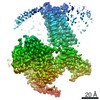
 Movie
Movie Controller


+ Open data

- Basic information
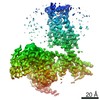
| Entry | Database: PDB / ID: 6k41 | |||||||||||||||||||||||||||||||||
|---|---|---|---|---|---|---|---|---|---|---|---|---|---|---|---|---|---|---|---|---|---|---|---|---|---|---|---|---|---|---|---|---|---|---|
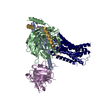
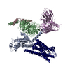
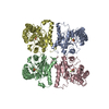
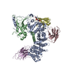
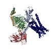
| Title | cryo-EM structure of alpha2BAR-GoA complex | |||||||||||||||||||||||||||||||||
 Components Components |
| |||||||||||||||||||||||||||||||||
 Keywords Keywords | MEMBRANE PROTEIN / GPCR / Complex / cryo-EM | |||||||||||||||||||||||||||||||||
| Function / homology |  Function and homology information Function and homology informationAdrenoceptors / Adrenaline signalling through Alpha-2 adrenergic receptor / Surfactant metabolism / Activation of the phototransduction cascade / Olfactory Signaling Pathway / alpha2-adrenergic receptor activity / Adrenaline signalling through Alpha-2 adrenergic receptor / Synthesis, secretion, and inactivation of Glucagon-like Peptide-1 (GLP-1) / Sensory perception of sweet, bitter, and umami (glutamate) taste / G beta:gamma signalling through PLC beta ...Adrenoceptors / Adrenaline signalling through Alpha-2 adrenergic receptor / Surfactant metabolism / Activation of the phototransduction cascade / Olfactory Signaling Pathway / alpha2-adrenergic receptor activity / Adrenaline signalling through Alpha-2 adrenergic receptor / Synthesis, secretion, and inactivation of Glucagon-like Peptide-1 (GLP-1) / Sensory perception of sweet, bitter, and umami (glutamate) taste / G beta:gamma signalling through PLC beta / Presynaptic function of Kainate receptors / alpha-2C adrenergic receptor binding / Prostacyclin signalling through prostacyclin receptor / G alpha (z) signalling events / Glucagon-type ligand receptors / G beta:gamma signalling through PI3Kgamma / G beta:gamma signalling through CDC42 / epinephrine binding / phospholipase C-activating adrenergic receptor signaling pathway / Adrenaline,noradrenaline inhibits insulin secretion / ADP signalling through P2Y purinoceptor 12 / Cooperation of PDCL (PhLP1) and TRiC/CCT in G-protein beta folding / Thromboxane signalling through TP receptor / G beta:gamma signalling through BTK / positive regulation of uterine smooth muscle contraction / Thrombin signalling through proteinase activated receptors (PARs) / Activation of G protein gated Potassium channels / Inhibition of voltage gated Ca2+ channels via Gbeta/gamma subunits / negative regulation of norepinephrine secretion / negative regulation of epinephrine secretion / G alpha (s) signalling events / Ca2+ pathway / G-protein activation / Extra-nuclear estrogen signaling / G alpha (12/13) signalling events / G alpha (q) signalling events / Vasopressin regulates renal water homeostasis via Aquaporins / GPER1 signaling / Glucagon-like Peptide-1 (GLP1) regulates insulin secretion / regulation of vascular associated smooth muscle contraction / heterotrimeric G-protein binding / ADP signalling through P2Y purinoceptor 1 / High laminar flow shear stress activates signaling by PIEZO1 and PECAM1:CDH5:KDR in endothelial cells / regulation of smooth muscle contraction / G alpha (i) signalling events / positive regulation of blood pressure / dopaminergic synapse / mu-type opioid receptor binding / : / corticotropin-releasing hormone receptor 1 binding / thioesterase binding / fear response / negative regulation of insulin secretion involved in cellular response to glucose stimulus / adenylate cyclase-inhibiting adrenergic receptor signaling pathway / norepinephrine binding / positive regulation of membrane protein ectodomain proteolysis / Adrenoceptors / G protein-coupled dopamine receptor signaling pathway / Adrenaline,noradrenaline inhibits insulin secretion / G alpha (z) signalling events / photoreceptor outer segment membrane / adrenergic receptor signaling pathway / spectrin binding / positive regulation of wound healing / G alpha (i) signalling events / alkylglycerophosphoethanolamine phosphodiesterase activity / regulation of heart contraction / parallel fiber to Purkinje cell synapse / phototransduction, visible light / photoreceptor outer segment / negative regulation of lipid catabolic process / regulation of vasoconstriction / viral release from host cell by cytolysis / postsynaptic modulation of chemical synaptic transmission / positive regulation of epidermal growth factor receptor signaling pathway / cellular response to hormone stimulus / adenylate cyclase-activating adrenergic receptor signaling pathway / peptidoglycan catabolic process / cardiac muscle cell apoptotic process / photoreceptor inner segment / positive regulation of neuron differentiation / adenylate cyclase-inhibiting serotonin receptor signaling pathway / G protein-coupled serotonin receptor binding / muscle contraction / presynaptic modulation of chemical synaptic transmission / positive regulation of cytokine production / female pregnancy / locomotory behavior / platelet activation / negative regulation of insulin secretion / GABA-ergic synapse / epidermal growth factor receptor signaling pathway / cell wall macromolecule catabolic process / G-protein beta/gamma-subunit complex binding / adenylate cyclase-modulating G protein-coupled receptor signaling pathway / adenylate cyclase-inhibiting G protein-coupled receptor signaling pathway / lysozyme / lysozyme activity / G beta:gamma signalling through PLC beta / Presynaptic function of Kainate receptors Similarity search - Function | |||||||||||||||||||||||||||||||||
| Biological species |  Homo sapiens (human) Homo sapiens (human)  Enterobacteria phage RB59 (virus) Enterobacteria phage RB59 (virus) | |||||||||||||||||||||||||||||||||
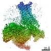
| Method | ELECTRON MICROSCOPY / single particle reconstruction / cryo EM / Resolution: 2.9 Å | |||||||||||||||||||||||||||||||||
 Authors Authors | Yuan, D. / Liu, Z. / Wang, H.W. / Kobilka, B.K. | |||||||||||||||||||||||||||||||||
 Citation Citation | Journal: Nat Chem Biol / Year: 2020 Title: Activation of the α adrenoceptor by the sedative sympatholytic dexmedetomidine. Authors: Daopeng Yuan / Zhongmin Liu / Jonas Kaindl / Shoji Maeda / Jiawei Zhao / Xiaoou Sun / Jun Xu / Peter Gmeiner / Hong-Wei Wang / Brian K Kobilka /    Abstract: The α adrenergic receptors (αARs) are G protein-coupled receptors (GPCRs) that respond to adrenaline and noradrenaline and couple to the Gi/o family of G proteins. αARs play important roles in ...The α adrenergic receptors (αARs) are G protein-coupled receptors (GPCRs) that respond to adrenaline and noradrenaline and couple to the Gi/o family of G proteins. αARs play important roles in regulating the sympathetic nervous system. Dexmedetomidine is a highly selective αAR agonist used in post-operative patients as an anxiety-reducing, sedative medicine that decreases the requirement for opioids. As is typical for selective αAR agonists, dexmedetomidine consists of an imidazole ring and a substituted benzene moiety lacking polar groups, which is in contrast to βAR-selective agonists, which share an ethanolamine group and an aromatic system with polar, hydrogen-bonding substituents. To better understand the structural basis for the selectivity and efficacy of adrenergic agonists, we determined the structure of the αAR in complex with dexmedetomidine and Go at a resolution of 2.9 Å by single-particle cryo-EM. The structure reveals the mechanism of αAR-selective activation and provides insights into Gi/o coupling specificity. | |||||||||||||||||||||||||||||||||
| History |
|
- Structure visualization
| Movie |
Movie viewer |
|---|---|
| Structure viewer | Molecule: MolmilJmol/JSmol |
- Downloads & links
-Download
| PDBx/mmCIF format | 6k41.cif.gz | 211.8 KB | Display | PDBx/mmCIF format |
|---|---|---|---|---|
| PDB format | pdb6k41.ent.gz | 158.7 KB | Display | PDB format |
| PDBx/mmJSON format | 6k41.json.gz | Tree view | PDBx/mmJSON format | |
| Others |  Other downloads Other downloads |
-Validation report
| Arichive directory | https://data.pdbj.org/pub/pdb/validation_reports/k4/6k41ftp://data.pdbj.org/pub/pdb/validation_reports/k4/6k41 | HTTPS FTP |
|---|
-Related structure data
| Related structure data | 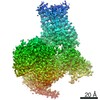 9911MC 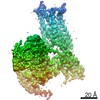 9912C 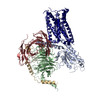 6k42C C: citing same article ( M: map data used to model this data |
|---|---|
| Similar structure data |

-Links
 PDBj
PDBj


- Assembly
| Deposited unit | 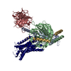
|
|---|---|
| 1 |
|
-Components
-Guanine nucleotide-binding protein ... , 3 types, 3 molecules ABG
| #1: Protein | Mass: 40100.500 Da / Num. of mol.: 1 Source method: isolated from a genetically manipulated source Source: (gene. exp.) Homo sapiens (human) / Gene: GNAO1 / Production host:  Spodoptera (butterflies/moths) / References: UniProt: P09471 Spodoptera (butterflies/moths) / References: UniProt: P09471 |
|---|---|
| #2: Protein | Mass: 38402.867 Da / Num. of mol.: 1 Source method: isolated from a genetically manipulated source Source: (gene. exp.) Spodoptera (butterflies/moths) / References: UniProt: P62874 |
| #3: Protein | Mass: 7861.143 Da / Num. of mol.: 1 Source method: isolated from a genetically manipulated source Source: (gene. exp.) Homo sapiens (human) / Gene: GNG2 / Production host: Spodoptera (butterflies/moths) / References: UniProt: P59768 |
-Protein / Antibody / Non-polymers , 3 types, 3 molecules RH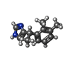
| #4: Protein | Mass: 58156.211 Da / Num. of mol.: 1 Source method: isolated from a genetically manipulated source Source: (gene. exp.) Enterobacteria phage RB59 (virus), (gene. exp.) Homo sapiens (human)Gene: ADRA2A, e, RB59_126, ADRA2B, ADRA2L1, ADRA2RL1 / Production host: Spodoptera (butterflies/moths)References: UniProt: Q28838, UniProt: A0A097J809, UniProt: P18089, lysozyme |
|---|---|
| #5: Antibody | Mass: 32898.781 Da / Num. of mol.: 1 Source method: isolated from a genetically manipulated source Source: (gene. exp.) Spodoptera (butterflies/moths) |
| #6: Chemical | ChemComp-CZX /  Mass: 200.280 Da / Num. of mol.: 1 / Source method: obtained synthetically / Formula: C13H16N2 / Comment: medication*YM Mass: 200.280 Da / Num. of mol.: 1 / Source method: obtained synthetically / Formula: C13H16N2 / Comment: medication*YM |
-Details
| Has protein modification | Y |
|---|
-Experimental details
-Experiment
| Experiment | Method: ELECTRON MICROSCOPY |
|---|---|
| EM experiment | Aggregation state: PARTICLE / 3D reconstruction method: single particle reconstruction |
- Sample preparation
| Component | Name: alpha2BAR-GoA complex / Type: COMPLEX / Entity ID: #1-#5 / Source: MULTIPLE SOURCES |
|---|---|
| Molecular weight | Value: 0.15 MDa / Experimental value: YES |
| Source (natural) | Organism: Spodoptera (butterflies/moths) |
| Buffer solution | pH: 7.5 |
| Specimen | Conc.: 10 mg/ml / Embedding applied: NO / Shadowing applied: NO / Staining applied: NO / Vitrification applied: YES |
| Specimen support | Grid material: GOLD / Grid mesh size: 400 divisions/in. / Grid type: Quantifoil R1.2/1.3 |
| Vitrification | Instrument: FEI VITROBOT MARK IV / Cryogen name: ETHANE / Humidity: 100 % |
- Electron microscopy imaging
| Experimental equipment |  Model: Titan Krios / Image courtesy: FEI Company |
|---|---|
| Microscopy | Model: FEI TITAN KRIOS |
| Electron gun | Electron source:  FIELD EMISSION GUN / Accelerating voltage: 300 kV / Illumination mode: SPOT SCAN FIELD EMISSION GUN / Accelerating voltage: 300 kV / Illumination mode: SPOT SCAN |
| Electron lens | Mode: BRIGHT FIELD |
| Image recording | Electron dose: 50 e/Å2 / Detector mode: SUPER-RESOLUTION / Film or detector model: GATAN K2 SUMMIT (4k x 4k) |
- Processing
| Software | Name: PHENIX / Version: 1.13_2998: / Classification: refinement |
|---|---|
| EM software | Name: PHENIX / Category: model refinement |
| CTF correction | Type: NONE |
| 3D reconstruction | Resolution: 2.9 Å / Resolution method: FSC 0.143 CUT-OFF / Num. of particles: 258283 / Symmetry type: POINT |
