 Movie
Movie Controller


+ Open data

- Basic information
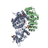
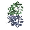
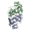
| Entry | Database: PDB / ID: 6i52 | ||||||
|---|---|---|---|---|---|---|---|
| Title | Yeast RPA bound to ssDNA | ||||||
 Components Components |
| ||||||
 Keywords Keywords | DNA BINDING PROTEIN / Complex / heterotrimer / DNA binding / OB-fold | ||||||
| Function / homology |  Function and homology information Function and homology informationheteroduplex formation / sporulation / DNA replication factor A complex / Gap-filling DNA repair synthesis and ligation in GG-NER / telomere maintenance via telomere lengthening / Removal of the Flap Intermediate / telomere maintenance via recombination / Translesion Synthesis by POLH / Activation of the pre-replicative complex / Translesion synthesis by REV1 ...heteroduplex formation / sporulation / DNA replication factor A complex / Gap-filling DNA repair synthesis and ligation in GG-NER / telomere maintenance via telomere lengthening / Removal of the Flap Intermediate / telomere maintenance via recombination / Translesion Synthesis by POLH / Activation of the pre-replicative complex / Translesion synthesis by REV1 / Translesion synthesis by POLK / Translesion synthesis by POLI / single-stranded telomeric DNA binding / Activation of ATR in response to replication stress / mitotic recombination / Termination of translesion DNA synthesis / reciprocal meiotic recombination / DNA unwinding involved in DNA replication / Gap-filling DNA repair synthesis and ligation in TC-NER / DNA topological change / Dual incision in TC-NER / telomere maintenance via telomerase / telomere maintenance / condensed nuclear chromosome / nucleotide-excision repair / double-strand break repair via homologous recombination / establishment of protein localization / site of double-strand break / single-stranded DNA binding / double-stranded DNA binding / DNA replication / sequence-specific DNA binding / chromosome, telomeric region / damaged DNA binding / protein ubiquitination / DNA repair / mRNA binding / nucleus / metal ion binding / cytoplasm / cytosol Similarity search - Function | ||||||
| Biological species |  synthetic construct (others) | ||||||
| Method | ELECTRON MICROSCOPY / single particle reconstruction / cryo EM / Resolution: 4.7 Å | ||||||
 Authors Authors | Yates, L.A. / Aramayo, R.J. / Zhang, X. | ||||||
| Funding support |  United Kingdom, 1items United Kingdom, 1items
| ||||||
 Citation Citation | Journal: Nat Commun / Year: 2018 Title: A structural and dynamic model for the assembly of Replication Protein A on single-stranded DNA. Authors: Luke A Yates / Ricardo J Aramayo / Nilisha Pokhrel / Colleen C Caldwell / Joshua A Kaplan / Rajika L Perera / Maria Spies / Edwin Antony / Xiaodong Zhang /  Abstract: Replication Protein A (RPA), the major eukaryotic single stranded DNA-binding protein, binds to exposed ssDNA to protect it from nucleases, participates in a myriad of nucleic acid transactions and ...Replication Protein A (RPA), the major eukaryotic single stranded DNA-binding protein, binds to exposed ssDNA to protect it from nucleases, participates in a myriad of nucleic acid transactions and coordinates the recruitment of other important players. RPA is a heterotrimer and coats long stretches of single-stranded DNA (ssDNA). The precise molecular architecture of the RPA subunits and its DNA binding domains (DBDs) during assembly is poorly understood. Using cryo electron microscopy we obtained a 3D reconstruction of the RPA trimerisation core bound with ssDNA (∼55 kDa) at ∼4.7 Å resolution and a dimeric RPA assembly on ssDNA. FRET-based solution studies reveal dynamic rearrangements of DBDs during coordinated RPA binding and this activity is regulated by phosphorylation at S178 in RPA70. We present a structural model on how dynamic DBDs promote the cooperative assembly of multiple RPAs on long ssDNA. | ||||||
| History |
|
- Structure visualization
| Movie |
Movie viewer |
|---|---|
| Structure viewer | Molecule: MolmilJmol/JSmol |
- Downloads & links
-Download
| PDBx/mmCIF format | 6i52.cif.gz | 108.5 KB | Display | PDBx/mmCIF format |
|---|---|---|---|---|
| PDB format | pdb6i52.ent.gz | 82 KB | Display | PDB format |
| PDBx/mmJSON format | 6i52.json.gz | Tree view | PDBx/mmJSON format | |
| Others |  Other downloads Other downloads |
-Validation report
| Summary document | 6i52_validation.pdf.gz | 375.1 KB | Display | wwPDB validaton report |
|---|---|---|---|---|
| Full document | 6i52_full_validation.pdf.gz | 382.6 KB | Display | |
| Data in XML | 6i52_validation.xml.gz | 10.5 KB | Display | |
| Data in CIF | 6i52_validation.cif.gz | 15.3 KB | Display | |
| Arichive directory | https://data.pdbj.org/pub/pdb/validation_reports/i5/6i52ftp://data.pdbj.org/pub/pdb/validation_reports/i5/6i52 | HTTPS FTP |
-Related structure data
| Related structure data | 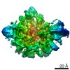 4410MC M: map data used to model this data C: citing same article ( |
|---|---|
| Similar structure data |


-Links
 PDBj
PDBj


- Assembly
| Deposited unit | 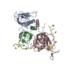
|
|---|---|
| 1 |
|
-Components
| #1: Protein | Mass: 13827.723 Da / Num. of mol.: 1 Source method: isolated from a genetically manipulated source Source: (gene. exp.) Gene: RFA3, YJL173C, J0506 / Plasmid: pRSF-Duet-1 / Production host:  |
|---|---|
| #2: Protein | Mass: 14809.870 Da / Num. of mol.: 1 Source method: isolated from a genetically manipulated source Source: (gene. exp.) Gene: RFA2, BUF1, YNL312W, N0368 / Plasmid: pRSF-duet-1 / Production host: |
| #3: Protein | Mass: 20643.072 Da / Num. of mol.: 1 Source method: isolated from a genetically manipulated source Source: (gene. exp.) Gene: RFA1, BUF2, RPA1, YAR007C, FUN3 / Plasmid: pRSF-duet-1 / Production host: |
| #4: DNA chain | Mass: 6038.899 Da / Num. of mol.: 1 / Source method: obtained synthetically / Source: (synth.) synthetic construct (others) |
-Experimental details
-Experiment
| Experiment | Method: ELECTRON MICROSCOPY |
|---|---|
| EM experiment | Aggregation state: PARTICLE / 3D reconstruction method: single particle reconstruction |
- Sample preparation
| Component |
| ||||||||||||||||||||||||
|---|---|---|---|---|---|---|---|---|---|---|---|---|---|---|---|---|---|---|---|---|---|---|---|---|---|
| Molecular weight | Value: 0.12 MDa / Experimental value: NO | ||||||||||||||||||||||||
| Source (natural) |
| ||||||||||||||||||||||||
| Source (recombinant) | Organism: | ||||||||||||||||||||||||
| Buffer solution | pH: 7.4 | ||||||||||||||||||||||||
| Buffer component |
| ||||||||||||||||||||||||
| Specimen | Conc.: 0.5 mg/ml / Embedding applied: NO / Shadowing applied: NO / Staining applied: NO / Vitrification applied: YES | ||||||||||||||||||||||||
| Specimen support | Grid material: COPPER / Grid mesh size: 300 divisions/in. / Grid type: Quantifoil R2/2 | ||||||||||||||||||||||||
| Vitrification | Instrument: FEI VITROBOT MARK IV / Cryogen name: ETHANE / Humidity: 100 % / Chamber temperature: 277 K Details: glow-discharged grid, wait time 30 seconds, blotting time 1 second |
- Electron microscopy imaging
| Experimental equipment |  Model: Titan Krios / Image courtesy: FEI Company |
|---|---|
| Microscopy | Model: FEI TITAN KRIOS |
| Electron gun | Electron source:  FIELD EMISSION GUN / Accelerating voltage: 300 kV / Illumination mode: FLOOD BEAM FIELD EMISSION GUN / Accelerating voltage: 300 kV / Illumination mode: FLOOD BEAM |
| Electron lens | Mode: BRIGHT FIELD / Cs: 2.7 mm |
| Specimen holder | Cryogen: NITROGEN / Specimen holder model: FEI TITAN KRIOS AUTOGRID HOLDER |
| Image recording | Electron dose: 2.1 e/Å2 / Detector mode: COUNTING / Film or detector model: GATAN K2 SUMMIT (4k x 4k) |
- Processing
| EM software |
| ||||||||||||||||||||||||
|---|---|---|---|---|---|---|---|---|---|---|---|---|---|---|---|---|---|---|---|---|---|---|---|---|---|
| CTF correction | Type: PHASE FLIPPING AND AMPLITUDE CORRECTION | ||||||||||||||||||||||||
| Symmetry | Point symmetry: C1 (asymmetric) | ||||||||||||||||||||||||
| 3D reconstruction | Resolution: 4.7 Å / Resolution method: FSC 0.143 CUT-OFF / Num. of particles: 340864 / Symmetry type: POINT | ||||||||||||||||||||||||
| Atomic model building | Protocol: FLEXIBLE FIT / Space: REAL |
