 Movie
Movie Controller

[English] 日本語
 Yorodumi
- PDB-6ehl: Model of the Ebola virus nucleoprotein in recombinant nucleocapsi... -
+ Open data

- Basic information
| Entry | Database: PDB / ID: 6ehl | ||||||||||||
|---|---|---|---|---|---|---|---|---|---|---|---|---|---|
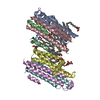
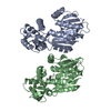
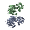
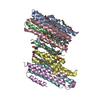
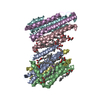
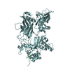
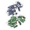
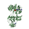
| Title | Model of the Ebola virus nucleoprotein in recombinant nucleocapsid-like assemblies | ||||||||||||
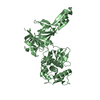
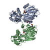
 Components Components | Nucleoprotein | ||||||||||||
 Keywords Keywords | VIRUS LIKE PARTICLE / nucleocapsid | ||||||||||||
| Function / homology | Ebola nucleoprotein / Ebola nucleoprotein / viral RNA genome packaging / helical viral capsid / viral nucleocapsid / host cell cytoplasm / ribonucleoprotein complex / RNA binding / Nucleoprotein Function and homology information Function and homology information | ||||||||||||
| Biological species |   Zaire ebolavirus Zaire ebolavirus | ||||||||||||
| Method | ELECTRON MICROSCOPY / subtomogram averaging / cryo EM / Resolution: 6.6 Å | ||||||||||||
 Authors Authors | Wan, W. / Kolesnikova, L. / Clarke, M. / Koehler, A. / Noda, T. / Becker, S. / Briggs, J.A.G. | ||||||||||||
| Funding support |  Germany, 3items Germany, 3items
| ||||||||||||
 Citation Citation | Journal: Nature / Year: 2017 Title: Structure and assembly of the Ebola virus nucleocapsid. Authors: William Wan / Larissa Kolesnikova / Mairi Clarke / Alexander Koehler / Takeshi Noda / Stephan Becker / John A G Briggs /   Abstract: Ebola and Marburg viruses are filoviruses: filamentous, enveloped viruses that cause haemorrhagic fever. Filoviruses are within the order Mononegavirales, which also includes rabies virus, measles ...Ebola and Marburg viruses are filoviruses: filamentous, enveloped viruses that cause haemorrhagic fever. Filoviruses are within the order Mononegavirales, which also includes rabies virus, measles virus, and respiratory syncytial virus. Mononegaviruses have non-segmented, single-stranded negative-sense RNA genomes that are encapsidated by nucleoprotein and other viral proteins to form a helical nucleocapsid. The nucleocapsid acts as a scaffold for virus assembly and as a template for genome transcription and replication. Insights into nucleoprotein-nucleoprotein interactions have been derived from structural studies of oligomerized, RNA-encapsidating nucleoprotein, and cryo-electron microscopy of nucleocapsid or nucleocapsid-like structures. There have been no high-resolution reconstructions of complete mononegavirus nucleocapsids. Here we apply cryo-electron tomography and subtomogram averaging to determine the structure of Ebola virus nucleocapsid within intact viruses and recombinant nucleocapsid-like assemblies. These structures reveal the identity and arrangement of the nucleocapsid components, and suggest that the formation of an extended α-helix from the disordered carboxy-terminal region of nucleoprotein-core links nucleoprotein oligomerization, nucleocapsid condensation, RNA encapsidation, and accessory protein recruitment. | ||||||||||||
| History |
|
- Structure visualization
| Movie |
Movie viewer |
|---|---|
| Structure viewer | Molecule: MolmilJmol/JSmol |
- Downloads & links
-Download
| PDBx/mmCIF format | 6ehl.cif.gz | 32.6 KB | Display | PDBx/mmCIF format |
|---|---|---|---|---|
| PDB format | pdb6ehl.ent.gz | 12.5 KB | Display | PDB format |
| PDBx/mmJSON format | 6ehl.json.gz | Tree view | PDBx/mmJSON format | |
| Others |  Other downloads Other downloads |
-Validation report
| Summary document | 6ehl_validation.pdf.gz | 766.5 KB | Display | wwPDB validaton report |
|---|---|---|---|---|
| Full document | 6ehl_full_validation.pdf.gz | 766.1 KB | Display | |
| Data in XML | 6ehl_validation.xml.gz | 11.8 KB | Display | |
| Data in CIF | 6ehl_validation.cif.gz | 16 KB | Display | |
| Arichive directory | https://data.pdbj.org/pub/pdb/validation_reports/eh/6ehlftp://data.pdbj.org/pub/pdb/validation_reports/eh/6ehl | HTTPS FTP |
-Related structure data
| Related structure data | 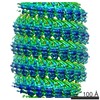 3869MC 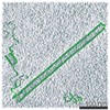 3870C 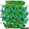 3871C 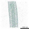 3872C 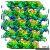 3873C 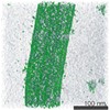 3874C 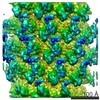 3875C 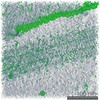 3876C 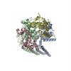 6ehmC M: map data used to model this data C: citing same article ( |
|---|---|
| Similar structure data |
-Links
 PDBj
PDBj
- Assembly
| Deposited unit | 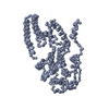
|
|---|---|
| 1 |
|
-Components
| #1: Protein | Mass: 83387.500 Da / Num. of mol.: 1 / Mutation: Truncation mutant (residues 1-450) Source method: isolated from a genetically manipulated source Source: (gene. exp.) Zaire ebolavirus (strain Mayinga-76) / Strain: Mayinga-76 / Gene: NP / Cell line (production host): HEK-293T / Production host:  Homo sapiens (human) / References: UniProt: P18272 Homo sapiens (human) / References: UniProt: P18272 |
|---|
-Experimental details
-Experiment
| Experiment | Method: ELECTRON MICROSCOPY |
|---|---|
| EM experiment | Aggregation state: HELICAL ARRAY / 3D reconstruction method: subtomogram averaging |
- Sample preparation
| Component | Name: Ebola virus - Mayinga, Zaire, 1976 / Type: VIRUS / Entity ID: all / Source: RECOMBINANT | |||||||||||||||||||||||||
|---|---|---|---|---|---|---|---|---|---|---|---|---|---|---|---|---|---|---|---|---|---|---|---|---|---|---|
| Source (natural) | Organism: Ebola virus - Mayinga, Zaire, 1976 | |||||||||||||||||||||||||
| Source (recombinant) | Organism: Homo sapiens (human) / Cell: HEK 293T | |||||||||||||||||||||||||
| Details of virus | Empty: NO / Enveloped: NO / Isolate: STRAIN / Type: VIRUS-LIKE PARTICLE | |||||||||||||||||||||||||
| Virus shell | Name: Nucleocapsid / Diameter: 280 nm | |||||||||||||||||||||||||
| Buffer solution | pH: 7.4 | |||||||||||||||||||||||||
| Buffer component |
| |||||||||||||||||||||||||
| Specimen | Embedding applied: NO / Shadowing applied: NO / Staining applied: NO / Vitrification applied: YES | |||||||||||||||||||||||||
| Specimen support | Grid material: COPPER / Grid mesh size: 300 divisions/in. / Grid type: C-flat 2/1 3C | |||||||||||||||||||||||||
| Vitrification | Instrument: FEI VITROBOT MARK II / Cryogen name: ETHANE / Humidity: 95 % |
- Electron microscopy imaging
| Experimental equipment |  Model: Titan Krios / Image courtesy: FEI Company |
|---|---|
| Microscopy | Model: FEI TITAN KRIOS |
| Electron gun | Electron source:  FIELD EMISSION GUN / Accelerating voltage: 300 kV / Illumination mode: FLOOD BEAM FIELD EMISSION GUN / Accelerating voltage: 300 kV / Illumination mode: FLOOD BEAM |
| Electron lens | Mode: BRIGHT FIELD / Nominal magnification: 81000 X / Nominal defocus max: 4500 nm / Nominal defocus min: 2000 nm / Cs: 2.7 mm / C2 aperture diameter: 50 µm / Alignment procedure: ZEMLIN TABLEAU |
| Specimen holder | Cryogen: NITROGEN / Specimen holder model: FEI TITAN KRIOS AUTOGRID HOLDER |
| Image recording | Average exposure time: 1 sec. / Electron dose: 2.4 e/Å2 / Detector mode: SUPER-RESOLUTION / Film or detector model: GATAN K2 QUANTUM (4k x 4k) / Num. of grids imaged: 1 |
| EM imaging optics | Energyfilter name: GIF Quantum LS / Energyfilter upper: 10 eV / Energyfilter lower: -10 eV |
| Image scans | Width: 3708 / Height: 3708 / Movie frames/image: 5 / Used frames/image: 1-5 |
- Processing
| EM software |
| ||||||||||||||||||||||||||||||||||||||||||||||||||||||||||||
|---|---|---|---|---|---|---|---|---|---|---|---|---|---|---|---|---|---|---|---|---|---|---|---|---|---|---|---|---|---|---|---|---|---|---|---|---|---|---|---|---|---|---|---|---|---|---|---|---|---|---|---|---|---|---|---|---|---|---|---|---|---|
| Image processing | Details: Frames were aligned using K2Align software, based off the MotionCorr algorithm. Tomograms were reconstructed with IMOD, using stripwise CTF-correction and weighted back projection. ...Details: Frames were aligned using K2Align software, based off the MotionCorr algorithm. Tomograms were reconstructed with IMOD, using stripwise CTF-correction and weighted back projection. Subtomogram averaging was performed using scripts derived from TOM, AV3, and DYNAMO. | ||||||||||||||||||||||||||||||||||||||||||||||||||||||||||||
| CTF correction | Details: CTF amplitude correction was performed during the wedge-weighted subtomogram averaging step. Type: PHASE FLIPPING ONLY | ||||||||||||||||||||||||||||||||||||||||||||||||||||||||||||
| Symmetry | Point symmetry: C1 (asymmetric) | ||||||||||||||||||||||||||||||||||||||||||||||||||||||||||||
| 3D reconstruction | Resolution: 6.6 Å / Resolution method: FSC 0.143 CUT-OFF / Num. of particles: 1 / Algorithm: BACK PROJECTION Details: Local resolution was estimated using moving window FSC calculations. Num. of class averages: 1 / Symmetry type: POINT | ||||||||||||||||||||||||||||||||||||||||||||||||||||||||||||
| EM volume selection | Details: Points along the helical axis were manually placed to define a spline. A cylindrical grid as defined at a given radius from the spline; grid spacing was chosen to provide ~4x oversampling. Num. of tomograms: 63 / Num. of volumes extracted: 488916 / Reference model: None | ||||||||||||||||||||||||||||||||||||||||||||||||||||||||||||
| Atomic model building | Protocol: FLEXIBLE FIT Details: Crystal structure was rigid-body fitted into density using Chimera. Missing regions of the structure was built using ideal secondary structures in coot. These were then flexibly fit using MDFF and NAMD. |
