Movie
Movie Controller

[English] 日本語
 Yorodumi
- PDB-6b3j: 3.3 angstrom phase-plate cryo-EM structure of a biased agonist-bo... -
+ Open data

- Basic information
| Entry | Database: PDB / ID: 6b3j | |||||||||||||||
|---|---|---|---|---|---|---|---|---|---|---|---|---|---|---|---|---|
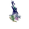
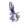
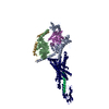
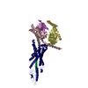
| Title | 3.3 angstrom phase-plate cryo-EM structure of a biased agonist-bound human GLP-1 receptor-Gs complex | |||||||||||||||
 Components Components |
| |||||||||||||||
 Keywords Keywords | SIGNALING PROTEIN / class B G protein-coupled receptor / agonist-receptor-G protein ternary complex / glucagon-like peptide 1 receptor / active-state G protein-coupled receptor / phase plate / phase contrast / single particle cryo-em | |||||||||||||||
| Function / homology |  Function and homology information Function and homology informationglucagon-like peptide 1 receptor activity / glucagon receptor activity / positive regulation of blood pressure / hormone secretion / post-translational protein targeting to membrane, translocation / response to psychosocial stress / regulation of heart contraction / adenylate cyclase-activating G protein-coupled bile acid receptor signaling pathway / adenylate cyclase-activating serotonin receptor signaling pathway / peptide hormone binding ...glucagon-like peptide 1 receptor activity / glucagon receptor activity / positive regulation of blood pressure / hormone secretion / post-translational protein targeting to membrane, translocation / response to psychosocial stress / regulation of heart contraction / adenylate cyclase-activating G protein-coupled bile acid receptor signaling pathway / adenylate cyclase-activating serotonin receptor signaling pathway / peptide hormone binding / regulation of skeletal muscle contraction / PKA activation in glucagon signalling / hair follicle placode formation / developmental growth / intracellular transport / D1 dopamine receptor binding / vascular endothelial cell response to laminar fluid shear stress / renal water homeostasis / activation of adenylate cyclase activity / Hedgehog 'off' state / adenylate cyclase-activating adrenergic receptor signaling pathway / cellular response to acidic pH / negative regulation of blood pressure / cellular response to glucagon stimulus / intracellular glucose homeostasis / adenylate cyclase activator activity / positive regulation of insulin secretion involved in cellular response to glucose stimulus / trans-Golgi network membrane / negative regulation of inflammatory response to antigenic stimulus / response to prostaglandin E / bone development / platelet aggregation / cognition / G-protein beta/gamma-subunit complex binding / positive regulation of insulin secretion / Olfactory Signaling Pathway / Activation of the phototransduction cascade / G protein-coupled acetylcholine receptor signaling pathway / G beta:gamma signalling through PLC beta / Presynaptic function of Kainate receptors / Thromboxane signalling through TP receptor / Activation of G protein gated Potassium channels / Inhibition of voltage gated Ca2+ channels via Gbeta/gamma subunits / G-protein activation / sensory perception of smell / Glucagon signaling in metabolic regulation / Prostacyclin signalling through prostacyclin receptor / G beta:gamma signalling through CDC42 / Synthesis, secretion, and inactivation of Glucagon-like Peptide-1 (GLP-1) / G beta:gamma signalling through BTK / photoreceptor disc membrane / ADP signalling through P2Y purinoceptor 12 / Glucagon-type ligand receptors / Sensory perception of sweet, bitter, and umami (glutamate) taste / transmembrane signaling receptor activity / Adrenaline,noradrenaline inhibits insulin secretion / Vasopressin regulates renal water homeostasis via Aquaporins / Glucagon-like Peptide-1 (GLP1) regulates insulin secretion / G alpha (z) signalling events / cellular response to catecholamine stimulus / ADP signalling through P2Y purinoceptor 1 / ADORA2B mediated anti-inflammatory cytokines production / G beta:gamma signalling through PI3Kgamma / adenylate cyclase-activating dopamine receptor signaling pathway / Cooperation of PDCL (PhLP1) and TRiC/CCT in G-protein beta folding / positive regulation of cold-induced thermogenesis / GPER1 signaling / cellular response to prostaglandin E stimulus / heterotrimeric G-protein complex / G alpha (12/13) signalling events / G-protein beta-subunit binding / Inactivation, recovery and regulation of the phototransduction cascade / extracellular vesicle / sensory perception of taste / Thrombin signalling through proteinase activated receptors (PARs) / adenylate cyclase-activating G protein-coupled receptor signaling pathway / signaling receptor complex adaptor activity / positive regulation of cytosolic calcium ion concentration / retina development in camera-type eye / GTPase binding / fibroblast proliferation / G protein activity / Ca2+ pathway / High laminar flow shear stress activates signaling by PIEZO1 and PECAM1:CDH5:KDR in endothelial cells / G alpha (i) signalling events / G alpha (s) signalling events / phospholipase C-activating G protein-coupled receptor signaling pathway / G alpha (q) signalling events / Hydrolases; Acting on acid anhydrides; Acting on GTP to facilitate cellular and subcellular movement / learning or memory / Ras protein signal transduction / cell surface receptor signaling pathway / Extra-nuclear estrogen signaling / cell population proliferation / G protein-coupled receptor signaling pathway / lysosomal membrane / GTPase activity / synapse / GTP binding / protein-containing complex binding Similarity search - Function | |||||||||||||||
| Biological species |  Homo sapiens (human) Homo sapiens (human) synthetic construct (others) | |||||||||||||||
| Method | ELECTRON MICROSCOPY / single particle reconstruction / cryo EM / Resolution: 3.3 Å | |||||||||||||||
 Authors Authors | Liang, Y.L. / Khoshouei, M. / Glukhova, A. / Furness, S.G.B. / Koole, C. / Zhao, P. / Clydesdale, L. / Thal, D.M. / Radjainia, M. / Danev, R. ...Liang, Y.L. / Khoshouei, M. / Glukhova, A. / Furness, S.G.B. / Koole, C. / Zhao, P. / Clydesdale, L. / Thal, D.M. / Radjainia, M. / Danev, R. / Baumeister, W. / Wang, M.W. / Miller, L.J. / Christopoulos, A. / Sexton, P.M. / Wootten, D. | |||||||||||||||
| Funding support |  Australia, 4items Australia, 4items
| |||||||||||||||
 Citation Citation | Journal: Nature / Year: 2018 Title: Phase-plate cryo-EM structure of a biased agonist-bound human GLP-1 receptor-Gs complex. Authors: Yi-Lynn Liang / Maryam Khoshouei / Alisa Glukhova / Sebastian G B Furness / Peishen Zhao / Lachlan Clydesdale / Cassandra Koole / Tin T Truong / David M Thal / Saifei Lei / Mazdak Radjainia ...Authors: Yi-Lynn Liang / Maryam Khoshouei / Alisa Glukhova / Sebastian G B Furness / Peishen Zhao / Lachlan Clydesdale / Cassandra Koole / Tin T Truong / David M Thal / Saifei Lei / Mazdak Radjainia / Radostin Danev / Wolfgang Baumeister / Ming-Wei Wang / Laurence J Miller / Arthur Christopoulos / Patrick M Sexton / Denise Wootten /     Abstract: The class B glucagon-like peptide-1 (GLP-1) G protein-coupled receptor is a major target for the treatment of type 2 diabetes and obesity. Endogenous and mimetic GLP-1 peptides exhibit biased agonism- ...The class B glucagon-like peptide-1 (GLP-1) G protein-coupled receptor is a major target for the treatment of type 2 diabetes and obesity. Endogenous and mimetic GLP-1 peptides exhibit biased agonism-a difference in functional selectivity-that may provide improved therapeutic outcomes. Here we describe the structure of the human GLP-1 receptor in complex with the G protein-biased peptide exendin-P5 and a Gα heterotrimer, determined at a global resolution of 3.3 Å. At the extracellular surface, the organization of extracellular loop 3 and proximal transmembrane segments differs between our exendin-P5-bound structure and previous GLP-1-bound GLP-1 receptor structure. At the intracellular face, there was a six-degree difference in the angle of the Gαs-α5 helix engagement between structures, which was propagated across the G protein heterotrimer. In addition, the structures differed in the rate and extent of conformational reorganization of the Gα protein. Our structure provides insights into the molecular basis of biased agonism. | |||||||||||||||
| History |
|
- Structure visualization
| Movie |
Movie viewer |
|---|---|
| Structure viewer | Molecule: MolmilJmol/JSmol |
- Downloads & links
-Download
| PDBx/mmCIF format | 6b3j.cif.gz | 222.9 KB | Display | PDBx/mmCIF format |
|---|---|---|---|---|
| PDB format | pdb6b3j.ent.gz | 169 KB | Display | PDB format |
| PDBx/mmJSON format | 6b3j.json.gz | Tree view | PDBx/mmJSON format | |
| Others |  Other downloads Other downloads |
-Validation report
| Arichive directory | https://data.pdbj.org/pub/pdb/validation_reports/b3/6b3jftp://data.pdbj.org/pub/pdb/validation_reports/b3/6b3j | HTTPS FTP |
|---|
-Related structure data
| Related structure data |  7039MC M: map data used to model this data C: citing same article ( |
|---|---|
| Similar structure data |


-Links
 PDBj
PDBj


- Assembly
| Deposited unit | 
|
|---|---|
| 1 |
|
-Components
-Guanine nucleotide-binding protein ... , 3 types, 3 molecules ABG
| #3: Protein | Mass: 45683.434 Da / Num. of mol.: 1 Mutation: S54N, G226A, E268A, N271K, N274D, R280K, T284D, I285T Source method: isolated from a genetically manipulated source Source: (gene. exp.) Homo sapiens (human) / Gene: GNAS, GNAS1, GSP / Production host:  Trichoplusia ni (cabbage looper) / References: UniProt: P63092 Trichoplusia ni (cabbage looper) / References: UniProt: P63092 |
|---|---|
| #4: Protein | Mass: 38534.062 Da / Num. of mol.: 1 Source method: isolated from a genetically manipulated source Source: (gene. exp.) Homo sapiens (human) / Gene: GNB1 / Production host: Trichoplusia ni (cabbage looper) / References: UniProt: P62873 |
| #5: Protein | Mass: 7861.143 Da / Num. of mol.: 1 Source method: isolated from a genetically manipulated source Source: (gene. exp.) Homo sapiens (human) / Gene: GNG2 / Production host: Trichoplusia ni (cabbage looper) / References: UniProt: P59768 |
-Protein / Protein/peptide / Antibody , 3 types, 3 molecules RPN
| #1: Protein | Mass: 56748.418 Da / Num. of mol.: 1 / Fragment: residues 24-463 Source method: isolated from a genetically manipulated source Source: (gene. exp.) Homo sapiens (human) / Gene: GLP1R / Production host: Trichoplusia ni (cabbage looper) / References: UniProt: P43220 |
|---|---|
| #2: Protein/peptide | Mass: 4228.670 Da / Num. of mol.: 1 / Source method: obtained synthetically / Source: (synth.) synthetic construct (others) |
| #6: Antibody | Mass: 15140.742 Da / Num. of mol.: 1 Source method: isolated from a genetically manipulated source Source: (gene. exp.)  |
-Details
| Has protein modification | Y |
|---|
-Experimental details
-Experiment
| Experiment | Method: ELECTRON MICROSCOPY |
|---|---|
| EM experiment | Aggregation state: PARTICLE / 3D reconstruction method: single particle reconstruction |
- Sample preparation
| Component | Name: Complex of a full-length, active-state glucagon-like peptide-1 receptor with exendin-P5 ligand, heterotrimeric Gs protein and nanobody 35. Type: COMPLEX / Entity ID: all / Source: RECOMBINANT |
|---|---|
| Molecular weight | Value: 0.15 MDa / Experimental value: NO |
| Source (natural) | Organism: Homo sapiens (human) |
| Source (recombinant) | Organism: Trichoplusia ni (cabbage looper) |
| Buffer solution | pH: 7.5 |
| Specimen | Conc.: 0.3 mg/ml / Embedding applied: NO / Shadowing applied: NO / Staining applied: NO / Vitrification applied: YES |
| Specimen support | Grid material: COPPER / Grid mesh size: 300 divisions/in. / Grid type: Quantifoil R1.2/1.3 |
| Vitrification | Instrument: FEI VITROBOT MARK IV / Cryogen name: ETHANE / Humidity: 100 % / Chamber temperature: 277 K |
- Electron microscopy imaging
| Experimental equipment |  Model: Titan Krios / Image courtesy: FEI Company |
|---|---|
| Microscopy | Model: FEI TITAN KRIOS |
| Electron gun | Electron source:  FIELD EMISSION GUN / Accelerating voltage: 300 kV / Illumination mode: OTHER FIELD EMISSION GUN / Accelerating voltage: 300 kV / Illumination mode: OTHER |
| Electron lens | Mode: BRIGHT FIELD / Calibrated magnification: 47170 X / Cs: 2.7 mm / C2 aperture diameter: 50 µm |
| Specimen holder | Cryogen: NITROGEN / Specimen holder model: FEI TITAN KRIOS AUTOGRID HOLDER |
| Image recording | Average exposure time: 8 sec. / Electron dose: 50 e/Å2 / Detector mode: COUNTING / Film or detector model: GATAN K2 SUMMIT (4k x 4k) / Num. of grids imaged: 1 / Num. of real images: 2793 |
| EM imaging optics | Energyfilter name: Gatan Quantum energy filter |
| Image scans | Width: 3838 / Height: 3710 / Movie frames/image: 50 |
- Processing
| Software | Name: PHENIX / Version: 1.12_2829: / Classification: refinement | ||||||||||||||||||||||||||||||||||||||||||
|---|---|---|---|---|---|---|---|---|---|---|---|---|---|---|---|---|---|---|---|---|---|---|---|---|---|---|---|---|---|---|---|---|---|---|---|---|---|---|---|---|---|---|---|
| EM software |
| ||||||||||||||||||||||||||||||||||||||||||
| CTF correction | Details: Phase plate CTF correction / Type: NONE | ||||||||||||||||||||||||||||||||||||||||||
| Symmetry | Point symmetry: C1 (asymmetric) | ||||||||||||||||||||||||||||||||||||||||||
| 3D reconstruction | Resolution: 3.3 Å / Resolution method: FSC 0.143 CUT-OFF / Num. of particles: 614883 / Symmetry type: POINT | ||||||||||||||||||||||||||||||||||||||||||
| Atomic model building | Details: 3C5T was rigid body fitted with minimal manual adjustments. The rest of the chain R and chains A,B,G,N were extensively remodeled. Chain P was built de novo. | ||||||||||||||||||||||||||||||||||||||||||
| Atomic model building | 3D fitting-ID: 1 / Source name: PDB / Type: experimental model
| ||||||||||||||||||||||||||||||||||||||||||
| Refine LS restraints |
|