 Movie
Movie Controller

[English] 日本語
 Yorodumi
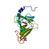
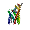
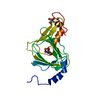
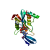
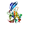
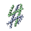
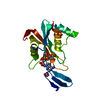
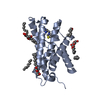
- PDB-3c5t: Crystal structure of the ligand-bound glucagon-like peptide-1 rec... -
+ Open data

- Basic information
| Entry | Database: PDB / ID: 3c5t | ||||||
|---|---|---|---|---|---|---|---|
| Title | Crystal structure of the ligand-bound glucagon-like peptide-1 receptor extracellular domain | ||||||
 Components Components |
| ||||||
 Keywords Keywords | Signaling protein/Signaling protein / ligand-bound G protein-coupled receptor extracellular domain / G-protein coupled receptor / Glycoprotein / Membrane / Transducer / Transmembrane / Amidation / Cleavage on pair of basic residues / Secreted / Signaling protein-Signaling protein COMPLEX | ||||||
| Function / homology |  Function and homology information Function and homology informationglucagon-like peptide 1 receptor activity / glucagon receptor activity / positive regulation of blood pressure / hormone secretion / post-translational protein targeting to membrane, translocation / response to psychosocial stress / regulation of heart contraction / peptide hormone binding / activation of adenylate cyclase activity / negative regulation of blood pressure ...glucagon-like peptide 1 receptor activity / glucagon receptor activity / positive regulation of blood pressure / hormone secretion / post-translational protein targeting to membrane, translocation / response to psychosocial stress / regulation of heart contraction / peptide hormone binding / activation of adenylate cyclase activity / negative regulation of blood pressure / hormone activity / regulation of blood pressure / transmembrane signaling receptor activity / Glucagon-type ligand receptors / Glucagon-like Peptide-1 (GLP1) regulates insulin secretion / toxin activity / positive regulation of cytosolic calcium ion concentration / adenylate cyclase-activating G protein-coupled receptor signaling pathway / G alpha (s) signalling events / learning or memory / cell surface receptor signaling pathway / extracellular region / membrane / plasma membrane Similarity search - Function | ||||||
| Biological species |  Homo sapiens (human) Homo sapiens (human) | ||||||
| Method |  X-RAY DIFFRACTION / SYNCHROTRON / MOLECULAR REPLACEMENT / Resolution: 2.1 Å X-RAY DIFFRACTION / SYNCHROTRON / MOLECULAR REPLACEMENT / Resolution: 2.1 Å | ||||||
 Authors Authors | Runge, S. | ||||||
 Citation Citation | Journal: J.Biol.Chem. / Year: 2008 Title: Crystal Structure of the Ligand-bound Glucagon-like Peptide-1 Receptor Extracellular Domain Authors: Runge, S. / Thogersen, H. / Madsen, K. / Lau, J. / Rudolph, R. | ||||||
| History |
|
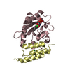
- Structure visualization
| Structure viewer | Molecule: MolmilJmol/JSmol |
|---|
- Downloads & links
-Download
| PDBx/mmCIF format | 3c5t.cif.gz | 46 KB | Display | PDBx/mmCIF format |
|---|---|---|---|---|
| PDB format | pdb3c5t.ent.gz | 31.3 KB | Display | PDB format |
| PDBx/mmJSON format | 3c5t.json.gz | Tree view | PDBx/mmJSON format | |
| Others |  Other downloads Other downloads |
-Validation report
| Arichive directory | https://data.pdbj.org/pub/pdb/validation_reports/c5/3c5tftp://data.pdbj.org/pub/pdb/validation_reports/c5/3c5t | HTTPS FTP |
|---|
-Related structure data
| Related structure data | 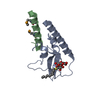 3c59SC S: Starting model for refinement C: citing same article ( |
|---|---|
| Similar structure data |

-Links
 PDBj
PDBj


- Assembly
| Deposited unit | 
| ||||||||
|---|---|---|---|---|---|---|---|---|---|
| 1 |
| ||||||||
| Unit cell |
|
-Components
| #1: Protein | Mass: 14325.841 Da / Num. of mol.: 1 Fragment: N-terminal extracellular domain, UNP residues 24-145 Source method: isolated from a genetically manipulated source Source: (gene. exp.) Homo sapiens (human) / Gene: Glucagon-like peptide-1 receptor(GLP1R) / Plasmid: pET15b / Species (production host): Escherichia coli / Production host:  |
|---|---|
| #2: Protein/peptide | Mass: 3373.765 Da / Num. of mol.: 1 / Fragment: UNP residues 56-86 / Source method: obtained synthetically Details: Exendin-4(9-39) was generated by chemical synthesis; this sequence occurs naturally in Heloderma suspectum. References: UniProt: P26349 |
| #3: Sugar | ChemComp-10M / 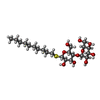  Type: D-saccharide / Mass: 498.628 Da / Num. of mol.: 1 Type: D-saccharide / Mass: 498.628 Da / Num. of mol.: 1Source method: isolated from a genetically manipulated source Formula: C22H42O10S / Comment: detergent*YM |
| #4: Water | ChemComp-HOH /  Mass: 18.015 Da / Num. of mol.: 126 / Source method: isolated from a natural source / Formula: H2O Mass: 18.015 Da / Num. of mol.: 126 / Source method: isolated from a natural source / Formula: H2O |
| Has protein modification | Y |
-Experimental details
-Experiment
| Experiment | Method: X-RAY DIFFRACTION / Number of used crystals: 1 |
|---|
- Sample preparation
| Crystal | Density Matthews: 4.12 Å3/Da / Density % sol: 70.18 % |
|---|---|
| Crystal grow | Temperature: 293 K / Method: vapor diffusion, hanging drop / pH: 8.5 Details: 0.1M Tris-HCl pH 8.5, 0.1M MgCl2, 0.4M MgTartrate, 9mM n-Decyl-beta-D-thiomaltoside, VAPOR DIFFUSION, HANGING DROP, temperature 293K |
-Data collection
| Diffraction | Mean temperature: 100 K |
|---|---|
| Diffraction source | Source: SYNCHROTRON / Site: MAX II  / Beamline: I911-3 / Wavelength: 1 Å / Beamline: I911-3 / Wavelength: 1 Å |
| Detector | Type: MARMOSAIC 225 mm CCD / Detector: CCD / Date: Nov 14, 2006 / Details: mirrors |
| Radiation | Protocol: SINGLE WAVELENGTH / Monochromatic (M) / Laue (L): M / Scattering type: x-ray |
| Radiation wavelength | Wavelength: 1 Å / Relative weight: 1 |
| Reflection | Resolution: 2.1→40 Å / Num. all: 17572 / Num. obs: 17273 / % possible obs: 98.3 % / Observed criterion σ(F): 0 / Observed criterion σ(I): 0 / Redundancy: 10.9 % / Rmerge(I) obs: 0.061 / Net I/σ(I): 27.1 |
| Reflection shell | Resolution: 2.1→2.2 Å / Redundancy: 11.1 % / Rmerge(I) obs: 0.395 / Mean I/σ(I) obs: 7.3 / Num. unique all: 2205 / % possible all: 97.8 |
- Processing
| Software |
| ||||||||||||||||||||||||||||||||||||||||||||||||||||||||||||||||||||||||||||||||||||||||||||||||||||||||||||||||||||||||||||||||||||||||||||||||||||||||||||||||||||||||||
|---|---|---|---|---|---|---|---|---|---|---|---|---|---|---|---|---|---|---|---|---|---|---|---|---|---|---|---|---|---|---|---|---|---|---|---|---|---|---|---|---|---|---|---|---|---|---|---|---|---|---|---|---|---|---|---|---|---|---|---|---|---|---|---|---|---|---|---|---|---|---|---|---|---|---|---|---|---|---|---|---|---|---|---|---|---|---|---|---|---|---|---|---|---|---|---|---|---|---|---|---|---|---|---|---|---|---|---|---|---|---|---|---|---|---|---|---|---|---|---|---|---|---|---|---|---|---|---|---|---|---|---|---|---|---|---|---|---|---|---|---|---|---|---|---|---|---|---|---|---|---|---|---|---|---|---|---|---|---|---|---|---|---|---|---|---|---|---|---|---|---|---|
| Refinement | Method to determine structure: MOLECULAR REPLACEMENT Starting model: PDB entry 3C59 without the ligand present Resolution: 2.1→36.5 Å / Cor.coef. Fo:Fc: 0.943 / Cor.coef. Fo:Fc free: 0.919 / SU B: 6.608 / SU ML: 0.093 / TLS residual ADP flag: LIKELY RESIDUAL / Isotropic thermal model: Isotropic / Cross valid method: THROUGHOUT / ESU R: 0.142 / ESU R Free: 0.14 / Stereochemistry target values: MAXIMUM LIKELIHOOD / Details: HYDROGENS HAVE BEEN ADDED IN THE RIDING POSITIONS
| ||||||||||||||||||||||||||||||||||||||||||||||||||||||||||||||||||||||||||||||||||||||||||||||||||||||||||||||||||||||||||||||||||||||||||||||||||||||||||||||||||||||||||
| Solvent computation | Ion probe radii: 0.8 Å / Shrinkage radii: 0.8 Å / VDW probe radii: 1.2 Å / Solvent model: MASK | ||||||||||||||||||||||||||||||||||||||||||||||||||||||||||||||||||||||||||||||||||||||||||||||||||||||||||||||||||||||||||||||||||||||||||||||||||||||||||||||||||||||||||
| Displacement parameters | Biso mean: 40.26 Å2
| ||||||||||||||||||||||||||||||||||||||||||||||||||||||||||||||||||||||||||||||||||||||||||||||||||||||||||||||||||||||||||||||||||||||||||||||||||||||||||||||||||||||||||
| Refinement step | Cycle: LAST / Resolution: 2.1→36.5 Å
| ||||||||||||||||||||||||||||||||||||||||||||||||||||||||||||||||||||||||||||||||||||||||||||||||||||||||||||||||||||||||||||||||||||||||||||||||||||||||||||||||||||||||||
| Refine LS restraints |
| ||||||||||||||||||||||||||||||||||||||||||||||||||||||||||||||||||||||||||||||||||||||||||||||||||||||||||||||||||||||||||||||||||||||||||||||||||||||||||||||||||||||||||
| LS refinement shell | Resolution: 2.1→2.155 Å / Total num. of bins used: 20
| ||||||||||||||||||||||||||||||||||||||||||||||||||||||||||||||||||||||||||||||||||||||||||||||||||||||||||||||||||||||||||||||||||||||||||||||||||||||||||||||||||||||||||
| Refinement TLS params. | Method: refined / Refine-ID: X-RAY DIFFRACTION
| ||||||||||||||||||||||||||||||||||||||||||||||||||||||||||||||||||||||||||||||||||||||||||||||||||||||||||||||||||||||||||||||||||||||||||||||||||||||||||||||||||||||||||
| Refinement TLS group |
|
