 Movie
Movie Controller

[English] 日本語
 Yorodumi
- PDB-6vi9: Observing a ring-cleaving dioxygenase in action through a crystal... -
+ Open data

- Basic information
| Entry | Database: PDB / ID: 6vi9 | |||||||||||||||
|---|---|---|---|---|---|---|---|---|---|---|---|---|---|---|---|---|
| Title | Observing a ring-cleaving dioxygenase in action through a crystalline lens - an alkylperoxo bound structure | |||||||||||||||
 Components Components | 3-hydroxyanthranilate 3,4-dioxygenase | |||||||||||||||
 Keywords Keywords | OXIDOREDUCTASE / In crystallo reaction / Extradiol dioxygenase / NAD+ biosynthesis / Intermediate | |||||||||||||||
| Function / homology |  Function and homology information Function and homology information3-hydroxyanthranilate 3,4-dioxygenase / 3-hydroxyanthranilate 3,4-dioxygenase activity / : / L-tryptophan catabolic process / NAD+ biosynthetic process / quinolinate biosynthetic process / ferrous iron binding Similarity search - Function | |||||||||||||||
| Biological species |  Cupriavidus metallidurans (bacteria) Cupriavidus metallidurans (bacteria) | |||||||||||||||
| Method |  X-RAY DIFFRACTION / SYNCHROTRON / MOLECULAR REPLACEMENT / Resolution: 2.31 Å X-RAY DIFFRACTION / SYNCHROTRON / MOLECULAR REPLACEMENT / Resolution: 2.31 Å | |||||||||||||||
 Authors Authors | Wang, Y. / Liu, F. / Yang, Y. / Liu, A. | |||||||||||||||
| Funding support |  United States, 4items United States, 4items
| |||||||||||||||
 Citation Citation | Journal: Proc.Natl.Acad.Sci.USA / Year: 2020 Title: Observing 3-hydroxyanthranilate-3,4-dioxygenase in action through a crystalline lens. Authors: Wang, Y. / Liu, K.F. / Yang, Y. / Davis, I. / Liu, A. | |||||||||||||||
| History |
|
- Structure visualization
| Structure viewer | Molecule: MolmilJmol/JSmol |
|---|
- Downloads & links
-Download
| PDBx/mmCIF format | 6vi9.cif.gz | 55.8 KB | Display | PDBx/mmCIF format |
|---|---|---|---|---|
| PDB format | pdb6vi9.ent.gz | 36.8 KB | Display | PDB format |
| PDBx/mmJSON format | 6vi9.json.gz | Tree view | PDBx/mmJSON format | |
| Others |  Other downloads Other downloads |
-Validation report
| Arichive directory | https://data.pdbj.org/pub/pdb/validation_reports/vi/6vi9ftp://data.pdbj.org/pub/pdb/validation_reports/vi/6vi9 | HTTPS FTP |
|---|
-Related structure data
| Related structure data |  6vi5C  6vi6C  6vi7C  6vi8C  6viaC  6vibC  6x11C  1yfuS S: Starting model for refinement C: citing same article ( |
|---|---|
| Similar structure data |


-Links
 PDBj
PDBj


- Assembly
| Deposited unit | 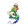
| ||||||||
|---|---|---|---|---|---|---|---|---|---|
| 1 | 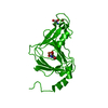
| ||||||||
| Unit cell |
|
-Components
| #1: Protein | Mass: 22590.377 Da / Num. of mol.: 1 Source method: isolated from a genetically manipulated source Source: (gene. exp.) Cupriavidus metallidurans (strain ATCC 43123 / DSM 2839 / NBRC 102507 / CH34) (bacteria)Strain: ATCC 43123 / DSM 2839 / NBRC 102507 / CH34 / Gene: nbaC, Rmet_5193 / Production host: References: UniProt: Q1LCS4, 3-hydroxyanthranilate 3,4-dioxygenase |
|---|---|
| #2: Chemical | ChemComp-FE2 /   Mass: 55.845 Da / Num. of mol.: 1 / Source method: obtained synthetically / Formula: Fe / Feature type: SUBJECT OF INVESTIGATION Mass: 55.845 Da / Num. of mol.: 1 / Source method: obtained synthetically / Formula: Fe / Feature type: SUBJECT OF INVESTIGATION |
| #3: Chemical | ChemComp-QXM / (  Mass: 237.955 Da / Num. of mol.: 1 / Source method: obtained synthetically / Formula: C7H4FeNO5 / Feature type: SUBJECT OF INVESTIGATION Mass: 237.955 Da / Num. of mol.: 1 / Source method: obtained synthetically / Formula: C7H4FeNO5 / Feature type: SUBJECT OF INVESTIGATION |
| #4: Chemical | ChemComp-TRS /   Mass: 122.143 Da / Num. of mol.: 1 / Source method: obtained synthetically / Formula: C4H12NO3 / Comment: pH buffer*YM Mass: 122.143 Da / Num. of mol.: 1 / Source method: obtained synthetically / Formula: C4H12NO3 / Comment: pH buffer*YM |
| #5: Water | ChemComp-HOH /  Mass: 18.015 Da / Num. of mol.: 97 / Source method: isolated from a natural source / Formula: H2O Mass: 18.015 Da / Num. of mol.: 97 / Source method: isolated from a natural source / Formula: H2O |
| Has ligand of interest | Y |
-Experimental details
-Experiment
| Experiment | Method: X-RAY DIFFRACTION / Number of used crystals: 1 |
|---|
- Sample preparation
| Crystal | Density Matthews: 2.47 Å3/Da / Density % sol: 50.19 % |
|---|---|
| Crystal grow | Temperature: 292 K / Method: vapor diffusion, sitting drop Details: 15% PEG8000, 0.1 M Tris-HCl, 0.2 M magnesium chloride, pH 8.5 |
-Data collection
| Diffraction | Mean temperature: 100 K / Serial crystal experiment: N |
|---|---|
| Diffraction source | Source: SYNCHROTRON / Site: APS / Beamline: 19-BM / Wavelength: 0.97919 Å |
| Detector | Type: DECTRIS PILATUS3 6M / Detector: PIXEL / Date: Nov 14, 2017 |
| Radiation | Monochromator: double crystal Si(111) / Protocol: SINGLE WAVELENGTH / Monochromatic (M) / Laue (L): M / Scattering type: x-ray |
| Radiation wavelength | Wavelength: 0.97919 Å / Relative weight: 1 |
| Reflection | Resolution: 2.3→50 Å / Num. obs: 11036 / % possible obs: 99.6 % / Redundancy: 16.7 % / Biso Wilson estimate: 26.38 Å2 / Rmerge(I) obs: 0.122 / Net I/σ(I): 6 |
| Reflection shell | Resolution: 2.3→2.34 Å / Redundancy: 17.4 % / Rmerge(I) obs: 0.996 / Num. unique obs: 478 / % possible all: 100 |
- Processing
| Software |
| ||||||||||||||||||||||||||||||||||||||||||||||||||||||||
|---|---|---|---|---|---|---|---|---|---|---|---|---|---|---|---|---|---|---|---|---|---|---|---|---|---|---|---|---|---|---|---|---|---|---|---|---|---|---|---|---|---|---|---|---|---|---|---|---|---|---|---|---|---|---|---|---|---|
| Refinement | Method to determine structure: MOLECULAR REPLACEMENT Starting model: PDB entry 1YFU Resolution: 2.31→30.55 Å / SU ML: 0.28 / Cross valid method: THROUGHOUT / σ(F): 1.35 / Phase error: 27.71
| ||||||||||||||||||||||||||||||||||||||||||||||||||||||||
| Solvent computation | Shrinkage radii: 0.9 Å / VDW probe radii: 1.11 Å | ||||||||||||||||||||||||||||||||||||||||||||||||||||||||
| Displacement parameters | Biso mean: 30.83 Å2 | ||||||||||||||||||||||||||||||||||||||||||||||||||||||||
| Refinement step | Cycle: LAST / Resolution: 2.31→30.55 Å
| ||||||||||||||||||||||||||||||||||||||||||||||||||||||||
| Refine LS restraints |
| ||||||||||||||||||||||||||||||||||||||||||||||||||||||||
| LS refinement shell |
|
