National Institutes of Health/National Institute of General Medical Sciences (NIH/NIGMS)
GM122510
United States
Swiss National Science Foundation
310030_144243
Switzerland
Citation
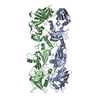
Journal: Structure / Year: 2017 Title: Refined Cryo-EM Structure of the T4 Tail Tube: Exploring the Lowest Dose Limit. Authors: Weili Zheng / Fengbin Wang / Nicholas M I Taylor / Ricardo C Guerrero-Ferreira / Petr G Leiman / Edward H Egelman / Abstract: The bacteriophage T4 contractile tail (containing a tube and sheath) was the first biological assembly reconstructed in three dimensions by electron microscopy at a resolution of ∼35 Å in 1968. A ...The bacteriophage T4 contractile tail (containing a tube and sheath) was the first biological assembly reconstructed in three dimensions by electron microscopy at a resolution of ∼35 Å in 1968. A single-particle reconstruction of the T4 baseplate was able to generate a 4.1 Å resolution map for the first two rings of the tube using the overall baseplate for alignment. We have now reconstructed the T4 tail tube at a resolution of 3.4 Å, more than a 1,000-fold increase in information content for the tube from 1968. We have used legacy software (Spider) to show that we can do better than the typical 2/3 Nyquist frequency. A reasonable map can be generated with only 1.5 electrons/Å using the higher dose images for alignment, but increasing the dose results in a better map, consistent with other reports that electron dose does not represent the main limitation on resolution in cryo-electron microscopy.
A: Tail tube protein gp19 R: Tail tube protein gp19 C: Tail tube protein gp19 B: Tail tube protein gp19 D: Tail tube protein gp19 E: Tail tube protein gp19 F: Tail tube protein gp19 G: Tail tube protein gp19 H: Tail tube protein gp19 I: Tail tube protein gp19 J: Tail tube protein gp19 K: Tail tube protein gp19 L: Tail tube protein gp19 M: Tail tube protein gp19 N: Tail tube protein gp19 O: Tail tube protein gp19 P: Tail tube protein gp19 Q: Tail tube protein gp19
Helical symmetry: (Circular symmetry: 6 / Dyad axis: no / N subunits divisor: 1 / Num. of operations: 4 / Rise per n subunits: 40.2 Å / Rotation per n subunits: 18.2 °)
-
Components
#1: Protein
Tailtubeproteingp19 / Gene product 19 / gp19
Mass: 18479.613 Da / Num. of mol.: 18 / Source method: isolated from a natural source Source: (natural) Enterobacteria phage T4 sensu lato (virus) References: UniProt: P13333
-
Experimental details
-
Experiment
Experiment
Method: ELECTRON MICROSCOPY
EM experiment
Aggregation state: FILAMENT / 3D reconstruction method: helical reconstruction
-
Sample preparation
Component
Name: Enterobacteria Phage T4 sensu lato tail tube protein gp19 filament Type: COMPLEX / Entity ID: all / Source: NATURAL
Molecular weight
Experimental value: NO
Source (natural)
Organism: Enterobacteria phage T4 sensu lato (virus)
Buffer solution
pH: 8
Specimen
Conc.: 1 mg/ml / Embedding applied: NO / Shadowing applied: NO / Staining applied: NO / Vitrification applied: YES
In the structure databanks used in Yorodumi, some data are registered as the other names, "COVID-19 virus" and "2019-nCoV". Here are the details of the virus and the list of structure data.
Jan 31, 2019. EMDB accession codes are about to change! (news from PDBe EMDB page)
EMDB accession codes are about to change! (news from PDBe EMDB page)
The allocation of 4 digits for EMDB accession codes will soon come to an end. Whilst these codes will remain in use, new EMDB accession codes will include an additional digit and will expand incrementally as the available range of codes is exhausted. The current 4-digit format prefixed with “EMD-” (i.e. EMD-XXXX) will advance to a 5-digit format (i.e. EMD-XXXXX), and so on. It is currently estimated that the 4-digit codes will be depleted around Spring 2019, at which point the 5-digit format will come into force.
The EM Navigator/Yorodumi systems omit the EMD- prefix.
Related info.:Q: What is EMD? / ID/Accession-code notation in Yorodumi/EM Navigator
Yorodumi is a browser for structure data from EMDB, PDB, SASBDB, etc.
This page is also the successor to EM Navigator detail page, and also detail information page/front-end page for Omokage search.
The word "yorodu" (or yorozu) is an old Japanese word meaning "ten thousand". "mi" (miru) is to see.
Related info.:EMDB / PDB / SASBDB / Comparison of 3 databanks / Yorodumi Search / Aug 31, 2016. New EM Navigator & Yorodumi / Yorodumi Papers / Jmol/JSmol / Function and homology information / Changes in new EM Navigator and Yorodumi
 Movie
Movie Controller


 Open data
 Basic information
 Components
 Keywords
 Function and homology information
 Enterobacteria phage T4 sensu lato (virus)
 Authors
 United States,
  Switzerland, 2items
  Citation
 Structure visualization
 Downloads & links
 Other downloads
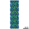


 PDBj
PDBj Assembly
 Sample preparation
 Electron microscopy imaging
 FIELD EMISSION GUN / Accelerating voltage: 300 kV / Illumination mode: FLOOD BEAM
 Processing