 Movie
Movie Controller


+ Open data

- Basic information
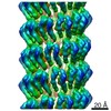
| Entry | Database: PDB / ID: 4ep0 | ||||||
|---|---|---|---|---|---|---|---|
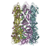
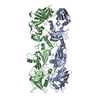
| Title | Structure of the bacteriophage C1 tail knob protein, gp12 | ||||||
 Components Components | Major tail protein | ||||||
 Keywords Keywords | STRUCTURAL PROTEIN / VIRAL PROTEIN | ||||||
| Function / homology | Distal tube protein, N-terminal / : / Caudoviral major tail protein N-terminus / Bacillus phage phi29, Tail knob protein gp9, C-terminal domain / identical protein binding / Major tail protein Function and homology information Function and homology information | ||||||
| Biological species |  Streptococcus phage C1 (virus) Streptococcus phage C1 (virus) | ||||||
| Method |  X-RAY DIFFRACTION / SYNCHROTRON / MOLECULAR REPLACEMENT / Resolution: 4 Å X-RAY DIFFRACTION / SYNCHROTRON / MOLECULAR REPLACEMENT / Resolution: 4 Å | ||||||
 Authors Authors | Aksyuk, A.A. / Rossmann, M.G. | ||||||
 Citation Citation | Journal: Proc Natl Acad Sci U S A / Year: 2012 Title: Structural investigations of a Podoviridae streptococcus phage C1, implications for the mechanism of viral entry. Authors: Anastasia A Aksyuk / Valorie D Bowman / Bärbel Kaufmann / Christopher Fields / Thomas Klose / Heather A Holdaway / Vincent A Fischetti / Michael G Rossmann /  Abstract: The Podoviridae phage C1 was one of the earliest isolated bacteriophages and the first virus documented to be active against streptococci. The icosahedral and asymmetric reconstructions of the virus ...The Podoviridae phage C1 was one of the earliest isolated bacteriophages and the first virus documented to be active against streptococci. The icosahedral and asymmetric reconstructions of the virus were calculated using cryo-electron microscopy. The capsid protein has an HK97 fold arranged into a T = 4 icosahedral lattice. The C1 tail is terminated with a ϕ29-like knob, surrounded by a skirt of twelve long appendages with novel morphology. Several C1 structural proteins have been identified, including a candidate for an appendage. The crystal structure of the knob has an N-terminal domain with a fold observed previously in tube forming proteins of Siphoviridae and Myoviridae phages. The structure of C1 suggests the mechanisms by which the virus digests the cell wall and ejects its genome. Although there is little sequence similarity to other phages, conservation of the structural proteins demonstrates a common origin of the head and tail, but more recent evolution of the appendages. | ||||||
| History |
|
- Structure visualization
| Structure viewer | Molecule: MolmilJmol/JSmol |
|---|
- Downloads & links
-Download
| PDBx/mmCIF format | 4ep0.cif.gz | 2.3 MB | Display | PDBx/mmCIF format |
|---|---|---|---|---|
| PDB format | pdb4ep0.ent.gz | 1.9 MB | Display | PDB format |
| PDBx/mmJSON format | 4ep0.json.gz | Tree view | PDBx/mmJSON format | |
| Others |  Other downloads Other downloads |
-Validation report
| Arichive directory | https://data.pdbj.org/pub/pdb/validation_reports/ep/4ep0ftp://data.pdbj.org/pub/pdb/validation_reports/ep/4ep0 | HTTPS FTP |
|---|
-Related structure data
| Related structure data | 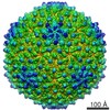 5445C 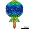 5446C  4eo2SC S: Starting model for refinement C: citing same article ( |
|---|---|
| Similar structure data |


-Links
 PDBj
PDBj- Assembly
| Deposited unit | 
| ||||||||||||||||||||||||||||||||||||||||||||||||||||||||||
|---|---|---|---|---|---|---|---|---|---|---|---|---|---|---|---|---|---|---|---|---|---|---|---|---|---|---|---|---|---|---|---|---|---|---|---|---|---|---|---|---|---|---|---|---|---|---|---|---|---|---|---|---|---|---|---|---|---|---|---|
| 1 | 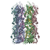
| ||||||||||||||||||||||||||||||||||||||||||||||||||||||||||
| 2 | 
| ||||||||||||||||||||||||||||||||||||||||||||||||||||||||||
| Unit cell |
| ||||||||||||||||||||||||||||||||||||||||||||||||||||||||||
| Noncrystallographic symmetry (NCS) | NCS domain:
NCS domain segments:
|