 Movie
Movie Controller

[English] 日本語
 Yorodumi
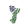
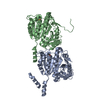
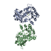
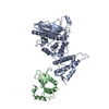
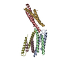
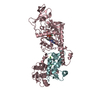
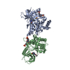
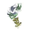
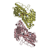
- PDB-3ktt: Atomic model of bovine TRiC CCT2(beta) subunit derived from a 4.0... -
+ Open data

- Basic information
| Entry | Database: PDB / ID: 3ktt | ||||||
|---|---|---|---|---|---|---|---|
| Title | Atomic model of bovine TRiC CCT2(beta) subunit derived from a 4.0 Angstrom cryo-EM map | ||||||
 Components Components | T-complex protein 1 subunit beta | ||||||
 Keywords Keywords | CHAPERONE / TRiC/CCT / CCT2(beta) / cryo-EM / Acetylation / ATP-binding / Cytoplasm / Nucleotide-binding | ||||||
| Function / homology |  Function and homology information Function and homology informationAssociation of TriC/CCT with target proteins during biosynthesis / RHOBTB2 GTPase cycle / RHOBTB1 GTPase cycle / zona pellucida receptor complex / scaRNA localization to Cajal body / positive regulation of telomerase RNA localization to Cajal body / chaperonin-containing T-complex / binding of sperm to zona pellucida / Neutrophil degranulation / Cooperation of PDCL (PhLP1) and TRiC/CCT in G-protein beta folding ...Association of TriC/CCT with target proteins during biosynthesis / RHOBTB2 GTPase cycle / RHOBTB1 GTPase cycle / zona pellucida receptor complex / scaRNA localization to Cajal body / positive regulation of telomerase RNA localization to Cajal body / chaperonin-containing T-complex / binding of sperm to zona pellucida / Neutrophil degranulation / Cooperation of PDCL (PhLP1) and TRiC/CCT in G-protein beta folding / chaperone-mediated protein complex assembly / Hydrolases; Acting on acid anhydrides; In phosphorus-containing anhydrides / positive regulation of telomere maintenance via telomerase / ATP-dependent protein folding chaperone / : / protein folding / cell body / microtubule / protein stabilization / ubiquitin protein ligase binding / ATP hydrolysis activity / ATP binding / metal ion binding Similarity search - Function | ||||||
| Biological species |  | ||||||
| Method | ELECTRON MICROSCOPY / single particle reconstruction / cryo EM / Resolution: 4 Å | ||||||
 Authors Authors | Cong, Y. / Baker, M.L. / Ludtke, S.J. / Frydman, J. / Chiu, W. | ||||||
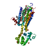
 Citation Citation | Journal: Proc Natl Acad Sci U S A / Year: 2010 Title: 4.0-A resolution cryo-EM structure of the mammalian chaperonin TRiC/CCT reveals its unique subunit arrangement. Authors: Yao Cong / Matthew L Baker / Joanita Jakana / David Woolford / Erik J Miller / Stefanie Reissmann / Ramya N Kumar / Alyssa M Redding-Johanson / Tanveer S Batth / Aindrila Mukhopadhyay / ...Authors: Yao Cong / Matthew L Baker / Joanita Jakana / David Woolford / Erik J Miller / Stefanie Reissmann / Ramya N Kumar / Alyssa M Redding-Johanson / Tanveer S Batth / Aindrila Mukhopadhyay / Steven J Ludtke / Judith Frydman / Wah Chiu /  Abstract: The essential double-ring eukaryotic chaperonin TRiC/CCT (TCP1-ring complex or chaperonin containing TCP1) assists the folding of approximately 5-10% of the cellular proteome. Many TRiC substrates ...The essential double-ring eukaryotic chaperonin TRiC/CCT (TCP1-ring complex or chaperonin containing TCP1) assists the folding of approximately 5-10% of the cellular proteome. Many TRiC substrates cannot be folded by other chaperonins from prokaryotes or archaea. These unique folding properties are likely linked to TRiC's unique heterooligomeric subunit organization, whereby each ring consists of eight different paralogous subunits in an arrangement that remains uncertain. Using single particle cryo-EM without imposing symmetry, we determined the mammalian TRiC structure at 4.7-A resolution. This revealed the existence of a 2-fold axis between its two rings resulting in two homotypic subunit interactions across the rings. A subsequent 2-fold symmetrized map yielded a 4.0-A resolution structure that evinces the densities of a large fraction of side chains, loops, and insertions. These features permitted unambiguous identification of all eight individual subunits, despite their sequence similarity. Independent biochemical near-neighbor analysis supports our cryo-EM derived TRiC subunit arrangement. We obtained a Calpha backbone model for each subunit from an initial homology model refined against the cryo-EM density. A subsequently optimized atomic model for a subunit showed approximately 95% of the main chain dihedral angles in the allowable regions of the Ramachandran plot. The determination of the TRiC subunit arrangement opens the way to understand its unique function and mechanism. In particular, an unevenly distributed positively charged wall lining the closed folding chamber of TRiC differs strikingly from that of prokaryotic and archaeal chaperonins. These interior surface chemical properties likely play an important role in TRiC's cellular substrate specificity. #1: Journal: To be PublishedTitle: To be published Authors: Cong, Y. / Baker, M.L. / Jakana, J. / Woolford, D. / Miller, E.J. / Reissmann, S. / Kumar, R.N. / Redding-Johanson, A.M. / Batth, T.S. / Mukhopadhyay, A. / Ludtke, S.J. / Frydman, J. / Chiu, W. | ||||||
| History |
|
- Structure visualization
| Movie |
Movie viewer |
|---|---|
| Structure viewer | Molecule: MolmilJmol/JSmol |
- Downloads & links
-Download
| PDBx/mmCIF format | 3ktt.cif.gz | 96.9 KB | Display | PDBx/mmCIF format |
|---|---|---|---|---|
| PDB format | pdb3ktt.ent.gz | 74.7 KB | Display | PDB format |
| PDBx/mmJSON format | 3ktt.json.gz | Tree view | PDBx/mmJSON format | |
| Others |  Other downloads Other downloads |
-Validation report
| Arichive directory | https://data.pdbj.org/pub/pdb/validation_reports/kt/3kttftp://data.pdbj.org/pub/pdb/validation_reports/kt/3ktt | HTTPS FTP |
|---|
-Related structure data
| Related structure data | 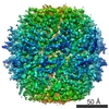 5145MC 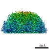 5148MC 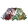 3iygC M: map data used to model this data C: citing same article ( |
|---|---|
| Similar structure data |
-Links
 PDBj
PDBj


- Assembly
| Deposited unit | 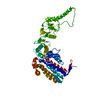
|
|---|---|
| 1 |
|
| Symmetry | Point symmetry: (Schoenflies symbol: D8 (2x8 fold dihedral)) |
-Components
| #1: Protein | Mass: 55107.234 Da / Num. of mol.: 1 / Source method: isolated from a natural source / Source: (natural) |
|---|---|
| Has protein modification | N |
-Experimental details
-Experiment
| Experiment | Method: ELECTRON MICROSCOPY |
|---|---|
| EM experiment | Aggregation state: PARTICLE / 3D reconstruction method: single particle reconstruction |
- Sample preparation
| Component | Name: CCT2(beta) subunit in the both-ring closed bovine TRiC complex Type: COMPLEX |
|---|---|
| Specimen | Embedding applied: NO / Shadowing applied: NO / Staining applied: NO / Vitrification applied: YES |
| Vitrification | Cryogen name: ETHANE / Details: vitrification using ethane as cryogen |
- Electron microscopy imaging
| Microscopy | Model: JEOL 3200FSC / Date: Aug 1, 2007 |
|---|---|
| Electron gun | Electron source:  FIELD EMISSION GUN / Accelerating voltage: 300 kV / Illumination mode: FLOOD BEAM FIELD EMISSION GUN / Accelerating voltage: 300 kV / Illumination mode: FLOOD BEAM |
| Electron lens | Mode: BRIGHT FIELD / Nominal magnification: 50000 X / Nominal defocus max: 3000 nm / Nominal defocus min: 1000 nm / Cs: 4.1 mm |
| Specimen holder | Temperature: 101 K / Tilt angle max: 0 ° / Tilt angle min: 0 ° |
| Image recording | Electron dose: 18 e/Å2 / Film or detector model: KODAK SO-163 FILM |
- Processing
| EM software |
| |||||||||||||||
|---|---|---|---|---|---|---|---|---|---|---|---|---|---|---|---|---|
| CTF correction | Details: FSC at 0.5 cut-off | |||||||||||||||
| Symmetry | Point symmetry: C2 (2 fold cyclic) | |||||||||||||||
| 3D reconstruction | Method: Projection matching / Resolution: 4 Å / Num. of particles: 101000 / Actual pixel size: 1.2 Å Details: Single particle 3D reconstruction using EMAN1.8+ with our recently developed 2-D fast rotation matching method (FRM2D) for the image alignment. Symmetry type: POINT | |||||||||||||||
| Atomic model building | Protocol: FLEXIBLE FIT / Space: REAL Details: METHOD--Local refinement, Flexible fitting REFINEMENT PROTOCOL--Local refinement, Flexible fitting | |||||||||||||||
| Atomic model building | Details: Homology model | |||||||||||||||
| Refinement step | Cycle: LAST
|
