[English] 日本語
 Yorodumi
- EMDB-9390: Structure of the assembled ATPase EscN from the enteropathogenic ... -
+ Open data
- Basic information
| Entry | Database: EMDB / ID: EMD-9390 | ||||||||||||
|---|---|---|---|---|---|---|---|---|---|---|---|---|---|
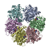
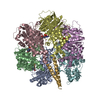
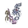
| Title | Structure of the assembled ATPase EscN from the enteropathogenic E. coli (EPEC) type III secretion system | ||||||||||||
 Map data Map data | Assembled ATPase EscN from the enteropathogenic E. coli (EPEC) type III secretion system | ||||||||||||
 Sample Sample |
| ||||||||||||
 Keywords Keywords | ATPase / Type III Secretion System / ADP / hexamer / HYDROLASE | ||||||||||||
| Function / homology |  Function and homology information Function and homology informationprotein-secreting ATPase / type III protein secretion system complex / protein secretion by the type III secretion system / proton-transporting ATPase activity, rotational mechanism / proton-transporting ATP synthase activity, rotational mechanism / ATP hydrolysis activity / ATP binding / metal ion binding / cytoplasm Similarity search - Function | ||||||||||||
| Biological species |   | ||||||||||||
| Method | single particle reconstruction / cryo EM / Resolution: 3.34 Å | ||||||||||||
 Authors Authors | Majewski DD / Worrall LJ | ||||||||||||
| Funding support |  Canada, Canada,  United States, 3 items United States, 3 items
| ||||||||||||
 Citation Citation |  Journal: Nat Commun / Year: 2019 Journal: Nat Commun / Year: 2019Title: Cryo-EM structure of the homohexameric T3SS ATPase-central stalk complex reveals rotary ATPase-like asymmetry. Authors: Dorothy D Majewski / Liam J Worrall / Chuan Hong / Claire E Atkinson / Marija Vuckovic / Nobuhiko Watanabe / Zhiheng Yu / Natalie C J Strynadka /   Abstract: Many Gram-negative bacteria, including causative agents of dysentery, plague, and typhoid fever, rely on a type III secretion system - a multi-membrane spanning syringe-like apparatus - for their ...Many Gram-negative bacteria, including causative agents of dysentery, plague, and typhoid fever, rely on a type III secretion system - a multi-membrane spanning syringe-like apparatus - for their pathogenicity. The cytosolic ATPase complex of this injectisome is proposed to play an important role in energizing secretion events and substrate recognition. We present the 3.3 Å resolution cryo-EM structure of the enteropathogenic Escherichia coli ATPase EscN in complex with its central stalk EscO. The structure shows an asymmetric pore with different functional states captured in its six catalytic sites, details directly supporting a rotary catalytic mechanism analogous to that of the heterohexameric F/V-ATPases despite its homohexameric nature. Situated at the C-terminal opening of the EscN pore is one molecule of EscO, with primary interaction mediated through an electrostatic interface. The EscN-EscO structure provides significant atomic insights into how the ATPase contributes to type III secretion, including torque generation and binding of chaperone/substrate complexes. | ||||||||||||
| History |
|
- Structure visualization
| Movie |
 Movie viewer Movie viewer |
|---|---|
| Structure viewer | EM map:  SurfView SurfView Molmil Molmil Jmol/JSmol Jmol/JSmol |
| Supplemental images |
- Downloads & links
-EMDB archive
| Map data |  emd_9390.map.gz emd_9390.map.gz | 3.5 MB |  EMDB map data format EMDB map data format | |
|---|---|---|---|---|
| Header (meta data) |  emd-9390-v30.xml emd-9390-v30.xml emd-9390.xml emd-9390.xml | 13.9 KB 13.9 KB | Display Display |  EMDB header EMDB header |
| FSC (resolution estimation) |  emd_9390_fsc.xml emd_9390_fsc.xml | 7.2 KB | Display |  FSC data file FSC data file |
| Images |  emd_9390.png emd_9390.png | 84.5 KB | ||
| Filedesc metadata |  emd-9390.cif.gz emd-9390.cif.gz | 6.3 KB | ||
| Archive directory |  http://ftp.pdbj.org/pub/emdb/structures/EMD-9390 http://ftp.pdbj.org/pub/emdb/structures/EMD-9390 ftp://ftp.pdbj.org/pub/emdb/structures/EMD-9390 ftp://ftp.pdbj.org/pub/emdb/structures/EMD-9390 | HTTPS FTP |
-Related structure data
| Related structure data |  6njoMC  9391C  6njpC C: citing same article ( M: atomic model generated by this map |
|---|---|
| Similar structure data |
- Links
Links
| EMDB pages |  EMDB (EBI/PDBe) / EMDB (EBI/PDBe) /  EMDataResource EMDataResource |
|---|---|
| Related items in Molecule of the Month |
- Map
Map
| File |  Download / File: emd_9390.map.gz / Format: CCP4 / Size: 30.5 MB / Type: IMAGE STORED AS FLOATING POINT NUMBER (4 BYTES) Download / File: emd_9390.map.gz / Format: CCP4 / Size: 30.5 MB / Type: IMAGE STORED AS FLOATING POINT NUMBER (4 BYTES) | ||||||||||||||||||||||||||||||||||||||||||||||||||||||||||||
|---|---|---|---|---|---|---|---|---|---|---|---|---|---|---|---|---|---|---|---|---|---|---|---|---|---|---|---|---|---|---|---|---|---|---|---|---|---|---|---|---|---|---|---|---|---|---|---|---|---|---|---|---|---|---|---|---|---|---|---|---|---|
| Annotation | Assembled ATPase EscN from the enteropathogenic E. coli (EPEC) type III secretion system | ||||||||||||||||||||||||||||||||||||||||||||||||||||||||||||
| Projections & slices | Image control
Images are generated by Spider. | ||||||||||||||||||||||||||||||||||||||||||||||||||||||||||||
| Voxel size | X=Y=Z: 1.02 Å | ||||||||||||||||||||||||||||||||||||||||||||||||||||||||||||
| Density |
| ||||||||||||||||||||||||||||||||||||||||||||||||||||||||||||
| Symmetry | Space group: 1 | ||||||||||||||||||||||||||||||||||||||||||||||||||||||||||||
| Details | EMDB XML:
CCP4 map header:
| ||||||||||||||||||||||||||||||||||||||||||||||||||||||||||||
-Supplemental data
- Sample components
-Entire : Homohexameric complex of ATPase EscN
| Entire | Name: Homohexameric complex of ATPase EscN |
|---|---|
| Components |
|
-Supramolecule #1: Homohexameric complex of ATPase EscN
| Supramolecule | Name: Homohexameric complex of ATPase EscN / type: complex / ID: 1 / Parent: 0 / Macromolecule list: #1 |
|---|---|
| Source (natural) | Organism:  |
-Macromolecule #1: Translocator EscN
| Macromolecule | Name: Translocator EscN / type: protein_or_peptide / ID: 1 / Number of copies: 6 / Enantiomer: LEVO |
|---|---|
| Source (natural) | Organism:  Strain: E2348/69 / EPEC |
| Molecular weight | Theoretical: 49.196566 KDa |
| Recombinant expression | Organism:  |
| Sequence | String: GSHMISEHDS VLEKYPRIQK VLNSTVPALS LNSSTRYEGK IINIGGTIIK ARLPKARIGA FYKIEPSQRL AEVIAIDEDE VFLLPFEHV SGMYCGQWLS YQGDEFKIRV GDALLGRLID GIGRPMESNI VAPYLPFERS LYAEPPDPLL RQVIDQPFIL G VRAIDGLL ...String: GSHMISEHDS VLEKYPRIQK VLNSTVPALS LNSSTRYEGK IINIGGTIIK ARLPKARIGA FYKIEPSQRL AEVIAIDEDE VFLLPFEHV SGMYCGQWLS YQGDEFKIRV GDALLGRLID GIGRPMESNI VAPYLPFERS LYAEPPDPLL RQVIDQPFIL G VRAIDGLL TCGIGQRIGI FAGSGVGKST LLGMICNGAS ADIIVLALIG ERGREVNEFL ALLPQSTLSK CVLVVTTSDR PA LERMKAA FTATTIAEYF RDQGKNVLLM MDSVTRYARA ARDVGLASGE PDVRGGFPPS VFSSLPKLLE RAGPAPKGSI TAI YTVLLE SDNVNDPIGD EVRSILDGHI VLTRELAEEN HFPAIDIGLS ASRVMHNVVT SEHLRAAAEC KKLIATYKNV ELLI RIGEY TMGQDPEADK AIKNRKLIQN FIQQSTKDIS SYEKTIESLF KVVA UniProtKB: protein-secreting ATPase |
-Macromolecule #2: ADENOSINE-5'-DIPHOSPHATE
| Macromolecule | Name: ADENOSINE-5'-DIPHOSPHATE / type: ligand / ID: 2 / Number of copies: 4 / Formula: ADP |
|---|---|
| Molecular weight | Theoretical: 427.201 Da |
| Chemical component information |  ChemComp-ADP: |
-Macromolecule #3: MAGNESIUM ION
| Macromolecule | Name: MAGNESIUM ION / type: ligand / ID: 3 / Number of copies: 4 / Formula: MG |
|---|---|
| Molecular weight | Theoretical: 24.305 Da |
-Macromolecule #4: ALUMINUM FLUORIDE
| Macromolecule | Name: ALUMINUM FLUORIDE / type: ligand / ID: 4 / Number of copies: 4 / Formula: AF3 |
|---|---|
| Molecular weight | Theoretical: 83.977 Da |
| Chemical component information |  ChemComp-AF3: |
-Macromolecule #5: water
| Macromolecule | Name: water / type: ligand / ID: 5 / Number of copies: 20 / Formula: HOH |
|---|---|
| Molecular weight | Theoretical: 18.015 Da |
| Chemical component information |  ChemComp-HOH: |
-Experimental details
-Structure determination
| Method | cryo EM |
|---|---|
 Processing Processing | single particle reconstruction |
| Aggregation state | particle |
- Sample preparation
| Concentration | 0.34 mg/mL |
|---|---|
| Buffer | pH: 7.5 |
| Grid | Details: unspecified |
| Vitrification | Cryogen name: ETHANE / Chamber humidity: 100 % / Instrument: FEI VITROBOT MARK IV |
- Electron microscopy
| Microscope | FEI TITAN KRIOS |
|---|---|
| Image recording | Film or detector model: GATAN K2 SUMMIT (4k x 4k) / Average electron dose: 57.67 e/Å2 |
| Electron beam | Acceleration voltage: 300 kV / Electron source:  FIELD EMISSION GUN FIELD EMISSION GUN |
| Electron optics | Illumination mode: FLOOD BEAM / Imaging mode: BRIGHT FIELD |
| Experimental equipment |  Model: Titan Krios / Image courtesy: FEI Company |
+ Image processing
-Atomic model buiding 1
| Initial model |
| ||||||
|---|---|---|---|---|---|---|---|
| Details | phenix.real_space_refine | ||||||
| Refinement | Space: REAL / Protocol: FLEXIBLE FIT | ||||||
| Output model |  PDB-6njo: |
 Movie
Movie Controller


 Z (Sec.)
 Y (Row.)
 X (Col.)


