 Movie
Movie Controller

[English] 日本語
 Yorodumi
- PDB-1gl7: Plasmid coupling protein TrwB in complex with the non-hydrolisabl... -
+ Open data

- Basic information
| Entry | Database: PDB / ID: 1gl7 | ||||||
|---|---|---|---|---|---|---|---|
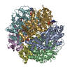
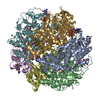
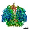
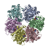
| Title | Plasmid coupling protein TrwB in complex with the non-hydrolisable ATP-analogue ADPNP. | ||||||
 Components Components | CONJUGAL TRANSFER PROTEIN TRWB | ||||||
 Keywords Keywords | COUPLING PROTEIN / TYPE IV SECRETION SYSTEM CONJUGATIVE COUPLING PROTEIN FROM PLASMID | ||||||
| Function / homology |  Function and homology information Function and homology informationvesicle-fusing ATPase / nucleotide binding / metal ion binding / identical protein binding / plasma membrane Similarity search - Function | ||||||
| Biological species |  | ||||||
| Method |  X-RAY DIFFRACTION / SYNCHROTRON / MOLECULAR REPLACEMENT / Resolution: 3 Å X-RAY DIFFRACTION / SYNCHROTRON / MOLECULAR REPLACEMENT / Resolution: 3 Å | ||||||
 Authors Authors | Gomis-Ruth, F.X. / Moncalian, G. / De La cruz, F. / Coll, M. | ||||||
 Citation Citation | Journal: Nature / Year: 2001 Title: The Bacterial Conjugation Protein Trwb Resembles Ring Helicases and F1-ATPase Authors: Gomis-Ruth, F.X. / Moncalian, G. / Perez-Luque, R. / Gonzalez, A. / Cabezon, E. / De La Cruz, F. / Coll, M. #1: Journal: J.Biol.Chem. / Year: 2002 Title: Conjugative Plasmid Protein Trwb, an Integral Membrane Type Iv Secretion System Coupling Protein: Detailed Structural Features and Mapping of the Active Site Cleft. Authors: Gomis-Ruth, F. / Moncalian, G. / De La Cruz, F. / Coll, M. | ||||||
| History |
|
- Structure visualization
| Structure viewer | Molecule: MolmilJmol/JSmol |
|---|
- Downloads & links
-Download
| PDBx/mmCIF format | 1gl7.cif.gz | 487.5 KB | Display | PDBx/mmCIF format |
|---|---|---|---|---|
| PDB format | pdb1gl7.ent.gz | 401.1 KB | Display | PDB format |
| PDBx/mmJSON format | 1gl7.json.gz | Tree view | PDBx/mmJSON format | |
| Others |  Other downloads Other downloads |
-Validation report
| Arichive directory | https://data.pdbj.org/pub/pdb/validation_reports/gl/1gl7ftp://data.pdbj.org/pub/pdb/validation_reports/gl/1gl7 | HTTPS FTP |
|---|
-Related structure data
| Related structure data | 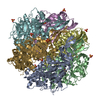 1e9rSC 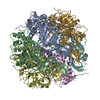 1e9sC  1gkiC  1gl6C S: Starting model for refinement C: citing same article ( |
|---|---|
| Similar structure data |


-Links
 PDBj
PDBj


- Assembly
| Deposited unit | 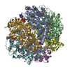
| ||||||||
|---|---|---|---|---|---|---|---|---|---|
| 1 |
| ||||||||
| Unit cell |
|
-Components
| #1: Protein | Mass: 48671.469 Da / Num. of mol.: 6 / Fragment: CYTOSOLIC FRAGMENT, RESIDUES 71-507 / Mutation: YES Source method: isolated from a genetically manipulated source Details: COMPLEX WITH ADPNP. / Source: (gene. exp.) #2: Chemical | ChemComp-ANP /   Mass: 506.196 Da / Num. of mol.: 6 / Source method: obtained synthetically / Formula: C10H17N6O12P3 / Comment: AMP-PNP, energy-carrying molecule analogue*YM Mass: 506.196 Da / Num. of mol.: 6 / Source method: obtained synthetically / Formula: C10H17N6O12P3 / Comment: AMP-PNP, energy-carrying molecule analogue*YM#3: Chemical | ChemComp-CL / |   Mass: 35.453 Da / Num. of mol.: 1 / Source method: obtained synthetically / Formula: Cl Mass: 35.453 Da / Num. of mol.: 1 / Source method: obtained synthetically / Formula: Cl#4: Water | ChemComp-HOH / |  Mass: 18.015 Da / Num. of mol.: 192 / Source method: isolated from a natural source / Formula: H2O Mass: 18.015 Da / Num. of mol.: 192 / Source method: isolated from a natural source / Formula: H2OCompound details | CHAIN A, B, D, E, F, G FIRST 70 RESIDUES DELETED | |
|---|
-Experimental details
-Experiment
| Experiment | Method: X-RAY DIFFRACTION / Number of used crystals: 1 |
|---|
- Sample preparation
| Crystal | Density Matthews: 2.9 Å3/Da / Density % sol: 57 % |
|---|---|
| Crystal grow | pH: 7.5 / Details: pH 7.50 |
-Data collection
| Diffraction | Mean temperature: 100 K |
|---|---|
| Diffraction source | Source: SYNCHROTRON / Site: EMBL/DESY, HAMBURG  / Beamline: BW7B / Wavelength: 1.05 / Beamline: BW7B / Wavelength: 1.05 |
| Radiation | Protocol: SINGLE WAVELENGTH / Monochromatic (M) / Laue (L): M / Scattering type: x-ray |
| Radiation wavelength | Wavelength: 1.05 Å / Relative weight: 1 |
| Reflection | Resolution: 2.91→52 Å / Num. obs: 67095 / % possible obs: 90 % / Redundancy: 3.5 % / Biso Wilson estimate: 61 Å2 / Rmerge(I) obs: 0.107 / Net I/σ(I): 5.3 |
| Reflection shell | Resolution: 2.91→3.07 Å / Redundancy: 3.5 % / Rmerge(I) obs: 0.453 / Mean I/σ(I) obs: 1.6 / % possible all: 66 |
- Processing
| Software |
| ||||||||||||||||||||||||||||||||||||||||||||||||||||||||||||
|---|---|---|---|---|---|---|---|---|---|---|---|---|---|---|---|---|---|---|---|---|---|---|---|---|---|---|---|---|---|---|---|---|---|---|---|---|---|---|---|---|---|---|---|---|---|---|---|---|---|---|---|---|---|---|---|---|---|---|---|---|---|
| Refinement | Method to determine structure: MOLECULAR REPLACEMENT Starting model: PDB ENTRY 1E9R Resolution: 3→50 Å / Isotropic thermal model: RESTRAINED / Cross valid method: THROUGHOUT / σ(F): 0 Details: FREE RFACTOR USED UNTIL PENULTIMATE REFINEMENT CYCLE. SAME INDICES AS IN 1GKI AND 1E9R USED. THERE IS NCS IN THE A.U. THE SIX PROTEIN CHAINS (A-G) ARE CHEMICALLY IDENTICAL. SOME N- AND C- ...Details: FREE RFACTOR USED UNTIL PENULTIMATE REFINEMENT CYCLE. SAME INDICES AS IN 1GKI AND 1E9R USED. THERE IS NCS IN THE A.U. THE SIX PROTEIN CHAINS (A-G) ARE CHEMICALLY IDENTICAL. SOME N- AND C-TERMINAL RESIDUES ARE DISORDERED AND HAVE NOT BEEN TRACED.
| ||||||||||||||||||||||||||||||||||||||||||||||||||||||||||||
| Displacement parameters | Biso mean: 64.3 Å2 | ||||||||||||||||||||||||||||||||||||||||||||||||||||||||||||
| Refinement step | Cycle: LAST / Resolution: 3→50 Å
| ||||||||||||||||||||||||||||||||||||||||||||||||||||||||||||
| Refine LS restraints |
|
