- EMDB-8279: Structure of RelA bound to ribosome in absence of A/R tRNA (Struc... -
+
データを開く
IDまたはキーワード:
読み込み中...
-
基本情報
登録情報
データベース: EMDB / ID: EMD-8279
タイトル
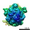
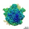
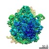
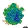
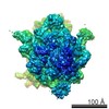
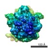
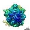
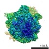
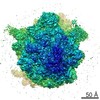
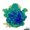
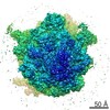
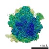
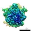
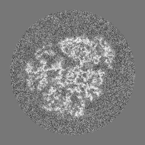
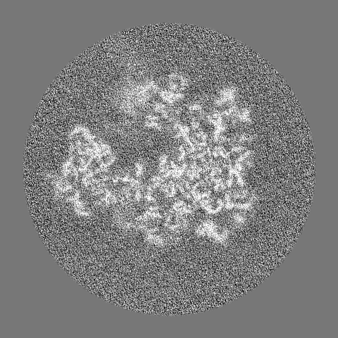
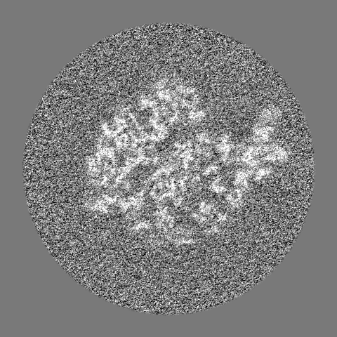
Structure of RelA bound to ribosome in absence of A/R tRNA (Structure I)
マップデータ
Ribosome*RelA structures reveal the mechanism of stringent response activation (Structure I)
試料
複合体: RelA bound to 70S ribosome in the absence of A/R tRNA
タンパク質・ペプチド: x 52種
RNA: x 6種
機能・相同性
機能・相同性情報
guanosine tetraphosphate metabolic process / GTP diphosphokinase activity / guanosine tetraphosphate biosynthetic process / GTP diphosphokinase / guanosine-3',5'-bis(diphosphate) 3'-diphosphatase activity / nucleobase-containing small molecule interconversion / negative regulation of cytoplasmic translational initiation / stringent response / response to starvation / mRNA base-pairing translational repressor activity ...guanosine tetraphosphate metabolic process / GTP diphosphokinase activity / guanosine tetraphosphate biosynthetic process / GTP diphosphokinase / guanosine-3',5'-bis(diphosphate) 3'-diphosphatase activity / nucleobase-containing small molecule interconversion / negative regulation of cytoplasmic translational initiation / stringent response / response to starvation / mRNA base-pairing translational repressor activity / ornithine decarboxylase inhibitor activity / transcription antitermination factor activity, RNA binding / misfolded RNA binding / Group I intron splicing / RNA folding / transcriptional attenuation / endoribonuclease inhibitor activity / RNA-binding transcription regulator activity / positive regulation of ribosome biogenesis / negative regulation of cytoplasmic translation / translational termination / four-way junction DNA binding / DnaA-L2 complex / translation repressor activity / negative regulation of translational initiation / negative regulation of DNA-templated DNA replication initiation / regulation of mRNA stability / translational initiation / ribosome assembly / mRNA regulatory element binding translation repressor activity / positive regulation of RNA splicing / assembly of large subunit precursor of preribosome / transcription elongation factor complex / DNA endonuclease activity / regulation of DNA-templated transcription elongation / cytosolic ribosome assembly / transcription antitermination / response to reactive oxygen species / regulation of cell growth / DNA-templated transcription termination / maintenance of translational fidelity / response to radiation / ribosomal large subunit assembly / mRNA 5'-UTR binding / large ribosomal subunit / ribosome biogenesis / ribosome binding / kinase activity / regulation of translation / ribosomal small subunit biogenesis / ribosomal small subunit assembly / small ribosomal subunit / small ribosomal subunit rRNA binding / transferase activity / 5S rRNA binding / large ribosomal subunit rRNA binding / cytosolic small ribosomal subunit / cytosolic large ribosomal subunit / tRNA binding / molecular adaptor activity / cytoplasmic translation / rRNA binding / negative regulation of translation / ribosome / structural constituent of ribosome / translation / phosphorylation / response to antibiotic / negative regulation of DNA-templated transcription / mRNA binding / GTP binding / DNA binding / RNA binding / zinc ion binding / ATP binding / membrane / plasma membrane / cytoplasm / cytosol 類似検索 - 分子機能
RelA/SpoT, AH and RIS domains / RelA/SpoT, AH and RIS domains / HD domain / RelA/SpoT family / RelA/SpoT, TGS domain / ACT domain / Region found in RelA / SpoT proteins / RelA/SpoT / Region found in RelA / SpoT proteins / TGS domain ...RelA/SpoT, AH and RIS domains / RelA/SpoT, AH and RIS domains / HD domain / RelA/SpoT family / RelA/SpoT, TGS domain / ACT domain / Region found in RelA / SpoT proteins / RelA/SpoT / Region found in RelA / SpoT proteins / TGS domain / ACT domain profile. / ACT domain / ACT-like domain / HD domain profile. / TGS domain profile. / TGS / TGS-like / HD domain / Ribosomal protein L10, eubacterial, conserved site / Ribosomal protein L10 signature. / Ribosomal protein L10 / : / Beta-grasp domain superfamily / Ribosomal protein S21, conserved site / Ribosomal protein S21 signature. / Ribosomal protein L25, short-form / Ribosomal protein S14, bacterial/plastid / Ribosomal protein L11, bacterial-type / Ribosomal protein L31 type A / Ribosomal protein S21 superfamily / Ribosomal protein S21 / Ribosomal protein S16, conserved site / Ribosomal protein S16 signature. / Ribosomal protein S21 / Ribosomal protein L31 signature. / Ribosomal protein L31 / Ribosomal protein L31 superfamily / Ribosomal protein L31 / Nucleotidyltransferase superfamily / Ribosomal protein L21, conserved site / Ribosomal protein L21 signature. / Ribosomal protein L11, conserved site / Ribosomal protein L11 signature. / Ribosomal protein L16 signature 1. / Ribosomal protein L10-like domain superfamily / : / Ribosomal protein L10 / Ribosomal protein L10P / Ribosomal protein L6, conserved site / Ribosomal protein L6 signature 1. / Ribosomal protein L16, conserved site / Ribosomal protein L16 signature 2. / Ribosomal protein L9 signature. / Ribosomal protein L9, bacteria/chloroplast / Ribosomal protein L9, C-terminal / Ribosomal protein L9, C-terminal domain / Ribosomal protein L9, C-terminal domain superfamily / Ribosomal protein L17 signature. / Ribosomal L25p family / Ribosomal protein L25 / Ribosomal protein L11, N-terminal / Ribosomal protein L11, N-terminal domain / Ribosomal protein L11/L12 / Ribosomal protein L11, C-terminal / Ribosomal protein L11, C-terminal domain superfamily / Ribosomal protein L11/L12, N-terminal domain superfamily / Ribosomal protein L11, RNA binding domain / Ribosomal protein L11/L12 / Ribosomal protein L36 signature. / Ribosomal protein L28/L24 superfamily / Ribosomal protein L25/Gln-tRNA synthetase, N-terminal / Ribosomal protein L25/Gln-tRNA synthetase, anti-codon-binding domain superfamily / Ribosomal protein L32p, bacterial type / Ribosomal protein L9, N-terminal domain superfamily / Ribosomal protein L9 / Ribosomal protein L9, N-terminal / Ribosomal protein L9, N-terminal domain / Ribosomal protein L28 / Ribosomal protein L35, conserved site / Ribosomal protein L35 signature. / Ribosomal protein L33, conserved site / Ribosomal protein L33 signature. / Ribosomal protein L35, non-mitochondrial / Ribosomal protein L5, bacterial-type / Ribosomal protein L18, bacterial-type / Ribosomal protein L6, bacterial-type / Ribosomal protein L19, conserved site / Ribosomal protein L19 signature. / Ribosomal protein S3, bacterial-type / Ribosomal protein S6, conserved site / Ribosomal protein S6 signature. / Ribosomal protein L36 / Ribosomal protein L36 superfamily / Ribosomal protein L36 / Ribosomal protein S19, bacterial-type / Ribosomal protein L9/RNase H1, N-terminal / Ribosomal protein L20 signature. / Ribosomal protein S7, bacterial/organellar-type / Ribosomal protein S11, bacterial-type / Ribosomal protein L27, conserved site 類似検索 - ドメイン・相同性
Small ribosomal subunit protein bS6 / Small ribosomal subunit protein uS7 / Large ribosomal subunit protein uL15 / Large ribosomal subunit protein uL10 / Large ribosomal subunit protein uL11 / Large ribosomal subunit protein bL19 / Large ribosomal subunit protein bL20 / Large ribosomal subunit protein bL27 / Large ribosomal subunit protein bL28 / Large ribosomal subunit protein uL29 ...Small ribosomal subunit protein bS6 / Small ribosomal subunit protein uS7 / Large ribosomal subunit protein uL15 / Large ribosomal subunit protein uL10 / Large ribosomal subunit protein uL11 / Large ribosomal subunit protein bL19 / Large ribosomal subunit protein bL20 / Large ribosomal subunit protein bL27 / Large ribosomal subunit protein bL28 / Large ribosomal subunit protein uL29 / Large ribosomal subunit protein bL31 / Large ribosomal subunit protein bL32 / Large ribosomal subunit protein bL33 / Large ribosomal subunit protein bL34 / Large ribosomal subunit protein bL35 / Large ribosomal subunit protein bL36A / Large ribosomal subunit protein bL9 / Small ribosomal subunit protein uS10 / Small ribosomal subunit protein uS11 / Small ribosomal subunit protein uS12 / Small ribosomal subunit protein uS13 / Small ribosomal subunit protein bS16 / Small ribosomal subunit protein bS18 / Small ribosomal subunit protein uS19 / Small ribosomal subunit protein bS20 / Small ribosomal subunit protein uS2 / Small ribosomal subunit protein uS3 / Small ribosomal subunit protein uS4 / Small ribosomal subunit protein uS5 / Small ribosomal subunit protein uS8 / Small ribosomal subunit protein uS9 / Large ribosomal subunit protein uL13 / Large ribosomal subunit protein uL14 / Large ribosomal subunit protein uL16 / Large ribosomal subunit protein uL23 / Small ribosomal subunit protein uS15 / GTP pyrophosphokinase / Large ribosomal subunit protein bL17 / Large ribosomal subunit protein bL21 / Large ribosomal subunit protein uL30 / Large ribosomal subunit protein uL6 / Small ribosomal subunit protein uS14 / Small ribosomal subunit protein uS17 / Large ribosomal subunit protein uL18 / Large ribosomal subunit protein uL2 / Large ribosomal subunit protein uL3 / Large ribosomal subunit protein uL24 / Large ribosomal subunit protein uL4 / Large ribosomal subunit protein uL22 / Large ribosomal subunit protein uL5 / Small ribosomal subunit protein bS21 / Large ribosomal subunit protein bL25 類似検索 - 構成要素
National Institutes of Health/National Institute of General Medical Sciences (NIH/NIGMS)
R01 GM106105
米国
Howard Hughes Medical Institute (HHMI)
米国
National Institutes of Health/National Institute of General Medical Sciences (NIH/NIGMS)
P01 GM62580
米国
引用
ジャーナル: Elife / 年: 2016 タイトル: Ribosome•RelA structures reveal the mechanism of stringent response activation. 著者: Anna B Loveland / Eugene Bah / Rohini Madireddy / Ying Zhang / Axel F Brilot / Nikolaus Grigorieff / Andrei A Korostelev / 要旨: Stringent response is a conserved bacterial stress response underlying virulence and antibiotic resistance. RelA/SpoT-homolog proteins synthesize transcriptional modulators (p)ppGpp, allowing ...Stringent response is a conserved bacterial stress response underlying virulence and antibiotic resistance. RelA/SpoT-homolog proteins synthesize transcriptional modulators (p)ppGpp, allowing bacteria to adapt to stress. RelA is activated during amino-acid starvation, when cognate deacyl-tRNA binds to the ribosomal A (aminoacyl-tRNA) site. We report four cryo-EM structures of E. coli RelA bound to the 70S ribosome, in the absence and presence of deacyl-tRNA accommodating in the 30S A site. The boomerang-shaped RelA with a wingspan of more than 100 Å wraps around the A/R (30S A-site/RelA-bound) tRNA. The CCA end of the A/R tRNA pins the central TGS domain against the 30S subunit, presenting the (p)ppGpp-synthetase domain near the 30S spur. The ribosome and A/R tRNA are captured in three conformations, revealing hitherto elusive states of tRNA engagement with the ribosomal decoding center. Decoding-center rearrangements are coupled with the step-wise 30S-subunit 'closure', providing insights into the dynamics of high-fidelity tRNA decoding.
 ムービー
ムービー コントローラー


 データを開く
 基本情報
基本情報 マップデータ
 試料
試料 機能・相同性情報
 データ登録者
 米国, 3件
  引用
引用 構造の表示
構造の表示
 ダウンロードとリンク
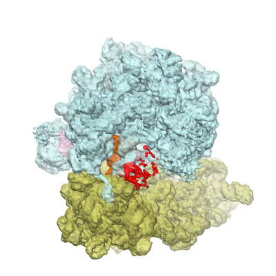 emd_8279.png
 http://ftp.pdbj.org/pub/emdb/structures/EMD-8279


 Z (Sec.)
 Y (Row.)
 X (Col.)


 試料の構成要素
 解析
解析 電子顕微鏡法
 FIELD EMISSION GUN
