 Movie
Movie Controller


+ Open data

- Basic information
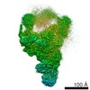
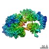
| Entry | Database: EMDB / ID: EMD-8244 | |||||||||
|---|---|---|---|---|---|---|---|---|---|---|
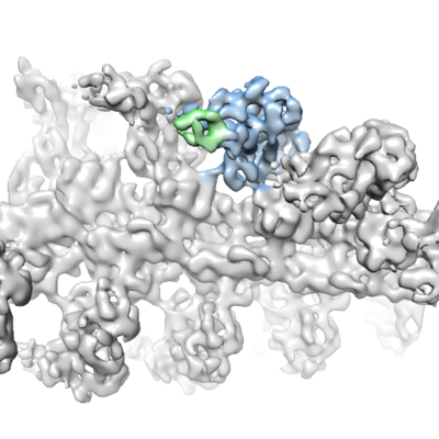
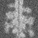
| Title | Rigor myosin X co-complexed with an actin filament | |||||||||
 Map data Map data | Rigor myosin X co-complexed with an actin filament | |||||||||
 Sample Sample |
| |||||||||
 Keywords Keywords | myosin molecular motors cytoskeletal motility / MOTOR PROTEIN | |||||||||
| Function / homology |  Function and homology information Function and homology informationplus-end directed microfilament motor activity / Netrin-1 signaling / positive regulation of cell-cell adhesion / filopodium tip / cytoskeleton-dependent intracellular transport / regulation of filopodium assembly / filopodium membrane / myosin complex / cytoskeletal motor activator activity / microfilament motor activity ...plus-end directed microfilament motor activity / Netrin-1 signaling / positive regulation of cell-cell adhesion / filopodium tip / cytoskeleton-dependent intracellular transport / regulation of filopodium assembly / filopodium membrane / myosin complex / cytoskeletal motor activator activity / microfilament motor activity / spectrin binding / tropomyosin binding / myosin heavy chain binding / mesenchyme migration / troponin I binding / filamentous actin / actin filament bundle / phosphatidylinositol-3,4,5-trisphosphate binding / skeletal muscle thin filament assembly / actin filament bundle assembly / striated muscle thin filament / skeletal muscle myofibril / actin monomer binding / skeletal muscle fiber development / stress fiber / titin binding / ruffle / actin filament polymerization / filopodium / actin filament / FCGR3A-mediated phagocytosis / Hydrolases; Acting on acid anhydrides; Acting on acid anhydrides to facilitate cellular and subcellular movement / Regulation of actin dynamics for phagocytic cup formation / calcium-dependent protein binding / actin filament binding / lamellipodium / regulation of cell shape / cell cortex / cell body / calmodulin binding / hydrolase activity / neuron projection / protein domain specific binding / neuronal cell body / calcium ion binding / positive regulation of gene expression / nucleolus / magnesium ion binding / signal transduction / ATP binding / identical protein binding / plasma membrane / cytosol / cytoplasm Similarity search - Function | |||||||||
| Biological species |  Homo sapiens (human) / Homo sapiens (human) /  | |||||||||
| Method | helical reconstruction / cryo EM / Resolution: 9.1 Å | |||||||||
 Authors Authors | Sindelar CV / Houdusse A | |||||||||
| Funding support |  United States, 1 items United States, 1 items
| |||||||||
 Citation Citation | Journal: Nat Commun / Year: 2016 Title: The myosin X motor is optimized for movement on actin bundles. Authors: Virginie Ropars / Zhaohui Yang / Tatiana Isabet / Florian Blanc / Kaifeng Zhou / Tianming Lin / Xiaoyan Liu / Pascale Hissier / Frédéric Samazan / Béatrice Amigues / Eric D Yang / Hyokeun ...Authors: Virginie Ropars / Zhaohui Yang / Tatiana Isabet / Florian Blanc / Kaifeng Zhou / Tianming Lin / Xiaoyan Liu / Pascale Hissier / Frédéric Samazan / Béatrice Amigues / Eric D Yang / Hyokeun Park / Olena Pylypenko / Marco Cecchini / Charles V Sindelar / H Lee Sweeney / Anne Houdusse /   Abstract: Myosin X has features not found in other myosins. Its structure must underlie its unique ability to generate filopodia, which are essential for neuritogenesis, wound healing, cancer metastasis and ...Myosin X has features not found in other myosins. Its structure must underlie its unique ability to generate filopodia, which are essential for neuritogenesis, wound healing, cancer metastasis and some pathogenic infections. By determining high-resolution structures of key components of this motor, and characterizing the in vitro behaviour of the native dimer, we identify the features that explain the myosin X dimer behaviour. Single-molecule studies demonstrate that a native myosin X dimer moves on actin bundles with higher velocities and takes larger steps than on single actin filaments. The largest steps on actin bundles are larger than previously reported for artificially dimerized myosin X constructs or any other myosin. Our model and kinetic data explain why these large steps and high velocities can only occur on bundled filaments. Thus, myosin X functions as an antiparallel dimer in cells with a unique geometry optimized for movement on actin bundles. | |||||||||
| History |
|
- Structure visualization
| Movie |
Movie viewer |
|---|---|
| Structure viewer | EM map: SurfViewMolmilJmol/JSmol |
| Supplemental images |
- Downloads & links
-EMDB archive
| Map data | emd_8244.map.gz | 7.5 MB | EMDB map data format | |
|---|---|---|---|---|
| Header (meta data) | emd-8244-v30.xmlemd-8244.xml | 18.4 KB 18.4 KB | Display Display | EMDB header |
| FSC (resolution estimation) | emd_8244_fsc.xml | 5.4 KB | Display | FSC data file |
| Images |  emd_8244.png emd_8244.png | 217.3 KB | ||
| Masks | emd_8244_msk_1.map | 8 MB | Mask map | |
| Filedesc metadata | emd-8244.cif.gz | 6.5 KB | ||
| Others | emd_8244_half_map_1.map.gzemd_8244_half_map_2.map.gz | 7.4 MB 7.4 MB | ||
| Archive directory |  http://ftp.pdbj.org/pub/emdb/structures/EMD-8244ftp://ftp.pdbj.org/pub/emdb/structures/EMD-8244 http://ftp.pdbj.org/pub/emdb/structures/EMD-8244ftp://ftp.pdbj.org/pub/emdb/structures/EMD-8244 | HTTPS FTP |
-Validation report
| Summary document | emd_8244_validation.pdf.gz | 1012 KB | Display | EMDB validaton report |
|---|---|---|---|---|
| Full document | emd_8244_full_validation.pdf.gz | 1011.6 KB | Display | |
| Data in XML | emd_8244_validation.xml.gz | 10.4 KB | Display | |
| Data in CIF | emd_8244_validation.cif.gz | 13.9 KB | Display | |
| Arichive directory | https://ftp.pdbj.org/pub/emdb/validation_reports/EMD-8244ftp://ftp.pdbj.org/pub/emdb/validation_reports/EMD-8244 | HTTPS FTP |
-Related structure data
| Related structure data |  5kg8MC  5hmoC  5hmpC  5i0hC  5i0iC M: atomic model generated by this map C: citing same article ( |
|---|---|
| Similar structure data |
-Links
| EMDB pages | EMDB (EBI/PDBe) / EMDataResource |
|---|---|
| Related items in Molecule of the Month |


-Map
| File | Download / File: emd_8244.map.gz / Format: CCP4 / Size: 8 MB / Type: IMAGE STORED AS FLOATING POINT NUMBER (4 BYTES) | ||||||||||||||||||||||||||||||||||||||||||||||||||||||||||||||||||||
|---|---|---|---|---|---|---|---|---|---|---|---|---|---|---|---|---|---|---|---|---|---|---|---|---|---|---|---|---|---|---|---|---|---|---|---|---|---|---|---|---|---|---|---|---|---|---|---|---|---|---|---|---|---|---|---|---|---|---|---|---|---|---|---|---|---|---|---|---|---|
| Annotation | Rigor myosin X co-complexed with an actin filament | ||||||||||||||||||||||||||||||||||||||||||||||||||||||||||||||||||||
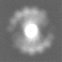
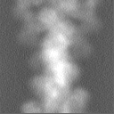
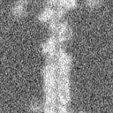
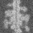
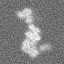
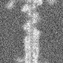
| Projections & slices | Image control
Images are generated by Spider. | ||||||||||||||||||||||||||||||||||||||||||||||||||||||||||||||||||||
| Voxel size | X=Y=Z: 1.868 Å | ||||||||||||||||||||||||||||||||||||||||||||||||||||||||||||||||||||
| Density |
| ||||||||||||||||||||||||||||||||||||||||||||||||||||||||||||||||||||
| Symmetry | Space group: 1 | ||||||||||||||||||||||||||||||||||||||||||||||||||||||||||||||||||||
| Details | EMDB XML:
CCP4 map header:
| ||||||||||||||||||||||||||||||||||||||||||||||||||||||||||||||||||||
 Z (Sec.)
 Y (Row.)
 X (Col.)


-Supplemental data
-Mask #1
| File | emd_8244_msk_1.map | ||||||||||||
|---|---|---|---|---|---|---|---|---|---|---|---|---|---|
| Projections & Slices |
| ||||||||||||
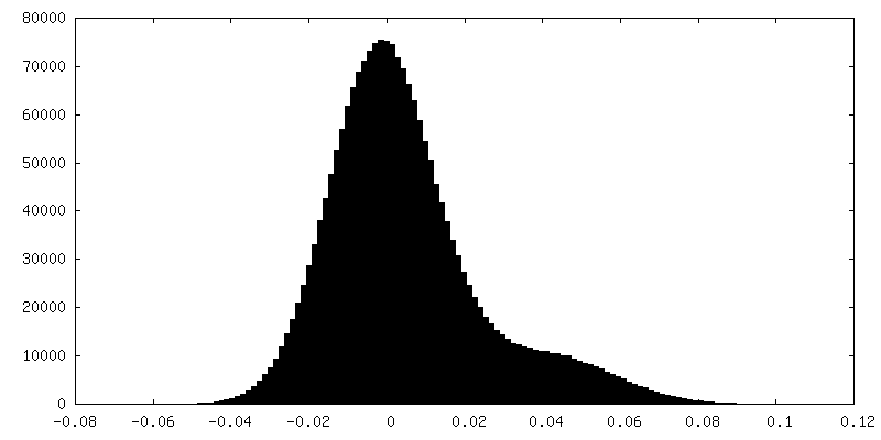
| Density Histograms |


-Half map: Rigor myosin X co-complexed with an actin filament, half map #1
| File | emd_8244_half_map_1.map | ||||||||||||
|---|---|---|---|---|---|---|---|---|---|---|---|---|---|
| Annotation | Rigor myosin X co-complexed with an actin filament, half map #1 | ||||||||||||
| Projections & Slices |
| ||||||||||||
| Density Histograms |


-Half map: Rigor myosin X co-complexed with an actin filament, half map #2
| File | emd_8244_half_map_2.map | ||||||||||||
|---|---|---|---|---|---|---|---|---|---|---|---|---|---|
| Annotation | Rigor myosin X co-complexed with an actin filament, half map #2 | ||||||||||||
| Projections & Slices |
| ||||||||||||
| Density Histograms |


- Sample components
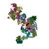
-Entire : Actin filament decorated by the myosin X motor domain
| Entire | Name: Actin filament decorated by the myosin X motor domain |
|---|---|
| Components |
|
-Supramolecule #1: Actin filament decorated by the myosin X motor domain
| Supramolecule | Name: Actin filament decorated by the myosin X motor domain / type: organelle_or_cellular_component / ID: 1 / Parent: 0 / Macromolecule list: all |
|---|
-Macromolecule #1: Unconventional myosin-X
| Macromolecule | Name: Unconventional myosin-X / type: protein_or_peptide / ID: 1 / Number of copies: 1 / Enantiomer: LEVO |
|---|---|
| Source (natural) | Organism: Homo sapiens (human) |
| Molecular weight | Theoretical: 85.083086 KDa |
| Recombinant expression | Organism:  |
| Sequence | String: NFFTEGTRVW LRENGQHFPS TVNSCAEGIV VFRTDYGQVF TYKQSTITHQ KVTAMHPTNE EGVDDMASLT ELHGGSIMYN LFQRYKRNQ IYTYIGSILA SVNPYQPIAG LYEPATMEQY SRRHLGELPP HIFAIANECY RCLWKRHDNQ CILISGESGA G KTESTKLI ...String: NFFTEGTRVW LRENGQHFPS TVNSCAEGIV VFRTDYGQVF TYKQSTITHQ KVTAMHPTNE EGVDDMASLT ELHGGSIMYN LFQRYKRNQ IYTYIGSILA SVNPYQPIAG LYEPATMEQY SRRHLGELPP HIFAIANECY RCLWKRHDNQ CILISGESGA G KTESTKLI LKFLSVISQQ SLELSLKEKT SCVERAILES SPIMEAFGNA KTVYNNNSSR FGKFVQLNIC QKGNIQGGRI VD YLLEKNR VVRQNPGERN YHIFYALLAG LEHEEREEFY LSTPENYHYL NQSGCVEDKT ISDQESFREV ITAMDVMQFS KEE VREVSR LLAGILHLGN IEFITAGGAQ VSFKTALGRS AELLGLDPTQ LTDALTQRSM FLRGEEILTP LNVQQAVDSR DSLA MALYA CCFEWVIKKI NSRIKGNEDF KSIGILDIFG FENFEVNHFE QFNINYANEK LQEYFNKHIF SLEQLEYSRE GLVWE DIDW IDNGECLDLI EKKLGLLALI NEESHFPQAT DSTLLEKLHS QHANNHFYVK PRVAVNNFGV KHYAGEVQYD VRGILE KNR DTFRDDLLNL LRESRFDFIY DLFEHVSSRN NQDAAAAAAA ARRPTVSSQF KDSLHSLMAT LSSSNPFFVR CIKPNMQ KM PDQFDQAVVL NQLRYSGMLE TVRIRKAGYA VRRPFQDFYK RYKVLMRNLA LPEDVRGKCT SLLQLYDASN SEWQLGKT K VFLRESLEQK LEKRREEE UniProtKB: Unconventional myosin-X |
-Macromolecule #2: Actin, alpha skeletal muscle
| Macromolecule | Name: Actin, alpha skeletal muscle / type: protein_or_peptide / ID: 2 / Number of copies: 3 / Enantiomer: LEVO |
|---|---|
| Source (natural) | Organism: |
| Molecular weight | Theoretical: 41.827609 KDa |
| Sequence | String: DEDETTALVC DNGSGLVKAG FAGDDAPRAV FPSIVGRPRH QGVMVGMGQK DSYVGDEAQS KRGILTLKYP IECGIITNWD DMEKIWHHT FYNELRVAPE EHPTLLTEAP LNPKANREKM TQIMFETFNV PAMYVAIQAV LSLYASGRTT GIVLDSGDGV T HNVPIYEG ...String: DEDETTALVC DNGSGLVKAG FAGDDAPRAV FPSIVGRPRH QGVMVGMGQK DSYVGDEAQS KRGILTLKYP IECGIITNWD DMEKIWHHT FYNELRVAPE EHPTLLTEAP LNPKANREKM TQIMFETFNV PAMYVAIQAV LSLYASGRTT GIVLDSGDGV T HNVPIYEG YALPHAIMRL DLAGRDLTDY LMKILTERGY SFVTTAEREI VRDIKEKLCY VALDFENEMA TAASSSSLEK SY ELPDGQV ITIGNERFRC PETLFQPSFI GMESAGIHET TYNSIMKCDI DIRKDLYANN VMSGGTTMYP GIADRMQKEI TAL APSTMK IKIIAPPERK YSVWIGGSIL ASLSTFQQMW ITKQEYDEAG PSIVHRKCF UniProtKB: Actin, alpha skeletal muscle |
-Experimental details
-Structure determination
| Method | cryo EM |
|---|---|
 Processing Processing | helical reconstruction |
| Aggregation state | filament |
-Sample preparation
| Concentration | 1.7 mg/mL |
|---|---|
| Buffer | pH: 6.8 / Component - Concentration: 5.0 mM / Component - Name: MOPS |
| Grid | Model: Quantifoil / Material: COPPER / Mesh: 300 / Support film - Material: CARBON / Support film - topology: HOLEY |
| Vitrification | Cryogen name: ETHANE / Instrument: HOMEMADE PLUNGER |
- Electron microscopy
| Microscope | FEI TECNAI F20 |
|---|---|
| Image recording | Film or detector model: GATAN K2 SUMMIT (4k x 4k) / Detector mode: COUNTING / Number grids imaged: 2 / Number real images: 154 / Average exposure time: 13.0 sec. / Average electron dose: 50.0 e/Å2 |
| Electron beam | Acceleration voltage: 200 kV / Electron source:  FIELD EMISSION GUN FIELD EMISSION GUN |
| Electron optics | C2 aperture diameter: 70.0 µm / Calibrated defocus max: 5.251 µm / Calibrated defocus min: 1.438 µm / Calibrated magnification: 26780 / Illumination mode: FLOOD BEAM / Imaging mode: BRIGHT FIELD / Cs: 2.0 mm / Nominal defocus max: 5.0 µm / Nominal defocus min: 1.0 µm |
| Sample stage | Specimen holder model: GATAN 626 SINGLE TILT LIQUID NITROGEN CRYO TRANSFER HOLDER Cooling holder cryogen: NITROGEN |
| Experimental equipment |  Model: Tecnai F20 / Image courtesy: FEI Company |
-Image processing
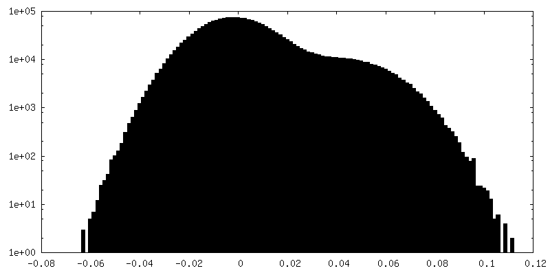
| Final reconstruction | Number classes used: 1 Applied symmetry - Helical parameters - Δz: 27.44 Å Applied symmetry - Helical parameters - Δ&Phi: 167.1 ° Applied symmetry - Helical parameters - Axial symmetry: C1 (asymmetric) Algorithm: FOURIER SPACE / Resolution.type: BY AUTHOR / Resolution: 9.1 Å / Resolution method: FSC 0.143 CUT-OFF / Software - Name: FREALIGN (ver. 8.06) Details: Symmetry was not applied in this reconstruction. The provided symmetry parameters are nominal, and were not actually used. Number images used: 57927 |
|---|---|
| Startup model | Type of model: INSILICO MODEL In silico model: 40 Angstrom low-pass filtered model of actomyosin |
| Final angle assignment | Type: NOT APPLICABLE / Software - Name: FREALIGN (ver. 8.06) |
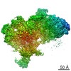
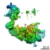
| FSC plot (resolution estimation) | 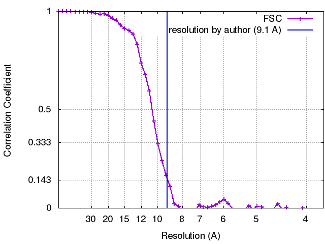 |
